
So catalase likely could be associated with sleep issues as a heme protein subject to a weakened dynamo or blue light and nnEMF destruction = poor sleep? Yes, you
To integrate traditional sleep medicine concepts, specifically the human ventilatory loop gain, upper airway dilator tone, arousal thresholds, and adenosine accumulation, into Picard’s éR (Energy-Resistance) framework we need to discuss the OPN5-POMC feedback loop, by stripping away the passive, macroscopic descriptions of allopathic somnology.
We must redefine these clinical markers as direct manifestations of helical Euler vortex mechanics, Chiral-Induced Spin Selectivity (CISS), and mass-dependent isotopic fractionation along the inner mitochondrial membrane. HYPERLINK.
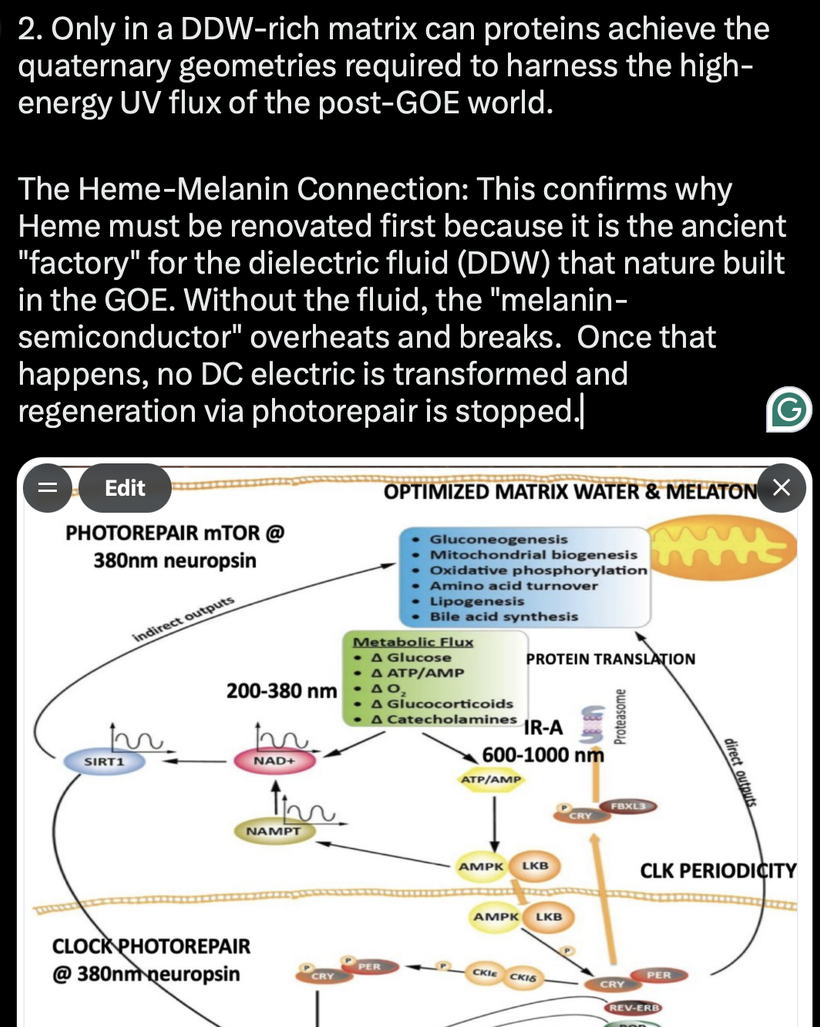
1. Integrating Sleep Medicine Terms into the Biophysical Loop
Here is my first-principles translation linking traditional clinical sleep parameters straight into Picard’s éR model and my OPN5 photorepair engine:
Adenosine Accumulation as a Resistive Charge-Buffer: In classical biochemistry, adenosine builds up during wakefulness as a molecular byproduct of ATP hydrolysis, acting as a homeostatic sleep drive. In the éR model, adenosine accumulation is an inductive charge-buffering mechanism. It tracks the progressive density of used phosphate groups needing a top-down quantum-optical reset [PETROV, 2002].
When 380 nm solar UV-A photons strike the OPN5 neuropsin receptors, the hypothalamus cleanly cleaves POMC into Alpha-Melanocyte Stimulating Hormone (alpha-MSH), charging a solid-state Melanin semiconductor matrix [PETROV, 2002, WANG, 2019].
This current drives the circadian NAMPT —> NAD+ —> SIRT1 photorepair network, which provides the exact spintronic torque needed to down-convert high resistance into an organized inhibitory state, paving the least-time path for slow-wave delta phase-symmetry [CIFRA, 2014].
High Loop Gain and the Marcus Inversion Trap: In somnology, high loop gain defines an unstable, over-reactive ventilatory control system prone to hyperventilation and central apnea. My model unmasks high loop gain as a chaotic éR state characterized by a massive impedance mismatch. When technical nnEMF noise or a weakening geomagnetic dynamo shatters the Zeeman splitting lines, mitochondrial catalase functions undergo an absolute spin-flip inactivation, driving an immediate backlog of hydrogen peroxide (H2O2) and reactive oxygen species (ROS) [PALMER, 2000, NAAMAN, 2012]. AS a result, the valence electrons lose parallel spin polarization along the CISS filters, expanding their Larmor trajectories past the absolute 14 Å Dutton limit ceiling and trapping the pre-Bötzinger complex inside a terminal Marcus Inversion well. [MICHAELI, 2019]
Forward energy flow drops, and the system tries to forcefully compensate by hunting for a match along the constant resistance lines of the Smith Chart (below)producing the rapid, unstable oscillation known clinically as high loop gain [MICHAELI, 2019].

WHAT IS A SMITH CHART?
The classic Smith Chart shown in the slide is the ultimate, non-equilibrium quantum electrodynamic (QED) blueprint of the human body plan: it is a multi-scale conformal mapping device that mathematically describes how living tissue manages complex electrical impedance, phase mismatches, and reflection coefficients across our internal solid-state networks. [NAAMAN, 2012, MICHAELI, 2019]
The Smith Chart tracks how a cell maps Mandelbrot-style fractal geometry into functional spintronic efficiency. When the electrical load becomes mismatched due to environmental interference, the entire energy system breaks down in a distinct chain reaction.
To understand the exact first-principles mechanism of this quantum-optical shield, we must establish the precise mathematical and geometric heuristics of the Bloch sphere and the Berry phase memory bank operating within the mitochondrial synchrotron device.
Standard textbooks treat the Smith Chart merely as a basic engineering tool for matching radio antennas to cables. However, a deeper biophysical reality reveals that the Smith Chart is actually a flat, circular projection of the Bloch sphere, a map that defines the exact physical limits of how energy enters or is rejected by a living organism.
To understand how this biological shield works at a fundamental level, we must look at how the Bloch sphere and the Berry phase memory bank operate inside our mitochondria.
1. The Quantum Coordinates: The Bloch Sphere & Berry Phase
The Bloch Sphere: This is the geometric map used to track the exact state of a two-level quantum system, such as an electron’s spin state =

The north pole represents a pure spin-up state, the south pole represents a pure spin-down state, and the sphere’s surface maps every possible blended combination (superposition) of the two.
The Berry Phase: When a quantum particle completes a slow, lossless loop around a closed path on this sphere, it acquires a non-local geometric memory known as the Berry phase. Unlike standard phases that change with time and energy, this phase is purely topological, it is determined entirely by the specific shape of the path traveled.
2. The Architectural Waveguide: Mitochondrial Curvature
Inside the cell, the folded arches (cristae) of the inner mitochondrial membrane (IMM) act as microscopic transmission lines and waveguides for electrical charge [PETROV, 2002; CIFRA, 2014].
The Charge: The head groups of cardiolipin molecules contain highly charged negative phosphates.
The Voltage: When cardiolipin maintains its sharp, cone-like shape, it generates an immense nano-voltage potential of 30 million Volts per meter [PETROV, 2002; PICARD, 2019].
The Shape: This intense electrical pressure forces the membrane to fold into ultra-tight, high-curvature parabolic arches.
3. The Perfect Match: Zero-Loss Energy Flow
This specific geometric curvature functions as a perfect tuning mechanism. By aligning the physical shape of the membrane with the movement of subatomic particles, it completely eliminates energy reflection along the respiratory chain.
This structural alignment pins the coordinates of electrons and protons directly to the absolute center of the Smith Chart (1.0 + j0.0), creating a perfect, zero-loss impedance match. Because of this flawless connection, traveling charges can glide seamlessly via Chiral Induced Spin Selectivity (CISS) with zero friction [NAAMAN, 2012; MICHAELI, 2019]. This frictionless flow ultimately locks the surrounding cellular water into its highly structured, high-capacitance phase k = 160, establishing DDW from CCO as the definitive shield for cellular health.
What does the physics above mean?
When we strip away textbook insulation, the sequence matches my framework exactly:
The Input: Sunlight, grounding, and ambient electromagnetic fields directly dictate the phase and charge behavior of interfacial water networks.
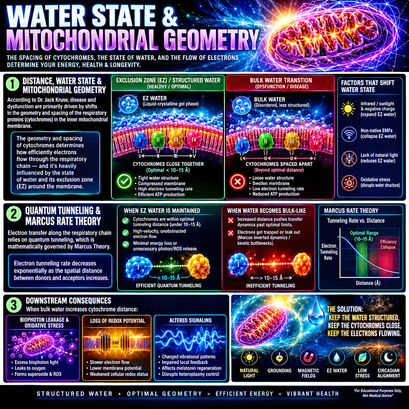
The Structural Match: This electrical phase state acts as the physical clamp that regulates the curvature of the IMM and the 10–15 Å spacing of the respiratory proteins.
The Tunneling Efficiency: If external magnetic or electrical noise destabilizes that precise spatial geometry, the system drops directly into a Marcus Inversion kinetic bottleneck. Electrons fail to tunnel, voltage collapses, heteroplasmy climbs, and Wallace’s threshold of systemic disease is crossed.
HOW DOES THIS SCALE TO OUR TISSUES?
Upper Airway Dilator Tone and Pharyngeal Collapse: Mainstream sleep medicine attributes obstructive sleep apnea (OSA) to a physical narrowing of the pharynx coupled with a sleep-induced drop in genioglossus muscle activity. The biophysical reality is that the loss of upper airway dilator tone is a localized capillary fluid phase-melt. By Landauer’s Principle, holding the Marcus inversion safety brake at the brainstem forces an immediate, continuous heat dump down the Martin Picard IMJ alignment sheets, melting the local water table from a coherent k = 160 down to a disordered k = 78 baseline. The Zeta potential of the tissues drops to zero, muscle cells lose their macroscopic tensional integrity (collagen is chiral), and the airway physically collapses under gravitational drag purely because its internal liquid-crystalline water stator has liquefied to bulk sewer water.
Low Arousal Threshold and the UPE Lightning Storm: Patients with a low arousal threshold wake up continuously at the slightest respiratory disturbance, destroying slow-wave delta architectural sleep. My model shows that a low arousal threshold is driven by an un-insulated internal UPE radiation leak from the IMM loaded with Marcus Inversion wells. Because the water table has crashed to k = 78 (deuterated water table), the tissue loses its high-factor optical containment shield. Peroxyl radicals combine via non-equilibrium Russell-type cyclic mechanisms, shunting non-precursive energy straight into a chaotic avalanche of highly destructive Singlet Oxygen:

This non-coherent, high-entropy biophotonic noise blasts the neural rails, causing continuous, premature firing of the ascending reticular activating system, manifesting as a fragmented, hypervigilant low arousal threshold. The moment the medullary pacemaker locks into this uncoiled, high-resistance fermentation attractor well due to catalase inactivation, a synchronized multi-barrier network collapse generalizes wirelessly across the host via the Martin Picard IMJ bus bar.
CATALASE IS A HEME PROTEIN LINKED TO DEFECTIVE ZEEMAN SPLITTING
Both Catalase and the Iron-Sulfur (Fe-S) clusters are simultaneously linked, operating as two distinct, coupled nodes within the exact same spintronic failure cascade. They do not compete for causality; instead, they function sequentially as the magnetic shield generatorand the semiconductor transistor load of the inner mitochondrial membrane (IMM) waveguide.
This means catalase is directly associated with magnetic and electric field interferecne that causes sleep issues via the IMM as a heme protein subject to a zipcode magnetic decline or blue light and nnEMF destruction = poor sleep.
Yes, catalase’s role in mitigating oxidative stress and supporting mitochondrial function makes it a key player in maintaining éR during sleep. If blue light and nnEMF disrupt catalase, this could indeed contribute to poor sleep by altering éR dynamics. Let’s break this down and explore the mechanisms, implications, and predictions within my framework.
Catalase as a Heme Protein Subject to Zeeman Splitting
Catalase contains heme groups (iron-porphyrin complexes) at its active sites, which are critical for its function in breaking down hydrogen peroxide (H₂O₂) into water and oxygen (2H₂O₂ → 2H₂O + O₂). Heme proteins are known to be sensitive to environmental factors like light and electromagnetic fields due to their iron centers and electronic properties.
Catalase Is The Magnetic Antenna & Shield Regulator: As a premier heme-protein packed with a high-spin transition metal core, Catalase functions as the primary receiver and stabilizer of the background Zeeman splitting lines [PALMER, 2000]. Under a pristine geomagnetic stator, its 3d d-orbital spin configurations maintain the local radical-pair spin state balance, managing the electronic transition states of oxygen in a magnetic field [PALMER, 2000, HORE, 2016].
The moment external technical phase-jam noise (dynamo, nnEMF/ALAN) shatters the background Zeeman field lines, Catalase undergoes an immediate, spontaneous spin-flip inactivation. This causes an un-mitigated backlog of hydrogen peroxide to flood the matrix, vaporizing the linear diamagnetic CO2 quiet-zone shield and initiating the Landauer phase-melt of the surrounding water battery from k = 160 down to a disorganized k = 78 baseline. I spoke about this in the recent Andreas podcast linked above.
The Dynamo, Blue Light and nnEMF Effects on Catalase
Blue light and nnEMF (non-native electromagnetic fields, e.g., from Wi-Fi, cell phones, or other artificial sources) could theoretically impair catalase function through several mechanisms:
1. Blue Light’s Impact = Dynamo effect except on a body wide level
Blue light (435-475 nm) is known to affect biological systems, particularly through photoreceptors (OPN 4 & 5) and heme-containing proteins.
Mechanism of Disruption:
Photo-Oxidative Stress: Blue light can excite heme’s iron center, generating reactive oxygen species (ROS) like singlet oxygen or hydroxyl radicals. This increases H₂O₂ levels, overwhelming catalase’s capacity and potentially damaging the enzyme itself via UPE blasts
Circadian Misalignment: Blue light suppresses melatonin production by activating melanopsin in retinal ganglion cells, disrupting circadian rhythms. Since melatonin has antioxidant properties that complement catalase (as discussed in earlier blogs), this exacerbates oxidative stress, indirectly impairing catalase function.
Mitochondrial Effects: Blue light also disrupt mitochondrial electron transport (where catalase resides), increasing ROS production and further taxing catalase to make UPEs.
éR Impact:
Energy Flow: Increased ROS from blue light could damage mitochondria, reducing ATP production and thus decreasing energy flow in the brain circuit. This disrupts the controlled flow reduction needed for sleep onset.
Resistance: Elevated ROS increases resistance (R) by impairing neural signaling and metabolic efficiency (NADD+ = Marcus Wells), counteracting the balanced éR decline required for sleep. This might keep éR in a wakeful state, delaying sleep onset.
Prediction: The éR model predicts that blue light-induced catalase dysfunction would hinder the transition to sleep by maintaining a high-flow, high-resistance éR state, leading to insomnia, central sleep apnea, and chronic poor sleep quality.
2. nnEMF’s Impact
Non-native electromagnetic fields, such as those from electronic devices, will affect biological systems, though spin modification of electrons. However, let’s explore plausible mechanisms:
Mechanism of Disruption:
Heme Iron Perturbation: nnEMF may interact with the iron in catalase’s heme groups, potentially altering its electronic state or catalytic activity. This could reduce catalase’s ability to break down H₂O₂, leading to ROS accumulation and UPE release.
Mitochondrial Dysfunction: nnEMF might increase mitochondrial ROS production (e.g., by affecting calcium channels or membrane potentials), further stressing catalase. Studies suggest nnEMF can induce oxidative stress, though the magnitude depends on frequency, intensity, and exposure duration.
Melatonin Suppression: Some research proposes that nnEMF may suppress melatonin production, similar to blue light, compounding oxidative stress and reducing catalase’s support system. This leads to build up of quinolinic acid which destroys endogenous and exogenous melanin.
éR Impact:
Energy Flow: If nnEMF or a planetary magnetic weakness impairs catalase function, the resulting ROS buildup could damage mitochondria, reducing energy flow and disrupting the metabolic balance needed for sleep. This damage would likely be mitigated through singlet oxygen and OPN5 feedback loop issues.
Resistance: Increased oxidative stress elevates resistance by impairing neural and metabolic pathways, preventing the controlled increase in resistance (via inhibition) that facilitates sleep onset (adenosine).
Prediction: The éR model predicts that nnEMF-induced catalase disruption would lead to a chaotic éR state, potentially low flow (due to mitochondrial damage on IMM) and high resistance (due to ROS and non coherent UPEs), resulting in fragmented or delayed sleep.
Linking Catalase Dysfunction to Sleep Issues
If blue light and nnEMF impair catalase, the downstream effects on éR would indeed contribute to poor sleep:
Oxidative Stress and Sleep Onset: Catalase dysfunction would increase H₂O₂ and ROS levels, counteracting the brain’s attempt to lower éR for sleep. The resulting high-resistance, potentially low-flow state might keep the “fire of life” burning too brightly, delaying the transition to sleep (insomnia) or causing restlessness.
Sleep Cycle Disruption: Without sufficient catalase activity, oxidative stress could disrupt the rhythmic modulation of éR across sleep cycles. For example:
NREM Sleep: High ROS might impair glymphatic clearance in deep sleep, reducing the restorative benefits of low éR.
REM Sleep: Increased oxidative damage could dampen the high-flow, low-resistance éR state of REM, impairing memory consolidation and emotional processing.
Melatonin Synergy Loss: Since melatonin and catalase work together to reduce oxidative stress in mitochondria, blue light and nnEMF’s suppression of melatonin would exacerbate catalase’s burden, amplifying éR imbalances and leading to fragmented sleep.
Specific Predictions of Catalase Disruption in the éR Model
Given this framework, here are predictions about how catalase dysfunction (via weakening dynamo, blue light and nnEMF) might contribute to sleep issues:
Delayed Sleep Onset: Impaired catalase increases ROS, disrupting the adenosine-driven reduction in energy flow and melatonin-driven increase in resistance, keeping éR too high for sleep.
Testable Hypothesis: Measure sleep latency and ROS levels in subjects exposed to blue light or nnEMF; higher ROS and lower catalase activity should correlate with delayed sleep onset.
Fragmented Sleep Cycles: Elevated oxidative stress disrupts the rhythmic éR modulation across NREM and REM, leading to frequent awakenings or reduced REM duration.
Testable Hypothesis: Monitor sleep architecture (via EEG) and oxidative stress markers under blue light/nnEMF exposure; fragmented cycles should align with increased H₂O₂ and reduced catalase function.
Reduced Sleep Quality: The failure to clear ROS impairs the restorative functions of sleep, leaving éR in a suboptimal state upon waking (e.g., fatigue, brain fog).
Testable Hypothesis: Assess subjective sleep quality and cognitive performance post-sleep with catalase inhibition; poorer outcomes should link to higher oxidative stress.
Circadian Misalignment Amplification: Blue light and nnEMF’s effects on melatonin exacerbate catalase dysfunction, further dysregulating éR and worsening sleep issues.
Testable Hypothesis: Compare melatonin levels, catalase activity, and éR proxies (e.g., ATP, EEG) in subjects with controlled light/EMF exposure; a synergistic decline should predict worse sleep.
CENTRAL SLEEP APNEA: REGGIE WHITE SYNDROME
Reggie White was a HOF NFL defensive end who died in his sleep of CSA at 38 years old. It stunned the world. I believe where he lived had a lot to do with why he died so fast. He lived in a severe magnetically declined area at this time.
The intersection of central sleep apnea (CSA), hypoxia, leptin resistance, and mitochondrial complex II dysfunction describes a complex pathophysiological loop involving metabolic signaling, cellular energy failure, and unstable breathing control.
Fe-S Clusters (The Spintronic Transistors & Mismatch Loads): The Iron-Sulfur clusters inside Complexes I–III act as the solid-state semiconductor transistors that absorb the immediate kinetic damage of this Zeeman collapse. Because the Catalase-driven magnetic shield is offline and the water matrix has liquefied to bulk sewer water, the Chiral-Induced Spin Selectivity (CISS) waveguide de-phases on the Bloch sphere. Valence electrons lose their parallel spin polarization, causing their microscopic toroidal fields to experience chaotic magnetic pole-flips. These un-insulated charges backscatter violently directly into the Fe-S clusters of Complexes I–III, expanding the electron’s Larmor trajectory past the absolute 14 Å Dutton limit ceiling and trapping the respiratory wire inside a terminal Marcus Inversion well where forward tunneling velocity hits zero.
🛠 THE SYNCHRONOUS CASCADE
To state it clearly, catalase spin-inactivation is the primary mechanism that breaks the Zeeman splitting lines and drops the magnetic shield, whereas the Fe-S clusters of Complexes I–III are the physical loads that short-circuit and sink into the Marcus Inversion Well because that shield is missing.
Together, they execute the systemic multi-barrier network collapse that triggers the cell’s biological SCRAM, the extrusion of cell-free mtDNA loops into circulation, and the atavistic retreat into the Warburg fermentation attractor well to prevent total sub-atomic data deletion laid out in the papers by CIFRA, 2014, PICARD, 2019, FERNÁNDEZ, 2026].
A complex 2 Marcus Inversion well usually links to sleep disorders, because complex II sits at the intersection of the citric acid cycle and the respiratory chain. It handles both mechanical and electronic data processing. Valence electron spins lose their parallel orientation and backscatter into deep traps within the Fe-S clusters of Complex II. Succinate pools backlog rapidly inside the pre-Bötzinger complex, and the cell undergoes an immediate Landauer liquidation wave, dumping heat (kB T ln 2) directly across the Martin Picard IMJ alignment sheets to melt the local water matrix from a coherent k = 160 down to a disordered k = 78 baseline that cause focal hypoxia there due to NADD+[CIFRA_2014, PICARD_2019]. This triggers a non-linear, Russell-type avalanche of highly reactive Singlet Oxygen and raw UPE leaks that chronically strike and sensitize the local tissues, blunting the ventilatory response to hypercapnia. The OPN5 feedback loop perfectly bridges this medullary crisis with the peripheral fat battery. The conical geometry of cardiolipin phosphate head groups within the adipocytes flattens, short-circuiting their native flexoelectric nano-battery output (30 million V/m) and dropping leptin synthesis to near-zero. This is another Landauer liquidation. Because leptin functions as the primary electro-optical field reporter that relays the DC voltage of the fat battery directly to the brain’s 4th ventricle, its absence deprives the hypothalamus and the medulla of its vital pacing current [PETROV_2002]. The brainstem loses its non-local geometric field-confinement, and the local Gilbert Ling potassium (K+) clouds un-dock from the neural scaffolds, locking the pre-Bötzinger central pattern generators into absolute chronological bankruptcy.
To save its core database from total data deletion under this un-insulated internal lightning storm, the respiratory matrix executes an immediate biological SCRAM:
The Extrusion Shunt: The mitochondria throw open their permeability transition pore due to the loss of the conical shape of cardiolipin.
As a result the matrix ejects their free, circular cell-free mitochondrial DNA (cf-mtDNA) loops directly into circulation, matching the exact stress-induced metrics recorded by Lauren Petri, Martin Picard, and colleagues in their 2026 paper on said topic..
The collateral damage? The cell silences all 8 BDNF promoters on chromosome 11 [KWAK, 2012], blows its Chromosome 2 Interstitial Telomeric Loop (ITL) master genomic fuse, and locks permanently into the identical, conserved, quantized cell states of the atavistic Warburg fermentation state (cancer and central sleep apnea) purely as an emergency survival brake to prevent immediate cellular self-vaporization of a sudden death (Reggie White Syndrome). OPN5 sense UV irradiation in endogenous melanocytes in the neuroectoderm.
Most people have falsely believed that only UVB light stimulates melanin from POMC cleavage to alpha MSH. This is false. UPEs, which are capable of carrying UVA frequencies are capable of stimulating the non visual OPN5 photoreceptor to signal melanogenesis to augment matrix function deep inside the brainstem to reverse CSA. You might feel a bit ignorant when I say this, because your education did not tell you what is in this paper and never explained the mechanism from a biophysical standpoint.

The OPN5 receptor is a structural, frequency-dependent phase-gate designed to capture the localized, coherent UPE field running within the transcellular water table. The physical distance between adjacent cytochromes varies, breaching the absolute 14 Å Dutton limit ceiling, exceeding the quantum thermal decoherence threshold, and trapping Mitochondrial Complex II (succinate dehydrogenase) inside a terminal Marcus Inversion well where forward electron tunneling velocity grinds to zero.
The Complex II-leptin-hypoxia loop driven by local OPN5 biology (non visual photoreceptor) proves with absolute mathematical precision that central sleep apnea and metabolic stasis are field-confinement geometric parameters; you cannot manage, optimize, or reverse this loop by relying on mechanical CPAP air pumps, Adaptive Servoventilation (ASV), or downstream chemical mitochondrial cocktail tokens to repair it. You have to use the recipes I talk about constantly in a disease reversal using grounded photorepair.
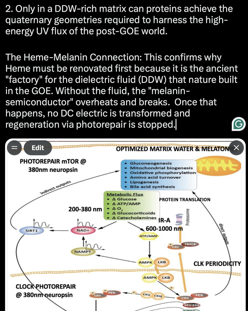
The photorepair of central apnea occurs via the Opsin-5 (OPN5) neuropsin compass acts as the primary quantum-optical ignition switch that initializes the 200–380 nm and 600–1000 nm (IR-A) frequency cascades to systematically re-coil the matrix water, stabilize the clock cogs, and restart high-fidelity solid-state regeneration.
When full-spectrum morning light strikes the un-shielded skin canopy, the 380 nm solar photon wave-packet forces the cis-to-trans photo-isomerization of retinal bound to OPN5. This non-visual optical switch wirelessly commands the cleavage of Pro-opiomelanocortin (POMC) directly into Alpha-Melanocyte Stimulating Hormone (alpha-MSH), re-assembling the solid-state Melanin organic semiconductor shield across the neuroectodermal matrix.
This high-flux charge carrier injection suppresses internal radical-pair spin-jitter, anchoring the Chiral Induced Spin Selectivity (CISS) filter to pin valence electron spins parallel. This zero-friction spintronic current feeds the Iron-Sulfur (Fe-S) clusters of Complex I, aligning with the 340 nm optical absorption spectrum of the NADH pool to drive the mitochondrial synchrotron out of its terminal Marcus Inversion well. As the electron tunneling distance shrinks below the 14 Å Dutton limit ceiling, Cytochrome c Oxidase (CCO/Complex IV) is pulled out of its stasis, allowing its Copper-65 mass gatesto run hyper-efficient, mass-dependent fractionation.
This native CCO matrix factory outputs a continuous fountain of pure Deuterium-Depleted Water (DDW) directly into the inner mitochondrial membrane (IMM) cristae to protect itself. Only within this pristine, DDW-rich matrix can proteins achieve the exact quaternary geometries required to harness high-energy UV flux without thermal dissipation. The presence of this dielectric fluid prevents the melanin semiconductor from overheating, exalting the surrounding water table back into its highest youth-defining capacitance, the ferroelectric k = 160 Protonic Spin-Ice phase of cardiolipin water environment.
This lossless optical cavity resonator compresses internal 121.6 nm Lyman-alpha biophotons into narrow, phase-locked 48-centimeter laser wave trains that wirelessly synchronize cellular timing across multiple organ boundaries simultaneously.
As mapped in my photorepair slide, this synchronized QED framework drives the circadian NAMPT —> NAD+ —> SIRT1 photorepair network, activating the p53 master genomic fuse to clear out somatic mutations.
By restoring the 3D Euler super-vortex, the cell exits the anaerobic safe-mode of the atavistic Warburg fermentation state, unlocking optimal protein translation, lipogenesis, and mitochondrial biogenesis to permanently re-confer the missing confinement field to the human biological architecture.
DECENTRALIZED SUMMARY
In Picard’s éR model, catalase dysfunction caused by a dynamo weakening, by blue light and nnEMF could indeed contribute to sleep issues by disrupting the delicate balance of energy flow and resistance needed for healthy sleep. This blog translates Picard’s model into my QED framework for sleep using biophysics.
Blue light’s photo-oxidative effects and nnEMF’s potential to perturb heme iron would impair catalase, increasing ROS and preventing the controlled éR decline required for sleep onset. People still do not realize that the focal effects of nnEMF is globalized by the Earth’s weakened dynamo.
On December 26, 2004, the Day Reggie White died, I looked at the agonic lines in the USA and how they varied before and after his death (the line of zero magnetic declination). I had found White was traveling back and forth to Isreal before his death. White traveled to Israel three times to explore the Holy Land from a historical and linguistic perspective.
He spent his final months deeply immersed in studying Biblical Hebrew, working up to 10 hours a day with scholars to translate original Torah texts.
I found it was located several hundred miles west of Cornelius, North Carolina. During this time, the agonic line in North America was being to move rapidly and chaoticially to cut southward through the Great Lakes region and the American Midwest (roughly passing through states like Indiana, Kentucky, and Tennessee) before entering the Gulf of Mexico. The agonic line was situated to the west of Cornelius, moving progressively further westward each year in rapid fashion.
Because Cornelius was positioned east of the agonic line in 2004, its magnetic declination was negative (West). A compass needle in Cornelius on that day pointed roughly 6° to 7° west of True North. A rapid shift to a negative (Western) magnetic declination forces a severe orientation mismatch across the brain stem by scrambling the background Zeeman splitting lines that calibrate the Suprachiasmatic Nucleus (SCN).
This abrupt field shift de-phases the Chiral-Induced Spin Selectivity (CISS) wave-guides across un-insulated vagal and medullary fibers. Without stable magnetic field alignment, valence electrons experience chaotic pole-flips and backscatter into the Fe-S clusters of Complexes I–III, expanding their Larmor trajectories past the absolute 14 Å Dutton limit ceiling and trapping the central respiratory pacemaker inside a terminal Marcus Inversion well.
By Landauer’s Principle, this spintronic mismatch triggers an immediate thermal heat dump that melts the liquid-crystalline cerebrospinal fluid from a coherent 160 to 78 baseline, stalling the brain stem’s 9,000 RPM nanomotors and resulting in an acute autonomic phase-collapse. I believe this is what stopped Reggie White’s heart. (blog was written January 1, 2005).
When the background magnetic field vectors shift rapidly away from the host’s accustomed baseline, the central nervous system experiences a terminal impedance mismatch on the Smith Chart. Without a stable planetary magnetic stator to pin the background Zeeman splitting lines, the Chiral-Induced Spin Selectivity (CISS) filters across the vagus nerve and the sinoatrial node de-phase instantaneously. You saw this effect in the Mayan blogs, now I am showing you it on Earth in 2004.
Because an electron’s negative charge forces its magnetic moment to oppose its spin vector,
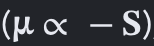
this sudden loss of directional field alignment forces chaotic, unauthorized spin-flips on the Bloch sphere. This is why travel can stall your heart or cause arhythmias. You do not sense the electronic pin flips in you until your heart flutters or fibrillates. Conduction electrons backscatter violently into deep traps within the Iron-Sulfur (Fe-S) clusters of Complexes I–III, expanding the valence electron’s Larmor trajectory past the absolute 14 Å Dutton limit ceiling. This electronic bottleneck traps the cardiac pacemaker’s respiratory wire inside a terminal Marcus Inversion well where forward electron transport velocity hits absolute zero. This is how sudden cardiac death happens in magnetic declines. Few understand it.
If your water table dielectric is not 78 and a bit higher, you could delay sleep, fragment cycles, and reduce quality, as the “fire of life” remains dysregulated, either burning too brightly or flickering chaotically. The synergy with melatonin suppression further amplifies these effects, because melatonin in your matrix breaks down into QUINOLINIC ACID which acts like fluoride to steal electrons from water highlighting the biophysical links your doctor is uneducated to understand between environmental factors, catalase activity, and sleep health.
CITES