QUANTUM ENGINEERING #20: THE OREXIN EYE PRISM

video
play-sharp-fill

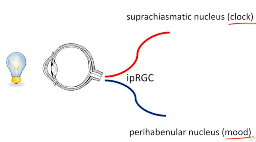
Your worldview has to be structured in principle on some specific doctrine that shapes how you function and view humanity. Embracing QED is akin to wearing steel-toed boots in a ballet-slipper world. Understanding that life is bigger than your set of challenges will help you overcome obstacles that will come your way from time to time. It’s impossible to increase the scope of our worldview when all that we are focused on is ourselves. In this way, life can be thought of as a prism. What you perceive depends on how you turn the glass. Your retina is a prism. It has photoreceptors for color vision, it has melanopsin for SCN function, it has orexin neurons, it has neuropsin, and it has Müller cells that are optical fibers (picture below) to refract the light.

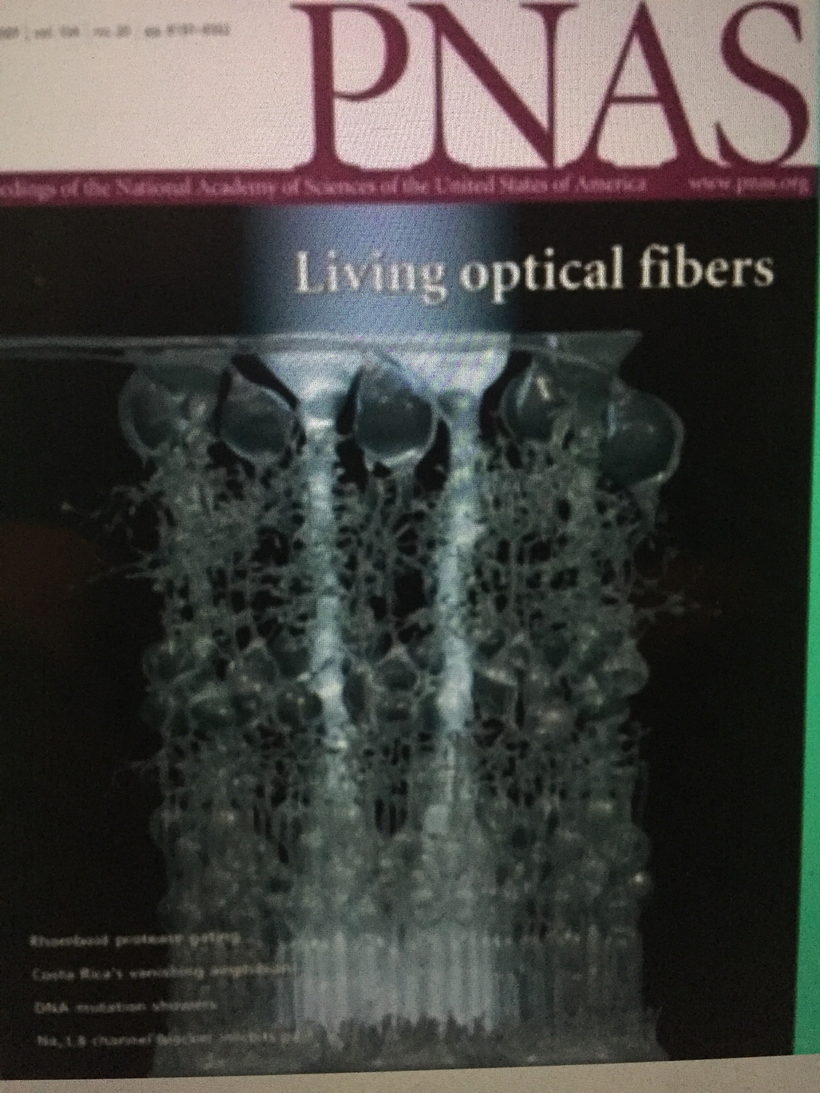
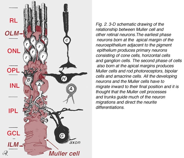
Müller cells are radial glial cells spanning the entire retinal thickness. Muller cells act as astrocytes do in forming the blood-brain barrier.  Müller cells have an extended funnel shape, a higher refractive index than their surrounding tissue, and are oriented along the direction of light propagation in the eye.  Müller cells provide trophic and anti-oxidative support for photoreceptors and neurons and regulate the tightness of the blood-retinal barrier. I believe IR-A light has a massive effect on cytochrome c oxidase to help regenerate damaged retina.

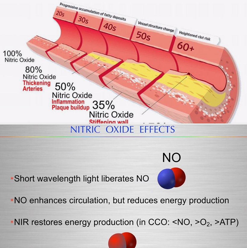
While the exact mechanism of PBM in treating retinal regeneration is not entirely clear in the literature I have reviewed, I believe I have found the likely target.  I believe that mitochondria in the Muller glial cells provide a massive source of red photons that are absorbed by the copper subunit of the terminal enzyme, cytochrome c oxidase (CCO).  PBM likely affects other parts of the electron transport chains of mitochondria like the Q-cycle as well. I believe the light absorbed by CCO enhances the ability of the mitochondria to catalyze the reduction of oxygen to produce ATP more efficiently.   It also augments the creation of water, which becomes a source of delocalized electrons and protons that can be used to regenerate damaged parts of the retina.

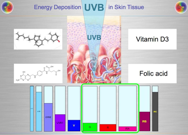
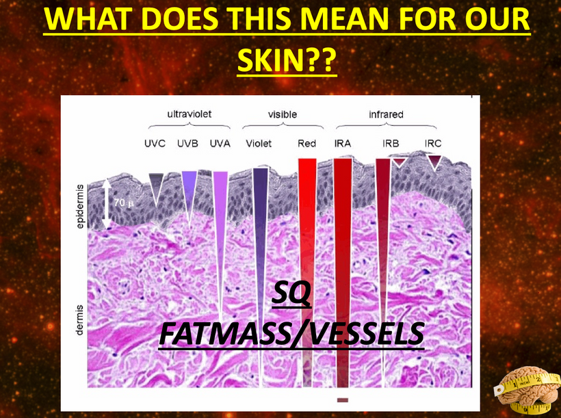
As CCO redox activity increases from PBM therapy, oxygen consumption also increases in the retina, leading to a rise in the rate of oxidative phosphorylation as well as cellular oxygen metabolism. Since neurons in the retina have an increased reliance on mitochondrial oxygen metabolism compared to most other cell types, PBM has already been shown to affect neuronal functions significantly by raising ATP production.  UV light seems like it would limit regeneration by this mechanism based on the slide above.

Traumatic stress of any type can elevate glutamate to abnormally high levels in the brain/retina. Light stress increases glutamate via the activation of orexin A in the retina.  A brain injury or stroke causes glutamate to flood the injured area.

Astrocytes make up the blood-brain barrier.  They release vitamin C to stress to alter metabolism to help NAD+ recycle faster to create ATP.  In the retina, Muller cells do the same thing for the neurons of the retina.  The glial Müller cells in the retina, regulate the glutamate cycle to prevent damage in oxidative stress conditions of the photoreceptors.

PBM likely increases the uptake of glutamate, helping the Müller cells to protect the retina.  They are more directly involved in the regulation of the synaptic activity in the inner retina.  They help organize the chaos that light creates in the retina.  Orexins seem evolved to deal with the chaos the sun creates and not any man-made light. Man-made light seems to unleash orexin A so that the PVN is chronically turned on to give us a syndrome that mimics adrenal fatigue.  It should not surprise you because light stress stimulates orexin A and this activates the PVN.  This also releases glutamate and an excessive amount of Vitamin C.  When Vitamin C is exhausted none of the catecholamine molecules associated with adrenal fatigue can be made.  This is why adrenal fatigue is not an adrenal disease.  It is a disease of the eyes, orexin, and the PVN due to excessive glutamate release and excessive use of Vitamin C so that dopamine, epinephrine, and noradrenaline can’t be made.

The light is broken down into its parts to impart information to the retina. In this way, Perception is a prism, and the retina is like shot silk – the quality of information it imparts depends on the quality and quantity where the light hits.

Counterintuitively, the retina of the vertebrate eye is inverted with respect to its optical function and light must pass through several tissue layers before reaching the light-detecting photoreceptor cells.  The reason for this I laid out in my Vermont 2017 talk on YouTube.

In mammals and humans, the extent of spontaneous repair after retina injury or disease is either non-existent or extremely limited (Karl and Reh, 2010). Rather than regenerate, damaged mammalian retinas commonly undergo reactive gliosis and scar formation (Bringmann et al., 2006). (See below)

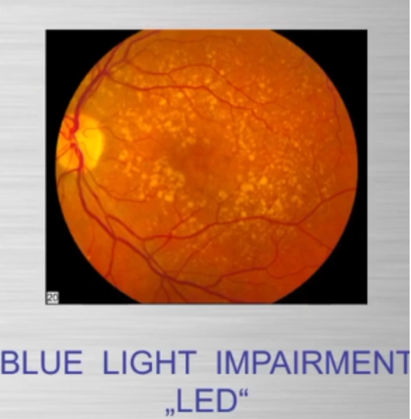
This lack of a complete regenerative response to damage directly limits the treatment options for retinal-based diseases such as age-related macular degeneration or Stargardt’s disease.  The use of PBM seems to help use the Muller cells to help regenerate damaged retinas.  I have had quite a bit of success with this in the last three years.  I read some papers on retinal damage in zebrafish and realized that Muller cells were the key to understanding how to regenerate damaged retinal tissues.

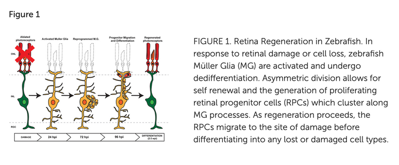
The retina forms from the central nervous system (CNS) and develops into a three-layered structure consisting of seven main types of cells and numerous other cell types identified by single-cell RNAseq (Macosko et al., 2015). The structure, function, cell types, and genes expressed in the retina are largely conserved among vertebrates, supporting the notion that information gained from models capable of spontaneous regeneration likely applies to mammals whose regenerative capacity varies.  It appears humans have a lot in common with zebrafish based on my recent experience with acute macular degeneration.

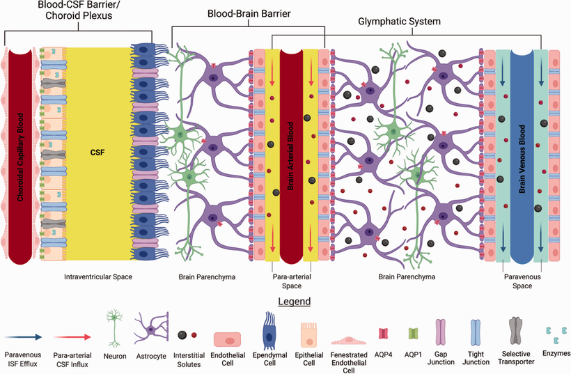
The information the retina creates is stored electromagnetically in coherent domains of water in cells like the Muller glia and in the CSF that surrounds the key areas of the brain that interact with the environment.  CSF is made by the choroid plexus in the brain. CSF is an ultrafiltrate of the blood.

OREXINS AS A PRISM

I’ve taught my readers about every single part of the retinal prism, but I’ve saved the orexin neurons for last.  They are intercalated deep into the retina of every vertebrate on planet Earth. They are the rare neurons in the entire human brain. Scarcity creates its value to signaling when it comes to light.  Light strikes things and changes them in ways our retina cannot sense.  This makes light the most powerful change stimulus behind the evolutionary adaptation.

Our prism can become our prison if we don’t understand how it operates and with what light it is designed to work.

With change being an inevitable element in our lives, we have only two options. Either embrace it and live life to the fullest or be stuck in the comfort zone of a compromised life.  Orexins make sure vertebrates never stay in their comfort zone of life.  They create stress for us to adapt to.  This response must be controlled.

Orexins are hypothalamic neuropeptides that were initially identified in the rat brain as endogenous ligands for a (previously) orphan G-protein-coupled receptor (GPCR). I spoke about them here over ten years ago. They are multitasking peptides involved in many physiological functions, including regulation of feeding behavior, wakefulness and autonomic/neuroendocrine functions, and sleep/wakefulness states in mammals. There are two isopeptides of orexin, orexin A and orexin B, which are produced from a common precursor peptide, pre-pro-orexin. UV and red light operate on these two orexins from sunlight.

The structures of orexins, as well as orexin genes, are highly conserved throughout mammalian species, suggesting strong evolutionary pressure has maintained these structures for hundreds of millions of years.  In this way, orexins are like DHA they haven’t changed in hundreds of millions of years of vertebrate evolution.  Note in Orexin -A below the disulfide bridges.  The photosensitivity of protein-bound cysteine residues has been shown to depend on the redox state of the microenvironment (Augenstein and Ghiron 1961; Dose and Rajewsky 1962; Augenstein and Riley 1964; Dose 1964, 1967; Fiore and Dose 1965; Grist et al. 1965; Risi et al. 1967; Koudelka and Augenstein 1968)

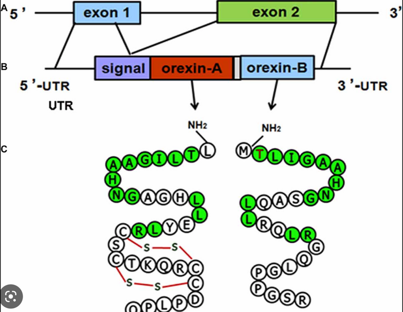
This data set correlates with the observation by Petersen et al. made in 1999, that cystine residues (cysteines involved in disulfide bridges) have a clear preference for aromatic residues as spatial neighbors.  Note that orexin has one tyrosine adjacent to its S-S bond.  Why is this important?  Recall that all cells release ultra-weak UV light.  Going further, vertebrate eukaryotes tend to release less ultraweak UV light unless the redox state is low.  When it is low eukaryotes release significantly more ultraweak UV.  The excessive ultraweak UV light then disrupts the S-S of orexin A and this changes the tertiary and quaternary protein folding of orexin and alters its physiologic signaling optically.  These photodynamic reactions allow for a temporally and spatially controlled and reversible modification of the surface of orexin A and, hence, can be used to produce functional interfaces that change signaling.

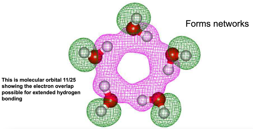
It has been reported that the UV illumination of Tryptophan and Tyrosine residues gives rise to delocalized electrons.  UV light can also liberate hydrogen from aromatic amino acids into the hydration shell.  The H-atom ejection from these amino acids was written about decades ago.  (Vladimirov et al. 1970; Feitelson 1971). The role of the hydrated electron in photo-reduction of cystine in the presence of indole (melatonin is an indole) led to the proposal of a possible mechanism behind this phenomenon over 50 years ago (Grossweiner and Usui 1970).  Few go back and read those papers to understand what orexins are really doing.  The liberation of these electrons and protons is what changes the hydrogen bonding network to become coherent domains.  Orexins are the gatekeepers that force water to take on a hexagonal form to become quantum coherent in the brain.

The connection of orexin to melatonin is more important to make.

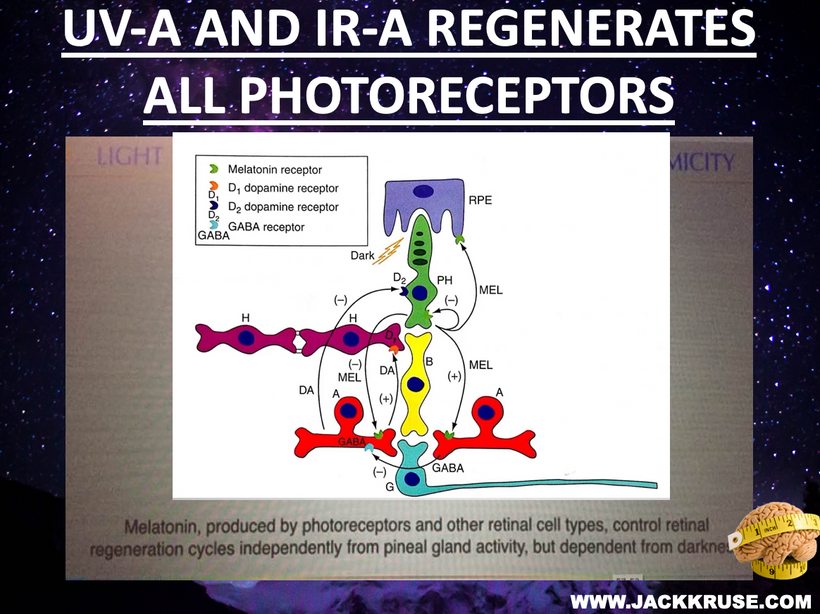
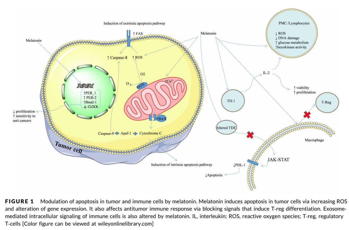
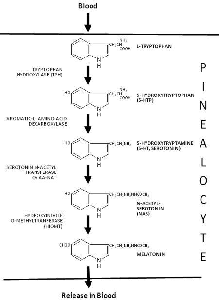
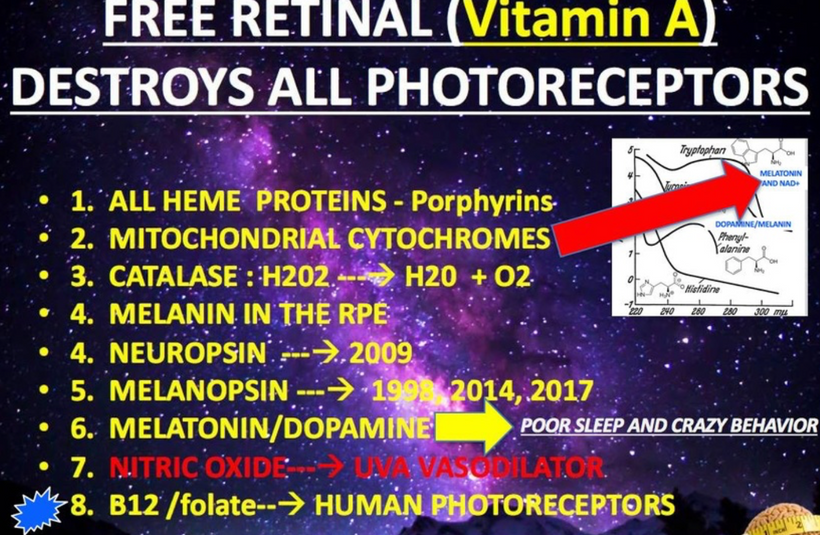
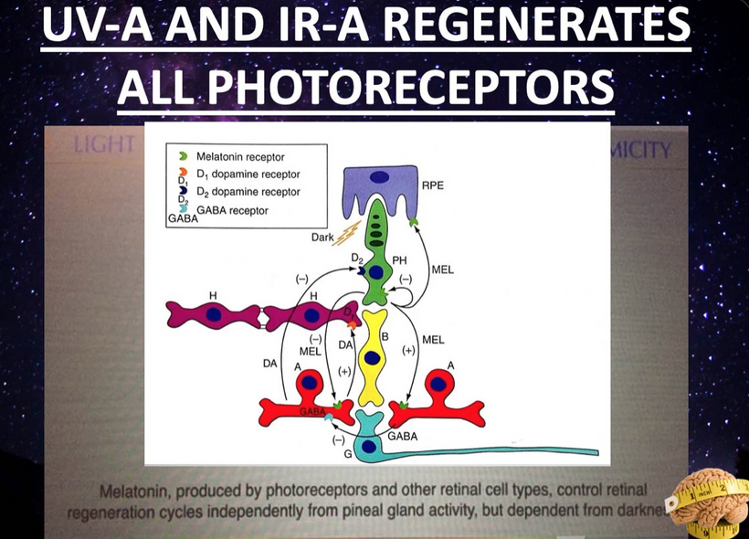
Melatonin and dopamine are known to be critical in regenerating all human photoreceptors (see below).  The problem is we do not know exactly why.  Melatonin has two receptors, MT1 and MT2.   The pathophysiological function of the melatonin MT1 and MT2 receptors has not yet been well-clarified.  The MT1 receptor is involved in the regulation of the circadian rhythm.  MT2 is not.  MT2 receptors are located in the reticular thalamus which is the non-REM sleep area.  Non-REM sleep disorders are also called arousal disorders. Non-REM parasomnias involve physical and verbal activity. You are not completely awake or aware during these events, are not responsive to others’ attempts to interact with you, and usually don’t remember or only partially remember the event the next day.

The MT1 receptor is in the locus ceruleus and lateral hypothalamus where REM areas exist.  This is the same area where orexin neurons project to.  When these orexin neurons are destroyed there we also see a REM sleep disorder develop called narcolepsy.  People with narcolepsy frequently enter REM sleep rapidly with sleep onset. This usually happens within 15 minutes of falling asleep day or night. Also, muscle weakness or dream activity that is associated with REM sleep can occur during wakefulness or be absent during sleep.  REM normally occurs in short periods and is characterized by a decrease in muscle tone and is associated with profound sympathetic activation (PVN), including increased heart rate, breathing, blood pressure, and temperature.

The lengths and structure of orexins suggested that orexin B is the ancestral form of the orexin neuropeptide. In mammals, orexins bind to two subtypes of GPCRs, i.e., orexin 1 receptor (OX1R) and orexin 2 receptor (OX2R).

I believe one orexin works best with UV light signaling and the other is optimized by red light.  One reacts best with fast-traveling red light (orexin B) and the other with slower-traveling UV light  (orexin A) in our tissues.   Phylogenetically, the orexin system is present exclusively in vertebrates so this puts them around 650 million years old.

Orexin A is a 33-amino-acid peptide with an N-terminal pyroglutamyl residue, two intrachain disulfide bonds, and C-terminal amidation. Strikingly, this structure is completely conserved among all mammalian species so far identified in the literature.  This means orexin A is the same in humans, gorillas, rats, mice, cows, pigs, sheep, dogs, seals, and dolphins.  Very few proteins have been found to have this long-term evolutionary lineage.

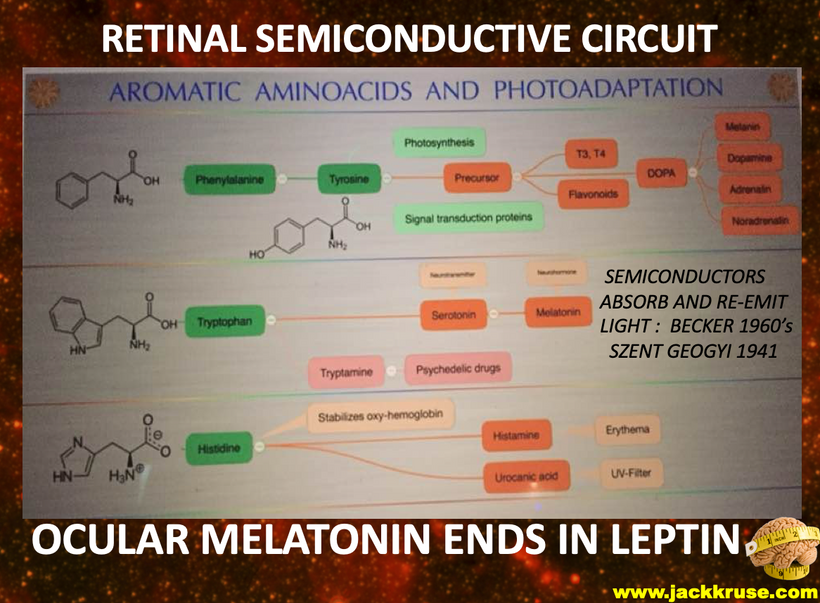
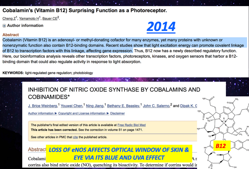
Most of you have learned from me there are a lot of photoreceptors in the eye like melanopsin, melatonin, neuropsin, B12, dopamine, melanin, cytochrome c oxidase,  nitric oxide, and heme proteins like catalase in RBCs etc. (see above and below)

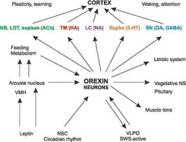
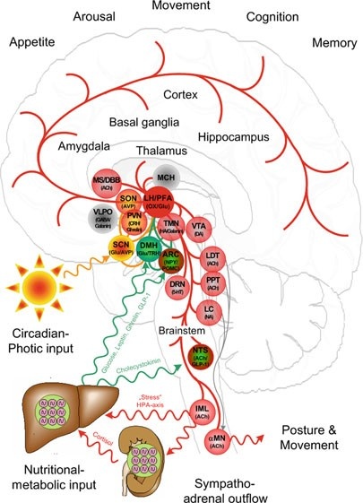
The central retinal pathways in the retina of all mammals contain orexin neurons that project to the areas of the brain adjacent to CSF pathways.  Orexin-producing neurons (orexin neurons), in the lateral hypothalamus, receive input from the forebrain structures including the extended amygdala and nucleus accumbens (NAc)—which are implicated in the processing of emotion and motivation—and send output to brain stem regions, which are implicated in the regulation of wakefulness. I told you in the cold thermogenesis series that sleep was our primordial state, and we evolved wakefulness.  I believe the orexin neurons were key in this transition.  Orexin made water more quantum coherent so more energy could be harvested to allow wakefulness to emerge.  Orexin neurons play an important role as a link between emotional states and wakefulness states.   Any acute stress activates orexins neurons, and this includes modern light stress.  This is why I have believed for 15 years that adrenal fatigue is a disease of the periventricular nucleus.  I believe orexins are the fuse that begins the process.

There are only 50,000–80,000 orexin-producing neurons in the human brain, located predominantly in the perifornical area and lateral hypothalamus. The hypothalamus sits adjacent to the third ventricle which is filled with CSF. CSF is 99.8% water created by mitochondria.  Let’s talk about the prism that your eye clock sits in.

Light transmission via the retina interacts with the amacrine cell level in the retina which in turn activates both orexin systems. The orexin systems modulate the stress response in mammals. The orexin system was initially suggested to be primarily involved in the stimulation of food intake, based on the finding that central administration of orexin-A and -B increased food intake. In addition, they stimulate wakefulness, regulate energy expenditure, and modulate the visceral function and brown fat activation.

Many studies support that the orexin neurons regulate brown adipose tissue (BAT) activity via the sympathetic nervous system to enhance energy expenditure.  This is the basis of my cold thermogenesis protocol.

Orexin promotes wakefulness. I believe inhibition of orexin function is how general anesthetics work.  Right now, most anesthesiologists still have no idea how the drugs they use daily work.  Studies have indicated that a major role of the orexin system is to integrate metabolic, circadian, and sleep debt influences to determine whether an animal should be asleep, or awake and active.  Orexin B are proteins that absorb the slowest form of light in tissue to maximize the fidelity of the signal. Orexin A neurons strongly excite various brain nuclei with important roles in wakefulness including the dopamine, norepinephrine, histamine (all made from aromatic amino acids), and acetylcholine systems, and appear to play an important role in stabilizing wakefulness and sleep (adenosine).  Adenosine is activated by IR-A light. The sleep-promoting action of adenosine can be reversed by orexin A applied to the lateral hypothalamus, by exciting glutamatergic input to orexin neurons via the action of orexin receptor 1.  I believe it is the ultraweak release of UV light that disrupts the S-S bond of Orexin-A that cause sympathetic outflow to increase. This leads to REM sleep disorders = narcolepsy.

The discovery that an orexin receptor mutation causes the sleep disorder canine narcolepsy in Doberman Pinschers subsequently indicated a major role for this system in sleep regulation. Narcolepsy is a REM sleep disorder. Genetic knockout mice lacking the gene for orexin were also reported to exhibit narcolepsy.

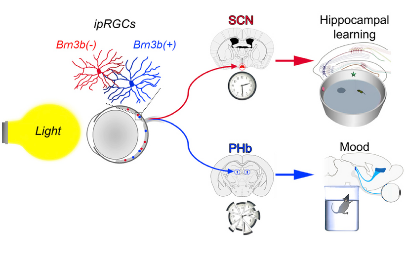
In humans, narcolepsy is associated with a specific variant of the human leukocyte antigen (HLA) complex. A lack of either red light or UV light is how narcolepsy begins.  It begins with poor light signaling in the prism of the retina. Light changes the charge density of the retina.  The proof is found in genome-wide analysis studies that show that, in addition to the HLA variant, people with narcolepsy also exhibit a specific genetic mutation in the T-cell receptor alpha locus. I spoke about the HLA complex in the brain-gut series when I explained how the chimp brain evolved into the homo sapien brain using the HLA complex.  I think orexins helped fuel the transition.

In conjunction, there are genetic anomalies that cause the immune system to attack and kill the critical orexin neurons.  Once they are destroyed, they do not come back.  Hence the absence of orexin-producing neurons in people with narcolepsy appears to be the result of an autoimmune disorder.  Can we see the effect of these changes elsewhere in the brain?  We do.  We see it in the hydrogen bonding networks of water in cells, in the blood, and in the CSF.

All paths can be seen, through the prism of fate.  Sunlight is the Windex our watery eyes require.  The periventricular nucleus controls our sympathetic response by how it reacts to the orexin signal from the central retinal pathways.  Moreover, the choroid plexus (CP) of the brain that creates CSF has recently been implicated in the regulation of circadian rhythmicity and therefore, it is now recognized that the functions of the CP cannot be limited to those listed in textbooks. Given the strong circadian influence over the sleep-wake cycle and orexinergic signaling, the role of the CP in modulating orexinergic signaling via circadian influence is worth further study. Furthermore, considering that the sleep-wake cycle modulates additional homeostatic processes such as amyloid-β and glymphatic clearance, the hydrogen bonding networks in the CP may also have a significant influence on the activity of these functions.  People forget that the S-S bond in orexin-A induces proteins to misfold due to the effect of UV light on this bond.  Brains with neurodegeneration all have low redox states and this means they will release a lot more ultra-weak UV light.  This light will destroy orexin-A and lead to protein misfolding at a log rate.  As this light stress develops, glutamate rises and the BBB becomes leaky.  The process progresses and gets worse.

The autonomic nervous system has two main arms, sympathetic and parasympathetic. The vagus is part of the parasympathetic system that lowers the stress response in the brain stem.  The periventricular nucleus is one of the key targets of the orexin neurons. Stressor activity increases chronically when the sympathetic system is constantly turned on. The sympathetic system outflow of the stress stimulus begins in the paraventricular nucleus (PVN). The vagus nerve is the major outflow tract that is the calming portion of the ANS and antagonizes the PVN to lower the stress response.  It has an amazing effect on the hexagonal prisms in CSF in the 4th ventricle.  This signal can be turned on and off by the light that comes through the prism of our retina.  This is why the light you allow is critical in signaling in how it changes hydrogen bonding in water.

Balancing both arms is critical in avoiding diseases, and creating an allostasis in optical signaling.  The light in the environment destroys that balance.  The autonomic nervous system tries to create order from the chaos in light.  It too is another prism.  The key to the orexin prism is found in its 33aminoacid peptide sequence that has two intrachain disulfide bridges formed by four cysteine residues (C6–C12 and C7–C14).  You might remember I important I told you cysteine was here.

Cysteine has something in common with orexin Cysteine is the rarest amino acid and being rare means it increases signal fidelity.  Eliminating correlated noise without changing other input properties substantially decreased the accuracy with which a cell’s spike outputs encoded light inputs in retinal ganglion cells. Thus covariation of excitatory and inhibitory inputs in the orexin system can be a critical determinant of the reliability of neural coding and computation in its target in the brain.

Orexin neurons widely project to all regions of the brain including the hypothalamus (de Lecea et al., 1998; Peyron et al., 1998), thalamus, cortex, brain stem, and spinal cord (van den Pol, 1999). There aren’t many of them, but their impact on water chemistry in us defines a non-linear stimulus where a small stimulus leads to a massive change.  

Orexin input to brain regions important for arousal, such as the locus ceruleus, help to regulate the response to a stressor (Hagan et al., 1999). Moreover, orexin-containing nerve terminals project to stress-sensitive centers such as the amygdala and bed nucleus of the stria terminalis (Date et al.,1999). Orexin neurons also project to the paraventricular nucleus of the hypothalamus (PVN) (Winsky-Sommerer et al., 2004), where neurons expressing corticotropin-releasing hormone (CRH = cortisol) initiate the Hypothalamic Pituitary Adrenal (HPA) Axis.   This forms half of the circadian mechanism of cortisol and melatonin. 

Further, orexin neurons densely project to the paraventricular nucleus of the thalamus (PVT) (Kirouac et al., 2005), which plays a role in regulating neuroendocrine and behavioral adaptations to severe or chronic stress (Hsu et al.,2014). Specifically, orexins may influence gene expression of the CRH type 1 receptor (CRH1R) in the paraventricular nucleus of the thalamus (Heydendael et al., 2012, 2011), and this brain region may then regulate the HPA axis via multisynaptic pathways through the BNST to the PVN. The orexin neuropeptides orexin A and orexin B interact with noradrenergic, cholinergic, serotonergic, histaminergic, and dopaminergic systems in the brain, in addition to the HPA axis (Sutcliffe and de Lecea, 2000). Thus, orexins have the potential to regulate the stress response through actions at multiple projection sites.  All mental disorders likely begin with orexin dysfunction.

CSF-contacting neurons are present in all vertebrates and are located mainly in the hypothalamic area and the spinal cord. The hypothalamus lies below the hypothalamic sulcus separating it from the thalamus above. Like the thalamus, a thin vertical space filled with CSF called the 3rd ventricle is positioned midline between the two halves of the hypothalamus thalamus. The blood-brain barrier is absent around the vagal vomiting center of the 4th ventricle so that it can monitor the blood for changes. The blood-brain barrier is also absent around the various areas in the hypothalamus where orexin targets land their signaling.  

The stimulus of light from the orexin prisms in our retina changes the CSF’s hydrogen bonding networks to a hexagonal arrangement that allows for another prism to be built in water that stores electromagnetic information to make the brain quantum coherent.  Full-spectrum sunlight reaching the brain creates more coherent domains in water. Normally water is only 40% quantum coherent.  From here on out you should realize that coherent domains should be regarded as long-range ensembles of electrons and protons of exclusion zones inside the water that has the superconductive ability at room temperature. This type of water is a plasma filled with electrons and protons.  Biological water has unique characteristics that make life possible.

SUMMARY

Water (H2O) is the third most common molecule in the Universe (following the H2 and CO molecule), and its standard chemical structure, based on the hydrogen bond, is actually confined by a simple scheme of charges interacting via static Coulomb forces; that is, it is totally reliant on electrostatics and omits all mention of electrodynamics and the consequent radiation field. It has been speculated that a large percentage of effects in condensed matter physics make use of the radiation field in one way or another but it still doesn’t seem to have found a place in much of basic chemistry. In biochemistry, no one seems to realize how important these effects are.  In biological systems almost all water is within a fraction of a micron or less from a surface or molecular backbone and so is interfacial water, which behaves in a quantum coherent manner. This occurs because as hydrogen bonds change, the Coulomb law of electrostatics does not apply to it. In these circumstances, similar charges become attractive making water react homogeneously. This water changes proteins into LED diodes capable of moving electrons around using light inside of tissues.  Biology itself depends on this, so as to allow the accumulation of tissues from negatively charged cell bodies.

Biological water is paramagnetic meaning that it holds a magnetic charge.

Water has a dipole moment and is, therefore, subject to paramagnetism. Chemical bonds (including covalent, ionic, hydrogen, and van der Waals types) have been commonly assumed to be dominating for biological organization and activity. However, these bonds represent forces acting at short distances in the nanometer region. Biological systems maintain coherence at every dimension scale because of how water is changed by our molecular protein prisms in cells. Long-range coherence, large-distance cooperation, and the whole-body control are significant properties of biological systems.

According to Giuliano Preparata, Emilio Del Giudice, and colleagues water water molecules at the interface surface act like a new chemical species and can change rapidly in cells. This clustering of hydrogen bonds imparts a crystalline likeproperty to the water. It become more like a liquid crystal.  In the bodies of living organisms, the clusters form hydration layers around biological molecules. Orexins are today’s example.  It is known from electronics that different patterns which contain information which result within a cluster depending upon its structure of the crystal.

Thus, depending on its structure, each molecule has an oscillatory pattern (resonance frequency) that can be determined by spectroscopy. It is known, through spectrographic analysis, that water and other dipole molecules are able to be entrained to exogenous oscillatory patterns by rearranging their cluster patterns. The cluster rearrangements then resonate with the entraining frequency of sunlight to do the things life can do.  This water is called a coherent domain.

Water becomes something unique when sunlight interacts with it in our cells.

Within the CD water, molecules oscillate between the ground state and an excited state close to the ionizing potential of water and, therefore, contain close to a million almost-free electrons. Those electrons are used to sculpt the physiology of life.

Quantum Electrodynamic (QED) field theory has produced a vision of water in a liquid state as a medium, which for a peculiarity of its molecular electronic spectrum reveals itself as an essential tool for long-range communications, being able to change its supra-molecular organization in the function of the interaction with the light in the environment.   Orexins have the biggest prism effect of making water the stage on which life performs on.

CITES

https://www.pnas.org/doi/10.1073/pnas.0611180104

Myung J, Schmal C, Hong S, et al. The choroid plexus is an important circadian clock component. Nat Commun2018; 9: 1–13.

Xie L, Kang H, Xu Q, et al. Sleep drives metabolic clearance from the adult brain. Science 2013; 342: 373–377.

Kang J, Lim M, Bateman R, et al. Amyloid-ℬ dynamics are regulated by orexin and the sleep-wake cycle. Science2009; 326: 1005–1007.

Spector R, Snodgrass S, Johanson C. A balanced view of the cerebrospinal fluid composition and functions: focus on adult humans. Exp Neurol 2015; 273: 57–68.

Richardson SJ, Wijayagunaratne RC, D’Souza DG, et al. Transport of thyroid hormones via the choroid plexus into the brain: the role of transthyretin and thyroid hormone transmembrane transporters. Front Neurosci 2015; 9: 66.

Myung J, Wu D, Simonneaux V, et al. Strong circadian rhythms in the choroid plexus: implications for sleep-independent brain metabolite clearance. J Exp Neurosci 2018; 12: 1179069518783762.


CPC #67: PULMONARY HYPERTENSION

I received the following email in May of 2014.

“Dear Dr. Kruse –

I am reaching out to you for your advice. 10 years ago, when we were 32, my friend was diagnosed with pulmonary hypertension, usually diagnosed in elderly patients. The doctors had never seen it anyone so young get this disease.

After a period where she was told she would die, she found a specialist who got her on a program that has been working well and she has been living with it since 2008.

Four months ago she started to decline and was in and out of the hospital with pneumonia. She was most recently admitted to UCLA for 3 weeks with inflammation levels over 100 in her entire body.

The PH has progressed and now the doctors also suspect PVOD.

Her pulmonary lymph nodes are all glowing on the scans they also suspect lymphoma, although she does not have any other markers for that.

However, my friend’s husband tells me that really the doctors have NO IDEA what is going on with her fundamentally they seem stumped. She is a complete mystery to them. I feel this will not be a mystery to your community.  This is why I am sending this email to you.  I hope that is OK.

They want to do a lung biopsy but fear she is too fragile. She has been sent home and is due back in the hospital in 4 weeks when they will re-test her inflammation levels.

I would appreciate any feedback you may have, Dr. Kruse.

Thank you in advance for your help.

She lives in Los Angeles, California her whole life and works at Qualcomm.”

My ANSWER was given on 5/31/2014:

The environment is everything you see, but more importantly, everything you don’t see but your cells sense.

UV, IR, other EMF both non-native and native, magnetism, air quality, water quality (we can’t see how high something is in deuterium), food quality. All of these radically affect the lung surface topology in your friend because of the fake light and nnEMF in LA. She and her husband really need to do a consult with me and consider becoming a patient of mine after I do the consult if they agree with my work up and plan of action.  I want them to both know LA is also the home of horrible atmospheric pollution that fills the basin DAILY. The amount of RF from the broadcast is off the chain and it has altered the Van Allen belts above their heads to limit her ability to make Vitamin D from the Sterols in her skin.  This has increased her risk of PH.  The nnEMF of LA and its population is problem number two.

It is no wonder to me why she has a serious lung disease. It also blocks the amount of UVA, UVB, and IR light from the sun that gets through the atmosphere in LA and SD to help her regenerate and this ruins both autophagy and apoptosis in mitochondrial in her lungs. Because of these atmospheric changes, even in sunny southern California, you may not get enough balanced UV and IR sunlight) on your body surfaces. The lung is one of the surfaces where topology and the topologic charge are massively important in the control of hydrogen flow in her body especially in mitochondria.  At night, her body may not even realize it’s night due to lack of connection to the earth and artificial lights, because NO ONE at UCLA even knew that melanopsin was present in the eye, skin/fat to cause chronic inflammatory damage. In fact, the skin damage she got from the blue light likely made her lungs worse daily. Why? All opsin are loosely bound to Vitamin A and as Vitamin A drops it causes circadian mismatches at all surfaces because melatonin levels drop and this ruins cell membrane function which the lung relies upon.

Add in makeup, her clothing, the use of sunscreen, makeup, and sunglasses that are trendy in LA and you see it all unfold in front of your eyes. It is not a rare problem in LA.

FOLLOW UP done yesterday:  This lady and her husband did many consults with me and they became clients of mine in El Salvador.  She called me and her latest follow-up at UCLA shows she has no evidence of PH 6 years later.  She no longer works for Qualcomm and she now resides in Central America at the 9th latitude.  She also told me she never got COVID and had no pulmonary flareups during the pandemic.

Artificial light exposure at night blunts melatonin secretion and melatonin is WHAT REPAIRS mtDNA and increases mitophagy which is what autophagy is called in mitochondria. Your friend needs this big time because she is getting ready to lose apoptosis next and she already might have if her nodes are lymphoma (wisdom of May 2018 webinar). This is worse for young children or people who live in cities with a big nightlife and with lots of people who use technology devices (LA). New parents and big city DWELLER should consider some sort of screen-time-restriction and blue blockers for their eyes and skin.  Vitamin D via your skin is crtical to reversing lung diseases.

CITES

https://twitter.com/DrJackKruse/status/1593636711514472449

https://twitter.com/DrJackKruse/status/1593638139863736320

https://twitter.com/DrJackKruse/status/1593637583656984579

https://twitter.com/heniek_htw/status/1593644489268133888

BTC #44: WAS SBF THE PROXY FOR MORGAN STANLEY’S CRYPTO TAKE OVER?

video
play-sharp-fill

You can kill a revolutionary but you can never kill the revolution.

Bitcoin must remain the most secure decentralized system in the world. It is Bitcoin’s decentralization that makes its users totally resistant to government censorship. If the fruits of your labor are stored within the Bitcoin network, no one can tell you how to use them, or take them away from you for arbitrary reasons.

This is what all Bitcoin users are looking for above all else. BTC is the last bastion of the virus that infected our colonist Founding Fathers. The latest iteration of our government is an embarrassing shell of our Constitutional Republic.

As long as Bitcoin has the fundamental properties it has today, not even the banning of governments can prevent its adoption by the general public. We The People still have a chance to make a more perfect union and BTC is the wedding ring to seal that deal.

Once a popular currency revolution like bitcoin is underway, there is no way to stop it. The powerful at the helm of the current system will have to accept it and adapt, or risk being totally overtaken in the future by the Bitcoin revolution.

So with this all said, who stands benefit from recent events?

CZ of Binance sits on the Twitter board and Jack Dorsey is one of the largest shareholders in the private takeover by Elon Musk.  Musk has tried for 25 years to disintermediate banks via his work at Paypal. Dorsey has convinced Must the using Bitcoin and the Lightning Network on Twitter is the best way to finish the job PayPal began.  Musk also owns over a billion dollars of Bitcoin.  Dorsey owns more than ten times that amount.  Musk cobbled together fiat financing through a variety of sources, including his own money from selling Tesla shares via his private account at Morgan Stanley.  MS wants to sell and custody Bitcoin for its clients.  Clearing a comprised exchange from the pavement makes a lot of sense. Musk raised $12.5 billion from banks to buy Twitter, with Morgan Stanley, Bank of America and Barclays each committing to $2.5 billion.  Did you know that 7 days after taking Twitter private Musk applied for a money transfer license?  Musk is not interested in free speech, he is interested in how money is going to change soon.

Most of us have been watching with rapt attention, but for those who missed the series of events, it went something like this:

  • Over the last month, SBF aroused the ire of Crypto Twitter with its pronouncements on industry regulation.
  • Last week, CoinDesk published an expose on SBF-aligned trading house Alameda that questioned its solvency.
  • Over the weekend, on Sunday, CZ of Binance announced that Binance would be dumping FTT token based on those recent concerns.
  • CZ’s comments initiated an exodus, as professional asset managers raced to get their assets off of FTX. SBF’s remarks on Monday that Binance was maliciously targeting FTX did nothing to stem the bleeding.
  • The bank run at FTX led to more than $6B in withdrawals from FTX in 24 hours before they stopped being able to process them.
  • By Tuesday midday, SBF shocked the non BTC “crypto world” by announcing that Binance had signed a non-binding LOI to acquire FTX.
  • By Wednesday afternoon, CZ had backed out of the deal and the WSJ was reporting that the hole on FTX’s balance sheet was $8B.  How convenient.
  • On Thursday, the Bahamas securities regulator – where the crypto exchange is officially headquartered – partially froze FTX’s assets and moved to appoint a liquidator for one of its affiliates.

The situation is still rapidly evolving, but the emotions being processed by the crypto industry are as immense as anything we’ve ever seen. Disbelief, shock, betrayal, apathy, disgust.

So ask yourself, who benefits from this SBF blowup?  Incentives determine outcomes. The answers are the 6 primary dealers who broker money/UST for the Federal Reserve stand to gain it a lot of regulatory power if SBF influence ends in Washington DC.

SUMMARY

You can kill a revolutionary but you can never kill the revolution.

So governments have chosen to go after media/VC created cypto revolutionaries, but they can’t kill the Bitcoin revolution. It has grown far too big now.  So now they want to control it.  They want to control the on and off ramps.  I will be discussing this and many more macro idea on November 15th for clients.  I hope some of you join my tribe at info@Kruseatdestin.com

People cannot give up on Bitcoin, because it is already their best option in every way. Bitcoin makes it easy for them to make everyday payments while protecting their wealth. Exchanges now can be made via Telegram or WhatsApp like what has happened in Nigeria to avoid possible checks by the Central Bank of any country.

Payment in fiat currency is then made via a simple bank transfer so that no mention is made of the cryptocurrency transaction.

Consider the current situation in Nigeria. Merchants also see Bitcoin as an incredible tool for trading across borders while avoiding some exorbitant taxes. As the most populous country in Africa, Nigeria also has a strong diaspora around the world. Nigerians simply want to use the most convenient and advantageous system for their commercial or personal exchanges.

Bitcoin is the most convenient system right now and today’s macro environment is highlighting BTC use to a government.

You can’t stop a revolution like Bitcoin that meets the expectations of an entire globe of people.

It is all the more difficult when the political powers in any country are not able to offer a real alternative to monetary failure.

Any attempt of the USA and its Wall Street bankers to control Bitcoin will only strengthen its existence and use. This is already happening elsewhere in the world when governments ban Bitcoin and tighten controls on its use.

This is a lesson for us all.

The events around FTX were always about control.

Like anything in life, truth has its price. Are you willing to pay it for your health? Your wealth?

The American public doesn’t know that an economic reset has been set by the power brokers, and the taxpayer doesn’t even know that they don’t know.  But they do know this epic blow up of SBF has implications. The bankers have been focused on COVID for 2 years by those helping the Fed by design. COVID was a compliance test for the greatest wealth transfer in human history. SBF was just another part in the bankers plan that got executed this week.  They had to fracture the relationship of VCs to Gary Gensler and Congress.  SBF was the lever.  The Fed needs their Wall Street proxies in control of how Bitcoin becomes the people’s money.  Be ready for the rest of the plan.  Fed banks will soon be custodian of BTC they sell to the masses.

Just like FTX, when ETHER collapses it will be obvious that the signals were everywhere.   Convert to BTC while and if you can.

There is a reason EO 6102 was placed in the Genesis block.

Remember, not your keys not your coins.

QUANTUM ENGINEERING #19: AUTISM/FOLATE & TERRESTRIAL SUNLIGHT

It has been estimated that approximately 30% of all cancer cases are attributable to diet and lifestyle.  This “belief” is tied to a misunderstanding of how our genome is affected by hypermethylation.  The process of methylation is one of the software programmed by sunlight that is a key epigenetic controller of DNA expression.   Folate is a natural water-soluble B vitamin.  That water comes from mitochondrial metabolism.  Solar exposure creates that water.  If you do not get sun you do not create water and your folate levels will suffer.

Folate helps reduce depressive symptoms in the brain. Natural folate is needed in the brain for the synthesis of norepinephrine, serotonin, and dopamine. Sunlight also increases melatonin production at the same time.  Melatonin is made from tryptophan.  All of them are substrates that have seasonal variations.

Folate is the naturally occurring form of vitamin B9. Before entering your bloodstream, your digestive system converts it to the biologically active form of vitamin B9 ⁠— 5-MTHF.

Folate’s synthetic form is folic acid.

Folic acid is a synthetic form of vitamin B9 that’s also known as pteroylmonoglutamic acid.

It’s used in supplements and added to processed food products, such as flour and breakfast cereals.

Unlike folate, not all of the folic acid you consume is converted into the active form of vitamin B9 — 5-MTHF — in your digestive system. Instead, it needs to be converted in your liver or other tissues.

Yet, this process is slow and inefficient in some people. After taking a folic acid supplement, it takes time for your body to convert all of it to 5-MTHF.  Folate doesn’t equal folic acid. Folic acid doesn’t operate on DNA like folate does.

Folate functions as a donor of one-carbon units and it has been implicated in the regulation of DNA methylation as well as DNA synthesis.  Folate is affected by UVB light you experience or do not experience.  It is destroyed by excessive amounts of UVB.  This makes it a very responsive “photoelectronic switch” to control the genome by changes that begin in the mitochondrial matrix.

Epidemiological studies suggest that suboptimal levels of folate and other B vitamins may affect DNA methylation rates and thereby influence genomic stability and cancer risk, although the exact underlying mechanisms have not been clarified.  Almost every study that used these two B vitamins (B12/folate) in cancer cases has failed to produce the effect the belief that dietary sources of these vitamins might prevent cancer has failed.

The Black Swan Mitochondriac perspective is quite different from these vitamins.  The proper amount of B12 and folate are built by the sun in the latitude you live and impacted by your skin color and your skin filters for sunlight by your SNP’s.

UV radiation may destroy blood folates in test tubes, but clinical data are scarce if it happens in life. Folate deficiency may increase the risk of cardiovascular diseases, colorectal carcinoma, megaloblastic anemia, pregnancy and birth complications, depression, and dementia.

Melanin pigments are important regulators for color and photochemistry of the evolution of essential functions of human skin. Melanin is the main ‘quencher’ of superoxide created at cytochrome one of the mitochondria to limit photoreceptor damage in cytochromes.  I believe this effect is critical in protection from diseases like Alzheimer’s & Parkinson’s disease.  See where melanin comes from:  aromatic amino acids with action spectra in the UV range.

The concentration of melanin, as well as its depth distribution, is strongly affected by ultraviolet radiation and its absorption.  You must have your skin and eye in the game to activate melanin, and if you are in blue light you must realize this destroys melanin.  Hence, why I said diabetes and PD are both blue light diseases.

In un-tanned skin, melanin pigments are found only in the basal layer of the epidermis, while in tanned skin it is distributed throughout the epidermis. In the eye is it found in the RPE.   So far, mainly the amount of melanin, and not its distribution, has been considered in view of skin photobiology. With an advanced radiative transfer model, investigations have shown how the depth distribution of melanin influences the amount of ultraviolet radiation that reaches living cells in the epidermis and thus can affect folate and B12 levels positively or negatively and affect repair or damage the DNA in the cells.

The radiative skin simulations have been performed for average pigmented skins (type III-IV). Researchers got surprised yet again because they are solar light ignorant.  This is why dementia continues to perplex researchers in Big Pharma.  Pills cannot build neural networks but the refinement of solar light can. Folate is created by photosynthesis and is found in food webs.  This is how light is refined to become folate.

They reported “a surprisingly large factor”, up to 12, is found between the ultraviolet protection of skin with melanin distributed throughout the epidermis, and skin with melanin only in the basal layer of the epidermis. This is how the skin can vary its own optical penetration to meet the mitochondrial redox needs of the tissue below.  Our system is very dynamic with respect to incident light coming through our skin.  These researchers do not realize how adaptative we are to terrestrial sunlight.

The clue was in their own work they showed that the synthesis of pre-vitamin D3, in the skin, can vary by more than 100% if the depth distribution of melanin is changed, while the degradation of folate in dermal blood is almost unaffected by variations in the melanin depth distribution.  This has huge implications for Black Swan’s understanding of how mitochondrial diseases manifest as heteroplasmy increases.

SUMMARY

An early review of potential problems with mass folic acid supplementation of the food supply was undertaken by Lucock and Yates. Here, they noted that a drastic increase in folates could lead to a selection for the previously rare MTHFR genetic substitution of T for C at area 677 (MTHFR C677T), and that if folic acid is supplemented at doses above 400 mcg that unmetabolized folic acid will circulate in the blood supply at a level largely consistent with the excess dose. In 2005, Lucock and Yates noted that high levels of folic acid in the blood does not generally occur as a result of ingesting natural folates and that “no work has been done so far to evaluate the biological and genetic consequences of excess long term exposure” to these circulating folic acids. After that review, there were two separate findings of unexpected increases in asthma and breathing problems associated with folic acid use.

It now appears clear that excessive methyl donor transfer has epigenetic effects in humans.  This work dovetailed with another review questioning the wisdom of mass folic acid supplementation published in 1996. Smith et al. pointed out that by supplementing the food supply; several hundred thousands of persons are exposed to greatly increased levels of folic acid.

These authors noted that prior research had shown that expectant mothers with low vitamin B-12 (vegans/vegetarians) AND high levels of folic acid were associated with offspring having an unexpected increased risk for insulin resistance and diseases associated with this condition.  Folate

Troen et al. found that some women past childbearing age subjected to high folic acid supplementation may be at risk for reduced immune system functioning causing inflammatory autoimmune conditions to spike.  I believe this link is big for disease risk like autism.

Decreased terrestrial sunlight also lowers serotonin levels. Given that the relationship between sunshine and serotonin is probably a multimediated phenomenon, one contributory facet may be the role of sunshine on human skin. Human skin has an inherent serotonergic system that appears capable of generating serotonin.

In addition to other body sites (e.g., brain, gut, platelets), serotonin is present in human cutaneous tissue. This conclusion is founded upon the discovery that the machinery of the serotonergic system is present in the skin. For example, tryptophan hydroxylase, the initial enzyme in the synthesis of serotonin, is found in human skin.  Likewise, serotonin and serotonin transporters have been detected in human keratinocytes, the predominant cell type (90%) in the epidermis. This leads to the deduction that mammalian skin can actually produce serotonin.  Stated in scientific prose, Slominski et al. posit that human skin expresses intrinsic serotonin biosynthetic pathways. Slominski et al also point out the common embryological ectodermal origin of the brain and the epidermis, which supports the presence in both of similar biological elements. These researchers even suggest that the cutaneous serotonergic system may be the evolutionary remnant of an ancestral system that operated primarily in the periphery.

In addition, terrestrial solar light has been reported to influence the binding of serotonin at the serotonin 1A receptor site, with lower light levels associated with lower binding levels in the cortical and subcortical limbic regions of the brain affecting psychiatric diseases.

SUNLIGHT reduces all these risks and it appears nature is trying to tell us that the sun lowers folate in foods in the summertime for a deep reason.  That reason is epigenetic hypermethylation which can lead to sleep apnea and cancer formations later in life.  It also alters how RBC can work within their circadian cycles with the innate immune system and TOLL receptors.

Folate is destroyed by strong sunlight with both UVA and UVC light.  Dark skin protects the stores we have, but there is now proof that folate levels are designed to be low when the solar radiation is strong in the local environment.  These days most people are eating food humans have engineered or altered in some way.  This throws off the normal variation of the natural folate cycle during seasons.  Today, people in developed countries are getting MASSIVE amounts of folates in the form of folic acid. Folates are now being ingested in three ways: as natural folates from food, as synthetic folic acid added to processed grains and synthetic vitamin supplements.

As a result of the supplementation, the circulating level of unmetabolized folic acid, as well as total folates, has greatly increased over the past generation, probably to levels largely unprecedented in human history.

Folic acid has been shown to be able to epigenetically alter the functioning of the genome and to have long term effects on gene expression as I mentioned above.

The Centers for Disease Control Vaccine Safety Datalink data set compared children with autism to control children on several variables. Many people who think the link of vaccines to autism might be shocked to find out that folic acid supplementation during gestation is associated with a serious increased risk for autism. This effect remains even when health-seeking behaviors and other variables are controlled.  This is information parents of kids with AUTISM need to know.  Autism, asthma, allergy, ectopy, eczema, diabetes T1D, T2D, and MODY,  auto-immunity, and spinal abnormalities have their lowest incidence is lowest in equatorial environments and it appears now we know why this is the case.

CITES

https://www.eurekalert.org/news-releases/678258

Slominski A, Wortsman J, Tobin DJ. The cutaneous serotoninergic/melatoninergic system: securing a place under the sun. FASEB J. 2005;19:176–194.

Spindelegger C, Stein P, Wadsak W, et al. Light-dependent alteration of serotonin-1A receptor binding in the cortical and subcortical limbic regions in the human brain. World J Biol Psychiatry. 2012;13:413–422.

Lambert GW, Reid C, Kaye DM, et al. Effect of sunlight and season on serotonin turnover in the brain. Lancet. 2002;360:1840–1842.

Cheng YS, Chen KC, Yang YK, et al. No seasonal variation in human midbrain serotonin transporter availability in Taiwan. Psychiatry Res. 2011;194:396–399.

Praschak-Rieder N, Willeit M, Wilson AA, et al. Seasonal variation in human brain serotonin transporter binding. Arch Gen Psychiatry. 2008;65:1072–1078.

THIS MONTHS LESSON IN CENTRALIZED HEALTHCARE: IS CODE LAW?

video
play-sharp-fill

Big tech has done a lousy job of solving healthcare problems by design because they were told to miss the fundamental point of how doctors and patients interact in a decentralized fashion.  Hospitals paid for the design of algorithms that mine the data they collect.

How that data is mined creates the reality between the doctor and patient.  That algorithm is a centralized medical tool.  When you speak to your doctor that information has no input from an algorithm.  The public has no idea how that one small thing affects their health.  Today’s blog is being written to explain to you how this happens.  I will also explain to you how I saw this impact patients’ care I was involved with recently and lead to a suboptimal outcome.

If you do not understand how centralized healthcare is harming you you will be impotent to avoid its collateral effects.

Physicians need to ask themselves the following question:  How did we get manipulated, my fellow doctors? Did we screw ourselves by allowing machine learning (ML), artificial intelligence (AI), and algorithms (algos) to get between us and our patients in the doctor-patient relationship?

From Google to Apple to 23andMe, many major tech companies have gotten into the health research space. Where is the physician’s role in this business? We have our patient’s best interests in mind and we should be the gatekeepers of research as a care ancillary at the point of care, implemented to benefit our patients well being and improve health outcomes. Shouldn’t we?

When hospitals gave Google’s AI the ability to mine tech-created data of electronic medical charts the battle was lost.  Physicians never realized that the EMR was medicine’s Trojan horse.

GOOGLE’s algorithm playbook was simple.  Copy and paste the Big Pharma business plan.  Create illness and solutions for sickness and you win with profits.

This algorithm data grab by Google was never an attempt to solve complex healthcare problems. Hubris and funding without contextual knowledge are a dangerous combination. Why do we keep waiting to get invited to this party when we should be the ones hosting it and driving the discussion?

THE KEY PROBLEM:

Individuals need a mechanism to ensure they have equal or stronger control over their personal information in the EMR.  Only the programmer who worked for the healthcare company knows for sure why the algo was created in the way it was and they have the code and you don’t.  Without the code and knowing what it does, you are blinded to what is most likely going to be recommended and will happen to you.

The public will come to realize that the secret of Data Science algorithms is not good. It was supposed to illuminate the truth, but the coders have written the software to remove all ideas, all concepts, in order to hide the truth to have a chance to profit from the deception. Right now data scientists are hiding this reality from everyone. The government has allowed healthcare companies to hire physicians so that healthcare companies could limit competition and then introduce the use of algorithms to replace proper medical advice. That is what is going on globally. Governments want physicians replaced by Artificial Intelligence because they believe it saves money. They could care less if it actually works.

Why?

All healthcare codes need to be fully audited by those who are subjected to their effect.  This idea is simple.  Every medical algo needs to have a proof of reserve audit before it can be approved for use by the FDA.  Today we are asking blockchain technology in crypto exchanges to use proof of reserve before they can sell any coins of value.  Why aren’t we doing the same in healthcare?

ASK THE FDA.

Who does the FDA work for?  It is not patients.  It is industry.  Specifically, centralized healthcare. 

Healthcare payers and Wall Street are 100% connected in profit creation.  When you see how they create the problems, you begin to see how the issues with algorithms can lead to poor outcomes via bad advice.  The FDA is charged with identifying these bad algos, but they have done a horrible job in creating an adequate regulatory audit trail to make sure the algo codes for accountability to the centralized healthcare players.  Public health is failing because of this lack of control. See COVID and vaccine mandates as a recent example.

We should be able to trace every transaction and follow ‘its’ path in an audit trail, same applies to healthcare but we never get to see those outside the clinical side where certification and rigid standards are the norms for medical record vendors.

In making this comparison, we have the same issues too with checking “new” algorithms in healthcare and working from the blindside with payers. One true fact that holds anywhere is that sometimes we don’t know exactly what some algorithms may do, like under a perfect storm scenario.

Usually, in healthcare, it leads to an insurance claim rejection or something along that line. Just as the trading houses are running data over many exchanges, we have the same thing happening in healthcare with data touching many areas and accountability is a top concern.

On Wall Street, they want brokers to audit the algorithms and in healthcare, first of all, we need to see them, and when it comes right down to it, they should be filed in a digital format so one can watch one operate with some fictitious sample data.

Actually, that’s a good idea for future laws to incorporate some of those. One state legislature in New Jersey is on to this and has a bill with statutes that would allow health insurance algorithms to be audited as to how they were put together (computer code) as well.

BITCOIN LEDGERS ON BLOCKCHAINS are fully transparent in this way.

Nothing in health care is.  There is ‘the problem.’

How does this change your health?

THE BLUEPRINT

Electronic medical records are really a billing device and a patient data aggregation sales device for hospitals. We should call them electronic billing records instead of EMRs. We should start telling patients their data is being sold without their knowledge. Doctors and patients are on the menu,  not at the table. This is how the centralized system cheats with algorithms.

Algorithms make patterns they don’t break them. This means they have no creative or innovative potential. The intent of the algo is found in its digital design. That design is done by human action. That action is based on math. That math doesn’t lie, but humans do. Only humans using algorithms nefariously leave their fingerprints in the design of the code.

To find out who screwed you, you have to understand the mathematical design of the algo. The system behind the design always will hide in the shadows because you have to understand the math and purpose of the algo to decipher intent. Criminals use this because the law does not have expertise in coding. This is why coders believe “code is law.

Healthcare corporations now use coding to enforce their beliefs on doctors using algos. They tell physicians that coding is designed to data mine in an evidence-based fashion.  the fact is, it is not.  It is designed to generate the most profit. What they never tell anyone is that the algos they built determine what becomes evidence-based. “Code is law” is a form of regulation whereby technology is used to enforce existing rules of the system who paid the coder. 

This raises the key questions:

1. As law and code converge, what is the responsibility of software developers?

2. What about those who paid the coders to give the result they want in an algo that sift patient data?

The social overlap between my legal friends and my blockchain colleagues, whether on the business or technical side, is remarkably small. It’s surprising given how closely related the two fields are. In fact, I can’t imagine a meaningful blockchain conversation that doesn’t quickly escalate into a regulatory or legal rabbit hole. However, there’s one conversation that reliably comes up in both circles: Is code is law?

My blockchain colleagues on Clubhouse, especially the more technical ones, use the phrase “code is law” to suggest that code — for example, software that usually underlies a smart contract — will one day in the future replace the law. They believe that code will one day be the final authority. Accordingly, if a code has an inadvertent glitch and performs in an unexpected, perhaps unfair way, they would shrug their shoulders and respond: “Well, code is the law.”

I have yet to find a lawyer or regulator who shares this view.

In medicine, physicians are beginning to understand how healthcare systems are gaming public health systems for their profit.  We really saw it during the pandemic.

Equitable treatment by the health care system really is a civil rights issue.  Very few in centralized healthcare see it this way.  Those of us who embrace decentralized medicine understand the risks of letting algos run our healthcare systems.  The COVID-19 pandemic has laid bare the many ways in which existing societal inequities produce healthcare inequities — a complex reality that humans can attempt to comprehend, but that is difficult to accurately reflect in an algorithm. The promise of AI in medicine was that it could help remove bias from a deeply biased institution and improve healthcare outcomes and save money; instead, it threatens to automate the bias in the system.  Here is where code is law fails those subject to a “software judge.”

If anything, the view in my medical-legal, and regulatory circles is the opposite. Legal practitioners and regulators, unsurprisingly, believe in the rule above all and cannot imagine a world where equities and circumstances are ignored.

The CFTC commissioner recently remarked in a speech, “I have heard some say that ‘the code is law,’ meaning that if the software code permits it, an action is allowed. I disagree with this fundamental premise. Case law, statutes, and regulations are the law. They apply to the code, just as they apply to other activities, contracts, or agreements.” This speech is cited below. He explained, “It is certainly possible that the software code does not represent the entirety of the participants’ agreement and must be interpreted in connection with traditional contract law concepts like good faith and fair dealing.” In other words, the rule of law trumps computer-generated code.

It was Lawrence Lessig, in his article of the same name and the book, Code and Other Laws of Cyberspace, who coined the phrase “code is law.” But when Lessig first used the phrase, he didn’t have in mind its contemporary usage. Lessig doesn’t argue that if software code permits an action, it is necessarily allowed. And he definitely doesn’t argue that software will replace the law.

Rather, when he wrote that “code is law,” Lessig was arguing that the internet should incorporate constitutional principles. Lessig astutely observed early on that the software that underlies the very architecture and infrastructure of the internet governs it as a whole. But who decides what the rules of code are? Who are the architects behind these code-based structures? There is an obvious and troublesome lack of transparency.

There are ways to undo it. Open-source software, if built correctly, can provide substantive protections such as freedom of speech on the internet. This is the path that Satoshi took in creating his algo.  Just like the U.S. Constitution has built-in checks on power to guarantee various freedoms, the internet should include built-in transparency measures to protect the freedoms of its users.

Though it admittedly sounds a bit futuristic, I can certainly imagine a future in which computers, software, the internet, artificial intelligence, and other machine learning technology replace today’s legal system, at least some aspects of it. Will software replace the law, our legal framework, and institutions, completely? It may happen, though likely not in our lifetime. Until then, perhaps part of the law could be automated through code in the near future.

As law and code converge, what is the responsibility of software developers? Should they take steps to protect our freedoms more intentionally? What do you think?

WHAT DO I THINK?

Artificial intelligence (AI) and algorithmic decision-making systems — algorithms that analyze massive amounts of data and make predictions about the future — are increasingly affecting Americans’ daily lives. People are compelled to include buzzwords in their resumes to get past AI-driven hiring software to get a job. Algorithms are deciding who will get housing or financial loan opportunities. And biased testing software is now forcing minority students and students with disabilities to grapple with increased anxiety that they may be locked out of their exams or flagged for cheating. But there’s another frontier of AI and algorithms that should worry us greatly: the use of these systems in centralized medical care and treatment.

Some algorithms used in the clinical space are severely under-regulated in the U.S. The U.S Department of Health and Human Services (HHS) and its subagency the Food and Drug Administration (FDA). The FDA is tasked with regulating medical devices — with devices ranging from a tongue depressor to a pacemaker and now, medical AI systems. While some of these medical devices (including AI) and tools that aid physicians in treatment and diagnosis are regulated, other algorithmic decision-making tools used in clinical, administrative, and public health settings — such as those that predict the risk of mortality, the likelihood of readmission, and in-home care needs — are not required to be reviewed or regulated by the FDA or any regulatory body.

This lack of oversight can lead to biased algorithms being used widely by hospitals and state public health systems, contributing to increased discrimination against patients.  Most physicians are unaware of what this healthcare algos were designed to do.

For example, in 2019, a bombshell study cited below found that a clinical algorithm many hospitals were using to decide which patients need care was showing racial bias.  Black patients had to be deemed much sicker than white patients at admission to be recommended for the same care. This happened because the algorithm had been trained on past data on healthcare spending, which reflects a history in which black patients had less to spend on their healthcare compared to white patients, due to longstanding wealth and income disparities. While this algorithm’s bias was eventually detected and corrected, the incident raises the question of how many more clinical and medical tools may be similarly discriminatory.

Another algorithm, created to determine how many hours of aid Arkansas residents with disabilities would receive each week, was criticized after making extreme cuts to in-home care. This is also cited below.  Some residents attributed extreme disruptions to their lives and even hospitalization to the sudden cuts. A resulting lawsuit found that several errors in the algorithm, errors in how it characterized the medical needs of people with certain disabilities, were directly to blame for inappropriate cuts made. The data they mined was from EMRs. Despite this outcry, the group that developed the flawed algorithm still creates tools used in health care settings in nearly half of U.S. states as well as internationally.

Another recent study cited below found that an AI tool trained on medical images, like x-rays and CT scans, had unexpectedly learned to discern patients’ self-reported race. It learned to do this even when it was trained only with the goal of helping clinicians diagnose patient images! This technology’s ability to tell patients’ race, even when their doctor cannot, could be abused in the future by insurers or employers, or unintentionally direct worse care to minority communities without detection or intervention.

REAL WORLD EXAMPLE

I was just involved in a case where the opinion of one surgeon was based upon a treatment algo for a brain tumor decision-making.  The recommendation was made to the patient and family not to proceed with surgery based on variables found in the patient’s EMR. When I was brought into the case I asked a simple question, why was surgery refused option for this patient?  The answer of the primary surgeon was, “that our algorithm analytics recommended against it.”  I then asked him, “what did your analytics tell you to recommend and you do in this case?  He said, I recommend and do what is evidenced based on our algorithm.”  I asked him if he had any input in the algos creation.  He said he did not.  I asked him, “then how do you know if it is evidence-based?”  He looked at me puzzled.  It never occurred to him that the coder was paid to give that answer.

I asked the young surgeon if he knew what happened to the patient since his advice was given.  He said he did not.  I told him that the patient spent the last 4 weeks in an ICU with many complications from inaction.  The 4 weeks of delay in treatment cost the insurance carrier over one million dollars. Moreover, the patient’s situation declined further in the ICU as a coma set in.  I told him I came in as a second opinion and I gave the advice to remove the tumor at once to limit the symptoms of the patient even though the surgical risks of surgery were substantial.  The time the patient spent in the ICU created massive risks for the patient as well.  It also created massive profits for the hospital and losses for the insurer.  I removed the tumor and the patient did not spend one night in the ICU.  the person was discharged to rehab in three days.

When I saw the young surgeon in the parking lot, I asked the young doctor if you knew about the case outcome once I came in.  He said he did.  I asked him about the algorithm he relied on.  I said, ‘do you know who created it and why it was created?’  He said no.  I told him it was created by a software engineer who was contracted by the hospital system to create the evidence he dispensed.  I then explained that the algo created a million-dollar bill for the profit of his employer, which I shared with him.  He responded to me, he did what the evidence-based algo told him to. He told me he did nothing wrong and he followed the protocols that were in the medical staff bylaws of the hospital.

This young physician was installed as department chair of neurosciences by the administrators of the hospital.  He told me that he was instructed when he was hired, as an employed physician by the healthcare system, that he had to follow evidenced-based “tools” the hospital had purchased for his specialty.  So, from his perspective, “he was just doing his job.”

Now, what do you think about “code is law?”

CITES

1. https://www.cftc.gov/PressRoom/SpeechesTestimony/opaquintenz16

2.https://www.science.org/doi/10.1126/science.aax2342

3.https://www.theverge.com/2018/3/21/17144260/healthcare-medicaid-algorithm-arkansas-cerebral-palsy

4.https://news.emory.edu/stories/2022/05/hs_ai_systems_detect_patient_race_27-05-2022/story.html

QUANTUM ENGINEERING # 18: BUILDING A CLOCK FROM THE SUN’S LIGHT

Here are a few thought-stimulating papers in this issue by Skutsch et al. They noted that COVID-19 deaths per million were higher in South America than in either EU & North America, Asia, Africa, & Oceania had death rates only a 1/5 of those in South America.  This data came out in  Vol 5 No 3 (2022): Melatonin Research.  Since the world just went through COVID I thought I’d use this publication to show you how important the creation and maintenance of the production of melatonin is in your mitochondria.  Many people are unaware that most melatonin in mammals is not made in the pituitary gland.  It is actually made in your mitochondria under the control of UV/IR-A light.

In 1958, an America Dermatologist was doing research removing cow pineal glands to see how melatonin levels would change. Obviously, there should be no melatonin left anywhere in the body if we believed in conventional wisdom.

Yet, the dermatologist found melatonin was still present in the blood!  (Cite 6)

Mitochondrial melatonin  (cite 15-17 below) also controls mitochondrial autophagy and apoptosis.  This means mitochondrial biology self-regulates itself using AM sunlight to regenerate every photoreceptor in your body.

Mother Nature put melatonin EVERYWHERE in our bodies where mitochondria are located.

Melatonin is made in your:

Eye (lens, retina, ciliary body, Inner ear)

Thymus

Immune cells (T cells have the highest concentration of any cell)

Gut (there is 400 times more melatonin in your gut than your pineal gland)

Microbiome

Ovaries and testes

Whatever organ you can name with mitochondria, melatonin has been found to be made there without the pineal gland present.

Getting back to the COVID story you can begin to see why so many people with low Vitamin D states and diseases that have low vitamin D levels would get this disease.  It also highlights why telling people to stay inside was a public health failure.

They found that the COVID-19 death rate was strongly associated with overweight & HIGH latitudes but not with the vaccine coverage percentage in these countries  = lack of sun was a big deal because these people had too little melatonin production.

In an attempt to explain differences, they hypothesized: (1) In overweight people there is less penetration of near-infrared radiation (NIR) to the depth of important organs; stimulation of these organs by NIR would result in elevated production of mitochondrial melatonin.  In overweight people, fatty tissue holds much of the body´s 25(OH)Vitamin D3 leaving less circulating in the blood making it less systemically protective.  The hypothesis advanced by Skutsch et al. receives support from an article by Zimmerman and Reiter published also in the issue I mentioned above.

They observed large quantities of melatonin, greater than 5 pg/ml min ramp rates for plasma and sweat melatonin, have been detected during strenuous exercise in sunlight as compared to 0.15 pg/ml min ramp rates for plasma melatonin under dim light melatonin onset conditions.

This difference is in excess of 30-fold! Sunlight contains high levels of NIR which likely stimulate mitochondrial melatonin production to help T-regulator cells fight disease.  High-latitude terrestrial light generally has less NIR irradiation and excessive weight restricts NIR penetration to important organs such as the lungs and heart involved in COVID.

Thus, both obesity and high latitude are factors that limit local melatonin production and compromise the protective effects of locally-produced melatonin in these important organs.  This is why we saw people with obesity and co-morbidities die from this disease.  Almost no one died from COVID unless they had co-morbidities. These observations not only apply to C-19 patients but to other disorders including diabetes, neurodegenerative diseases, and seasonal depression.

This new data implies that if you have better mitochondria, then you make more melatonin! Not only do you stave off pretty much every disease known to man, you also get better sleep! This also explains how people with conventionally defined mitochondrial diseases have poor sleep (cite 18). This would imply that “high heteroplasmy rates” (see above: many poorly functioning mitochondria = disease) lead to poor sleep. So, if you do things for your mitochondria you’re doing big things for sleep!  This is why poor sleep is a proxy for mitochondrial damage.

Another implication of this relationship between melatonin as an antioxidant is that being under oxidative stress would enhance melatonin production. The most important example is ultraviolet (UV) light which triggers the eventual production of melatonin by triggering anti-oxidant defense mechanisms (cite 19).

This brings us to a secondary role of melatonin, it is the conductor of the circadian rhythm trained by UV light in the eye. Melatonin not only resets your oxidation status in the central retinal pathways in front of the SCN and our habenular nucleus, but it also resets the “clocks” throughout your body. Long story short, it acts on genes and proteins involved in your molecular circadian clocks (cite 20). Melatonin production in your mitochondria is controlled by the original clock: the SUN!

UV/IR-A light in your eye determines your circadian rhythm in your SCN and your moods due to the connections in the habenular nucleus link to your production of melatonin in both of these brain regions connected to your eye!!!

Look at the biochem chart below:

Melatonin production starts with the amino acid tryptophan. The more light energy absorbed from the sun tryptophan has, the faster it can make it to melatonin.

UV light gives tryptophan more energy (cite 21)

UV light also sets the “rhythm” to produce melatonin via neuropsin (OPN5) (cite 22).

In the short term, this leads to more serotonin available (tryptophan goes to serotonin before melatonin) (cite 23). In fact, natural light exposure in someone’s eye increases serotonin by itself likely for this mechanism.

Altogether, these set the stage for your “nighttime melatonin casserole” in your brain.

UV light in your eye helps you make a bunch of serotonin (the ingredients) and set the circadian rhythm (set the temperature and time in the oven). The result is that at nighttime, your melatonin-making enzymes in your pineal gland turn on and this allows for the massive conversion of serotonin to melatonin!

Hence, melatonin is “made” in your eye in the morning via UV/IR-A light.

That’s why you must get UV light on your eyeballs every single day to make the most melatonin possible.  This is why I tell people to act like the Sphinx at sunrise.  Face the east and look toward the sun while grounding your limbs allows you to absorb a ton of light energy to activate tryptophan.

It’s simple when you see all the basics laid out.

Go outside between 7 AM and Noon (if you want to be fancy, UV light is available as soon as the UV index = 1)

Exposure your naked eye to unfiltered sunlight (no glasses, contacts, windows, cats, etc.)

Get at least 3-5 minutes, but more is better to activate tryptophan.  Below is how light activates it.  Note the amount of rings melatonin has compared to leptin!

SUMMARY

Mitochondriacs need to step up their game on their understanding of what melatonin is, does, and how we use it.  This hormone controlled most other hormones in eukaryotes before leptin evolved.  Its function changed once leptin showed up after the Cambrian explosion.

It is a hormone much like testosterone and cortisol. This means that melatonin is made and then has actions elsewhere but also locally in tissues.

It is both soluble in fat and water. That means that melatonin will go through cell membranes (which have a lot of fat) and then into your blood (which is mainly water = 93%). The implication of this is that as soon as melatonin is made is going to travel elsewhere in your body where it is needed.

It is not sticking around where it was made UNLESS it is being used up locally by tissues under oxidative stress.

First and foremost, melatonin is an anti-oxidant. Long ago during the Sun’s midlife crisis, its light increased in strength by releasing 10% more UV light, and subsequently, this fueled the creation of more oxygen on planet Earth. This changed Earth’s surface from a relatively “harsh” primordial soup to an updated environment. Just a change in light changed things.  Life adapted, by adding leptin biology to the melatonin story you see here in this blog. With a stronger sun, living things were getting more oxidized by oxygen and UV light. Life had to come up with a kick-butt anti-oxidant to offset this new environment. As evolution would have it, melatonin was made to do the job. (cite 1)

Melatonin is the “Old Zeus” of endogenous anti-oxidants.  Leptin became the next generation of quantum upgrades. You can see all the rings it has compared to melatonin to deal with the change in sunlight. Melatonin and leptin together help ameliorate almost everything that’s oxidizing you.  Oxidation = rust.   Your mitochondria produce a ton of endogenous oxidants.  taking exogenous antioxidants lowers endogenous production.  We do not want that.  Mitochondrial also produce superoxide, hydrogen peroxide, hydroxyl radical, nitric oxide, and peroxynitrite, free radicals. Luckily melatonin can control the “fire” caused by all these oxidants. (cite -2) And, melatonin is far more effective than vitamin C, Vitamin A, Vitamin E, and glutathione as an anti-oxidant (cites 3-5).  In part, the optimization of mitochondrial biology was so good that humans stopped making Vitamin C.  When humans get really sick they can still derive benefits from IV Vitamin C but that is not how we want to treat people if we can avoid it.

There are some major implications for this science. You make the optimal amount of melatonin when you get good sleep and have a circadian rhythm that helps you produce your own melatonin. Thus, the best “anti-oxidant” you can take is 7-9 hours of AWESOME sleep. Not some supplement a centralized physician wants to sell you.

CITES

1- Tan DX, Hardeland R, Manchester LC, et al. The changing biological roles of melatonin during evolution: from an antioxidant to signals of darkness, sexual selection, and fitness. Biol Rev Camb Philos Soc. 2010;85(3):607-23.

2- Reiter RJ, Rosales-corral S, Tan DX, Jou MJ, Galano A, Xu B. Melatonin as a mitochondria-targeted antioxidant: one of evolution’s best ideas. Cell Mol Life Sci. 2017;74(21):3863-3881.

3- Lopez-Burillo, S., Tan, D. X., Mayo, J. C., Sainz, R. M., Manchester, L. C., and Reiter, R. J. Melatonin, xanthurenic acid, resveratrol, EGCG, vitamin C and alpha-lipoic acid differentially reduce oxidative DNA damage induced by Fenton reagents: a study of their individual and synergistic actions. J Pineal Res 2003;34(4):269-277.

4- Montilla-Lopez P, Munoz-Agueda, MC, Lopez M, et al. Comparison of melatonin versus vitamin C on oxidative stress and antioxidant enzyme activity in Alzheimer’s disease induced by okadaic acid in neuroblastoma cells. Eur J Pharmacol. 9-20-2002;451(3):237-243.

5- Mayo JC, Tan DX, Sainz RM, Natarajan M, Lopez-Burillo S, Reiter RJ. Protection against oxidative protein damage induced by metal-catalyzed reaction or alkylperoxyl radicals: comparative effects of melatonin and other antioxidants. Biochimica et Biophysica Acta (BBA) – General Subjects. https://doi.org/10.1016/S0304-4165(02)00527-5….

6- Chowdhury I, Sengupta A, Maitra SK. Melatonin: fifty years of the scientific journey from the discovery of the bovine pineal gland to the delineation of functions in humans. Indian J Biochem Biophys. 2008;45(5):289-304.

7- Ostrin LA. Ocular and systemic melatonin and the influence of light exposure. Clin Exp Optom. 2019;102(2):99-108.

8- Fujimoto C, Yamasoba T. Oxidative stresses and mitochondrial dysfunction in age-related hearing loss. Oxid Med Cell Longev. 2014;2014:582849.

9- Ren W, Liu G, Chen S, et al. Melatonin signaling in T cells: Functions and applications. J Pineal Res. 2017;62(3)

10- Carrillo-vico A, Lardone PJ, Alvarez-sánchez N, Rodríguez-rodríguez A, Guerrero JM. Melatonin: buffering the immune system. Int J Mol Sci. 2013;14(4):8638-83.

11- Chen CQ, Fichna J, Bashashati M, Li YY, Storr M. Distribution, function and physiological role of melatonin in the lower gut. World J Gastroenterol. 2011;17(34):3888-98.

12- Li Y, Hao Y, Fan F, Zhang B. The Role of Microbiome in Insomnia, Circadian Disturbance, and Depression. Front Psychiatry. 2018;9:669.

13- Tamura H, Takasaki A, Taketani T, et al. The role of melatonin as an antioxidant in the follicle. J Ovarian Res. 2012;5:5.

14- Acuña-castroviejo D, Escames G, Venegas C, et al. Extrapineal melatonin: sources, regulation, and potential functions. Cell Mol Life Sci. 2014;71(16):2997-3025.

15- Kerenyi NA, Sotonyi P, Somogyi E (1975) Localizing acetylserotonin transferase by electron microscopy. Histochemistry 46:77–80 127.

16- Kerenyi NA, Balogh I, Somogyi E, Sotonyi P (1979) Cytochemical investigation of acetylserotonin-transferase activity in the pineal gland. Cell Mol Biol Incl Cyto Enzymol 25:259–262

17- Sakaguchi K, Itoh MT, Takahashi N, Tarumi W, Ishizuka B (2013) The rat oocyte synthesizes melatonin. Reprod Fertil Rev 25:674–682

18- Ramezani RJ, Stacpoole PW. Sleep disorders associated with primary mitochondrial diseases. J Clin Sleep Med. 2014;10(11):1233-9.

19- Kleszczyński K, Hardkop LH, Fischer TW. Differential effects of melatonin as a broad range UV-damage preventive dermato-endocrine regulator. Dermatoendocrinol. 2011;3(1):27-31.

20- Kandalepas PC, Mitchell JW, Gillette MU. Melatonin Signal Transduction Pathways Require E-Box-Mediated Transcription of Per1 and Per2 to Reset the SCN Clock at Dusk. PLoS ONE. 2016;11(6):e0157824.

21- Fraikin GY, Strakhovskaya MG, Ivanova EV, Rubin AB. Near-UV activation of enzymatic conversion of 5-hydroxytryptophan to serotonin. Photochem Photobiol. 1989;49(4):475-7.

22- Buhr ED, Yue WW, Ren X, et al. Neuropsin (OPN5)-mediated photoentrainment of local circadian oscillators in mammalian retina and cornea. Proc Natl Acad Sci USA. 2015;112(42):13093-8.

23- Miyamoto H, Nakamaru-ogiso E, Hamada K, Hensch TK. Serotonergic integration of circadian clock and ultradian sleep-wake cycles. J Neurosci. 2012;32(42):14794-803.

24- GW Lambert, C Reid, DM Kaye, GL Jennings, MD Esler, Effect of sunlight and season on serotonin turnover in the brain, The Lancet, Volume 360, Issue 9348, 2002, Pages 1840-1842, ISSN 01406736