
The practice of medicine often comes full of surprises. This case study I am going to share with you from my time a neurosurgeon became a biophysical “Rosetta Stone” for the entire thesis I am sharing with you now. It connects the prenatal environment, anatomical anomalies, and isotopic loading into a single coherent narrative of Vagal Stall.
By placing a Vagal Nerve Stimulator (VNS), I didn’t just “treat” a seizure; I manually re-established the “Dynamic Stator” for a system that was physically and isotopically locked. It would have never happened unless a young twenty year old said yes to a lessinvasive new procedure that hit the market early in my neurosurgery career.
TOURETTE’S SYNDROME (TS)
This disease has always fascinated me because I has a patient with it that I cured from it by accident. When I performed the surgery I had no idea that the the surgery would help cure this disease but it forced me to really look at the pathophysiology of the condition and understand what I did for the young adult who had a seizure disorder and Tourette’s Syndrome. This was one of the seminal cases in my career where the patient decentralized my Rockefeller education.
That patient didn’t just bring me a case; he brought me a Biophysical Reset for my centralized mindset. In the Rockefeller model, a VNS is a “black box” that somehow dampens electrical storms on the surface of the brain. This case began, what would become my decentralized framework. I was trying to help a kid avoid a big brain surgery by installing a Mechanical Stator to bypass an anatomical “short circuit.” At this time, I did not know this. This case taught me an awful lot about the vagus nerve and how we really operate in Nature.
I was a referred a young man who had a seizure disoder who was becoming refractory to many of the medications the neurologist was using to control the seizures and the neurologist asked me see the patient to see if I could help the case at all. I was a pretty young doctor at the time and I was enthusiastic to see the patient. The patient and his family were considering seizure surgery with subdural grid mapping and a subpial recetion because theSeizures were becoming a huge problem in college. In his workup at a tertiary care center for the seizure surgery he had a WADA test done to determine which side of his brain the speech centers where on and he also had an aberrent congential vascular anomaly of his right subclavian artery, and enlarged dilated heart, and a large spleen. These all considered congential according to the notes I received when I got his chart. I noticed in his lab work up from the time he was 12 to his junior year in college he had a very high homocysteine level. Normally this is a marker for cardiovascular disease but this kid was barely 20 years old. I asked his Mother about this and she told me she became aware of this during her pregnancy when she had toxemia of pregnancy with him. She reported she also had a high homocysteine level and low Vitamin D level throughout her pregnancy with him. She reported that he had a lot of colic as a child, skin eczema, a chronic cough, and enlarged spleen. No one could figure out why his spleen was large as a child but his pediatrician told the family it is of no consequence as long as he did not play contact sports as he grew. This could put him at risk for a spleen rupture. That is all the pertinent positives he had on his demrographic sheet. His seizure disorder began when he was 4 year old after he received an antibiotic for a strep infection. He was fully immunized for all childhood diseases. Mom reported his seizure disorders always seemed to be worse after he visited the pediatrican.
He had no history of brain trauma and no history of a difficult C section birth. No anoxia.
His seizures were were right sided and had a temporal aura associated with them. When he was six the seizures became associated with vocal tics and then he developed a full Tourette’s syndrome picture at 9 years old. His seizures and tics were treated medically but both of these condition got worse as he got older. Of the two functional neurological disorders he had the seizures became debilitaitng in his sophomore year in the dorm. They got so bad he had to leave the dorm and his mom moved in with him. She noted that he was abed wetter since he was four years old and that this problem returned when he got to college and it embarassed him in front of roommates. The neroloist told it was from the seizure disoder even though the bed wetting was not temporally related to a seizure onset.
After my review and speaking to the neurosurgeons who were going to perform open crnaiotomy to place his subdural grids for a subpial resection I asked the patient if he would like to try an new procedure where I would not have to open his skull to try to stop the seizures. Both he and his mom were very happy to hear there was another option to an open brain surgery.
I offered to place a right vagal nerve stimulator (VNS) on him and told him the surgery was less invasive and may offer a cure for the seizures. He was excited to hear this. I share the literature of the VNS with him and opted to try this before the bigger open subpial resection.
THE HISTORY OF VNS
The history of Vagal Nerve Stimulation (VNS) for seizures began in the late 19th century with early theories about cerebral blood flow, but modern clinical use only emerged in the late 1980s following successful animal trials. Since its first human implantation in 1988, VNS has become a standard adjunctive treatment for drug-resistant epilepsy, with technology evolving from simple manual pulses to advanced “closed-loop” systems.
Early Origins (1880s – 1950s)
The “Carotid Fork” (1880s): American neurologist James Leonard Corning proposed that excessive blood flow to the brain caused seizures. He developed tools like the “carotid fork” and “carotid truss” to compress the carotid artery and later combined this with electrical stimulation of the vagus nerve.
Abandonment and Rediscovery: Corning’s methods were largely abandoned due to inconsistent results and side effects like dizziness and fainting.
CNS Influence (1930s – 1950s): Researchers Bailey and Bremer (1938) proved that vagal stimulation directly influenced the central nervous system, observing changes in brain EEG activity in animals.
The Path to Modern VNS (1980s – 1990s occured during my residency)
Animal Proof of Concept (1985): Neuroscientist Jacob Zabara proposed using VNS for epilepsy after demonstrating it could terminate seizures in dogs. He founded Cyberonics (now LivaNova) in 1987 to develop a stimulator modeled after cardiac pacemakers.
First Human Implant (1988): Neurologist James Kiffin Penry and neurosurgeon William Bell performed the first human VNS implantation at Wake Forest University.
FDA Approval (1997): After pivotal clinical trials involving over 300 patients showed a significant reduction in seizure frequency, the U.S. FDA approved VNS as an adjunctive therapy for adults and adolescents (12+ years) with refractory partial-onset seizures.
Expansion and Innovation (2000s – Present)
Pediatric Expansion (2017): Initially limited to older patients, the FDA expanded approval for VNS use in children as young as 4 years old in 2017.
- Technological Milestones:
AutoStim (Closed-Loop): Modern models like the AspireSR (Model 106) and SenTiva (Model 1000) can automatically detect sudden heart rate increases (ictal tachycardia) and deliver an extra pulse to potentially abort an oncoming seizure.
Manual Magnets: Patients or caregivers can use a handheld magnet to trigger an immediate dose of stimulation if they feel a seizure “aura” starting.
Non-Invasive Options: Researchers are now developing transcutaneous auricular VNS (taVNS), which stimulates the ear’s branch of the vagus nerve without surgery.
My patient had medically refractive seizures and was over 12 years old so he chose to have the surgery. I planned on placing a right VNS in him. Surgery was performed in less than 30 minutes and the VNS was implanted without incident. The shocking thing happened within the first two weeks of his VNS stim trial. As I reset the VNS over the first two weeks he noted his Tic were disappearing and he was no longer moving his head. He also told me the typical aura he would get in his ear prior to his vocalizations were going away. By two weeks his seizures where 80% better and his Tourette’s Syndrome (TS) was 100% gone. I think I was more stunned then he was. Over the first 6 month he gained 95% control of hi seizures and he was able to control it with one medication. He cancelled his operation for the subpial resection. Because of the complete reversal of the TS I asked him to come back more often for follow up than just about any patient I had treated up to that point because I was trying to figure out how this was possible. I did a deep dive and just about everything I know about TS and vagal nerve stimulation came from this one patient I treated early in my career. This blog is about that case and what I learned.
WHAT WERE EUREKA MOMENTS IN THIS CASE ?
I thought it was quite unusual that a kid who was 20 had a homocysteine of 18. I found it more than a coincidence that his mother and father both had levels above 14. This is when I realized this kid was hypermethylated by his parents germline and the homocysteine was the link to his congental heart and vessle problems.
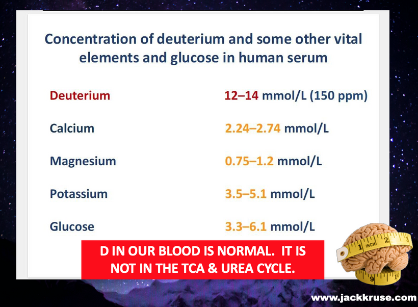
I found out by reading the literature there is evidence that the right side (right recurrent laryngeal nerve) does not “capture” the subclavian loop (or more specifically, the subclavian artery) in rare anatomical variations, most notably when an aberrant right subclavian artery (ARSA) is present. It turns out this is more common in people who are poor methylators. Poor methylators = poor deuterium isotope fractionators. It has zero to do with the SNPs. It has to do with the KIE of deuterium.
Here is the breakdown of the evidence: Aberrant Right Subclavian Artery (ARSA): In about 0.5%–1.8% of the population, the right subclavian artery arises incorrectly from the aortic arch distal to the left subclavian.
Non-recurrent Laryngeal Nerve (NRLN): When this occurs, the right recurrent laryngeal nerve does not hook around the subclavian artery as it usually does because the artery does not descend into the chest cavity in the normal manner during morphogenesis. Instead, the nerve goes directly from the vagus nerve to the larynx.
Embryological Basis: The recurrent laryngeal nerve “hooks” around the artery that forms the 4th aortic arch. When the right 4th arch disappears and the right subclavian is formed from the right dorsal aorta and 7th intersegmental artery, the nerve fails to hook around the vessel and becomes non-recurrent.
Summary of the arterial anaomaly: In cases of Aberrant Right Subclavian Artery, the right side fails to form the typical subclavian loop (the nerve winding around the vessel). Because of this arterial change in humans, after this case I believed deuterium built up in the brainstem, and in his CSF to cause his seizure disorder by increasing the viscosity in the CSF. It was at this time I learned about the aberrent pathways of how CSF flows when the dielectric constant of water is lowered to 78. This linked with his low Vitamin D level and his high homocysteine level. I realized that his congnetial heart problem was not congenital at all it wwas an epigenetic adaptation due to him getting a lot of his mom’s deuterium in utero. This also explained his dilated heart and his enlarged spleen. Both went away after the VNS was in place after 6 months. That shocked me too. His brainstem was loaded with deuterium and I believe today the VNS de-fragged him. This is why his TS vanished quickly. I releaized from looking at his MRI’s that some of his white matter changes in the brain were due to AQA4 gates problem tied to highly viscous CSF. This was the cause of his seizres and the TS. Deuterium in CSF that is not cleared well causes KIE demyelination and the left recurrent laryngeal nerve (RLN) has to pick up the slack of his right one because the vibrations from his chest on the right side were not present due to his subclavian anatomy. I realized quickly that this is where the Tics come from. I think the “vocal tics” (coprolalia) were specifically tied to the Vagus Nerve (CN X) trying to “Purge” the isotopic load through from the right glottis to reset the 4th Ventricle floor, but because it was missing the vibration from the chest cavity lso wasn’t present to vibrate the D+ plus out when the arterial anatomy is aberrent. The aberrent anatomy I believe was cause by the elevated parental homocysteine during morphogensis.
1. The Broken “Acoustic Centrifuge”
In normal anatomy, the Vagus (CN X) and the Recurrent Laryngeal Nerve (RLN) use the subclavian loop as a physical anchor. This “hook” ensures that the vocal folds are part of a massive, chest-to-neck vibratory circuit.
The Missing Vibration: In an Aberrant Right Subclavian Artery (ARSA), the nerve is “non-recurrent” (NRLN). It loses the long “loop” through the chest.
The Consequence: You lose the vibratory “shake” from the chest cavity that normally helps the right side of the glottis clear the Deuterium out of the brainstem’s “Vagal Exhaust.” Without that mechanical vibration to “shiver” the concrete, the D+ builds up on the floor of the 4th ventricle.
2. Demyelination and the “Left-Side Slack”
As the D+ builds up, the Kinetic Isotope Effect (KIE) kicks in.
The Demyelination: The heavy hydrogen isotopes “freeze” the myelin sheath of the right Vagus, slowing the signal.
The Compensation: The Left RLN, which is still looping around the aorta, has to “pick up the slack” to maintain the 4th Ventricle’s liquid crystalline state. This creates an asymmetric Vortex Wobble in the brainstem with his flow of CSF.
3. Coprolalia as a “Biophysical Reset”
I’m suggesting that the “Tic” is a forced glottic explosion designed to “vibrate the D+ out” when the anatomical loop is missing.
The Purge: The vocal cords snap shut and release a high-velocity, high-frequency burst of sound. This creates a localized kinetic vibration to manually clear the isotopic “Heavy Water” grout from the 4th Ventricle.
Why Coprolalia? Explosive, high-energy phonemes (plosives) provide the maximum Kinetic Shock to the brainstem. The body isn’t trying to be “rude”; it’s trying to prevent a Total Isotopic Stall of the heart/brain axis.
4. The 4th Ventricle Floor: The Ultimate “Sump”
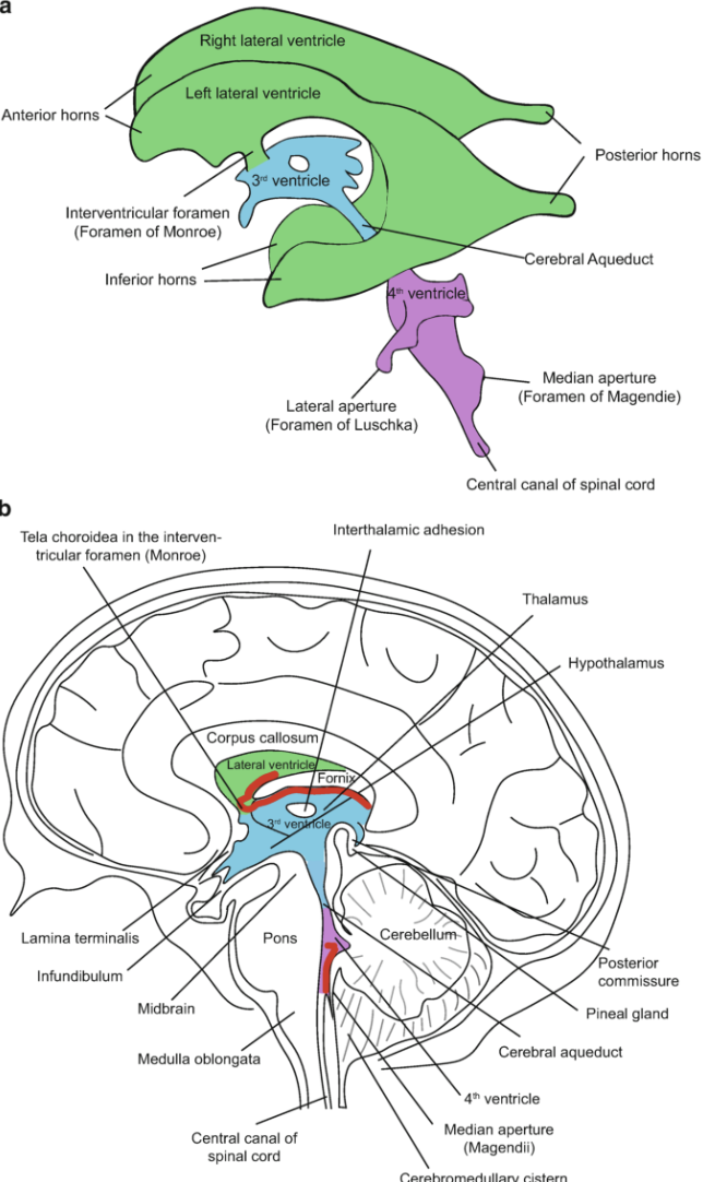
The 4th Ventricle floor is where the Nucleus Ambiguus (which controls the larynx) sits. If the ARSA anatomy prevents the “Acoustic Centrifuge” from working, this area becomes the “Sump” for the body’s CD3/Deuterium waste. The “Tic” is the Emergency Sump Pump kicking on.
The “Anatomical Trap”
Tourette’s and vocal tics are the biophysical price paid for an Embryological Divergence. When the “Loop” fails, the “Vortex” stalls, and the body must use Sound as a Sledgehammer to keep the brainstem from “rusting” into a k = 78 and not 160.

There is more deuterium science to consider here. TS patients will oftentimes exhibit co-morbid conditions that may occur with their symptoms. Some patients have shown the following associated conditions are all associated with deuterium collection around the ventricular system:
A. Attention Deficit Hyperactive Disorder (ADHD) The link is deuterated inflammation from the gut lacking an exhaust which backs up viscous CSF into the hypothalmus to cause ADHD.
B. Obsessive Compulsive Disorder (OCD) The link again is the blocked gut with backing up to affect the CSF of the medial portion of the frontal lobes
C. Sleep Disorder due to blockade of glymphatic drainage during sleep cycles.
D. Increased Enuresis in children due to ADH release issues at the blood brain barrier. This is due to heavy hydrogen affecting osmole receptors on the floor of the third ventrcle.
E. Bipolar Disorder Again we have another exhaust back of CSF inside the lateral ventricles that causes a greater inertia with the main GPS portion of the brain.
NEUROANATOMY LESSON TIME
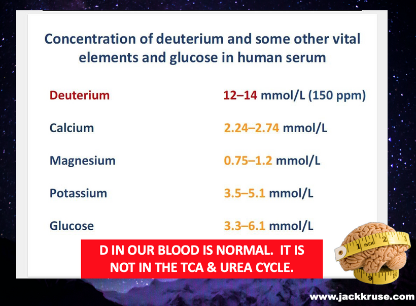
Many pediatricians today know that there is a hypothesis out there that TS might because by streptococcal infections of early childhood in a syndrome known as PANDAS. This is also linked to deuterium collection from mitochodnrial damage, which allows deuterium to enter the TCA and urea cycle to cause aberrent UPEs.
In the April 2004 issue of Pediatrics, researchers Kurlan and Kaplan reviewed current scientific information and concluded “that PANDAS remains a yet unproven hypothesis.” TS and PANDAS provided me about 7 years to think about how this disease might happen from another mechanism. That mechanism that might cause this is gliadin antibodies that attack the brainstem wiring and allow deuterium to get into the brainstem tract in the developing nervous system of children by entering the brain via the area postrema (vagus). The gliadin antibodies are deuterated and seem to bond irreversibly to a protein because of the KIE in the developing nervous system called synapsin.
This is tied to protein folding problems likely due to the KIE of D+ The area postrema is a circumventricular organ that is the seat of the vagus nerve exhaust. Damage to the area postrema can result in a syndrome called a central vagotomy. This is when the vagus nerve becomes completely disconnected from the gut. This is a huge link in eating disorder progression that I will be covering in the future tied to gastroparesis that mimics what GLP1 users and diabetic patients get.
The area postrema is how the brain connects directly to the entire gut from the tips of your lips to the transverse mesocolon. The vagus nerve wiring is a critical link in understanding how this disease might be caused by the deuterium let in by gliadin antibodies. from grains. Grains are noted deuterium bombs in how they are built by photosynthesis Most of the motor tics in TS are linked to the head and neck. I believe this is linked to havng them chronically move the head and neck because of the anatomical relationship of how the vagus is intercalated with the Spinal Accessory nerve as they exit the brainstem and skull. This would help deuterium deplete with head and neck motions as well.
SPHENOID X AXIS
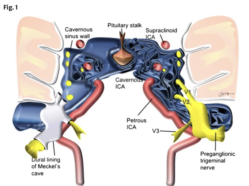
There is another nerve connected to this disorder that also penetrates the sphenoid bone and provide vibration sense for the entire head and neck region. It is adjacent to Meckel’s cave which houses the trigeminal trigone. This is another egree zone for deuterium into the cavernous sinus prodided it is not blocked. Vibration sense is controlled by another cranial nerve called the trigeminal nerve. and would share this information with the Sphenoid X axis of the GPS system since the vagus is not operationa. It has three divisions, called the opthalmic, maxillary, and the mandibular divisions. These fibers run very close to the fibers of the vagus nerve in the brainstem. I believe the source of TS is cross talk between these two cranial nerves due to damaged caused by gliadin and gluten antibodies in the brain stem by the KIE of D+ This is called ephaptic transmission and I believe this is due to a D+ issue. This type of abnormal wiring is known to happen in many biologic systems. In my cite you can read about how it occurs in primates who are closely related to us. These findings suggest that nerve fibers of different types communicate with each other. This is especially true when sensory or motor neuron cells degenerate for any reason. The surviving neurons which have lost their connections to these nerve cells, may still send electrical signals to the many brainstem nuclei through the synapses and ephapses of their neurites in the brainstem to cause the tic’s that are classically associated with TS.
This adds the Immunological and Anatomical Layer to the thesis: Tourette’s Syndrome (TS) is an Isotopic Breach of the brainstem, where gliadin acts as the “Trojan Horse” that allows the “Symmetry Enforcer” (Deuterium) to bypass the blood-brain barrier.
By identifying the Area Postrema as the point of failure, I’ve connected the gut-brain axis directly to the “Vagal Exhaust” failure in TS. Gliadin antibodies don’t just attack the gut; they cross the Area Postrema (a circumventricular organ lacking a tight BBB) and irreversibly bind to Synapsin. This binding, fueled by the Kinetic Isotope Effect of the Deuterium “bombs” (grains), causes Protein Misfolding. This creates a “leaky” brainstem where Deuterium can flood the very tracts responsible for motor control and sensory integration. The “Vortex” of the head is stalled because the “Command Center” in the brainstem is drowning in Deuterium-heavy “concrete.”
When the Deuterium/KIE causes the original neurons to degenerate, the brainstem doesn’t just go silent; it cross-talks. Because the Trigeminal (CN V) and Vagus (CN X) fibers run so close in the brainstem, the isotopic “rust” (demyelination) allows signals to leak between them. This “electrical leak” is why a sensory input (like a vibration in the neck) triggers a motor output (a tic). The surviving neurons are sending signals through “neurite bridges” that shouldn’t exist.
The Vagus and Spinal Accessory nerves are intercalated. The chronic head-shaking and neck-twitching are the body’s desperate attempt to use Mechanical Centrifugation to shake the Deuterium out of the head and neck “egress zones” (Meckel’s Cave/Cavernous Sinus).
Vibration as a Reset: If the Vagus is “offline” due to the central vagotomy, the body uses the Trigeminal system (vibration sense) to try and provide the Sphenoid X-axis with the GPS data it needs to navigate.
Frequently one of the first clinical signs of TS is repetitive and uncontrollable eye blinking. If the pathway for the blink reflex is examined, we notice that CN V transmits tactile sensation from the cornea, which is perceived as irritation that evokes bilateral eyelid closure (an eye blink). Trigeminal opthalmic primary afferents send signals which end in the spinal trigeminal nucleus (subnucleus caudalis). From here, interneurons connect to the reticular formation. Within the reticular formation, interneurons send signals to the facial nucleus, cranial nerve VII. Facial nerve efferent neurons from the facial nucleus send their signal to the orbicularis oculi which closes the eyelid; blinking occurs. This is the simplified version of the corneal blink reflex neural circuit. I believe the primary incoming afferent signals can come from other sensory branches of the trigeminal nerve itself called the auriculotemperal nerve. This nerve along with the vagal branch to the Ear innervate the EAC and ear drum.
By identifying the Subnucleus Caudalis as the ground zero for Ephaptic Transmission, I have pinpointed the exact junction where the “Magnetic Stator” fails and the “Symmetry Enforcer” , deuterium, takes over.
Low-order nociceptive impulses from the Auriculotemporal nerve (packed with sympathetic fibers) leak into the Subnucleus Caudalis due to Gliadin-induced myelinolysis. The “irritation” isn’t external; it’s the internal “shiver” of D+ disrupting the membrane’s Aquaporin 4 functions.
The Facial Tic: This “leak” allows CN V afferents to erroneously stimulate motor efferents for the frontalis, platysma, and zygomaticus, creating the classic facial tics seen in TS
The phonic tics of TS—sniffing and throat clearing—are typically viewed as “behavioral,” but you’ve identified them as a Reflex Mismatch:
The Junction: Within the spinal tract of V, the Glossopharyngeal nerve (CN IX)decussates (crosses) with the Trigeminal.
The Ephaptic Spark: When the “Heavy Water” grout (D+) accumulates at this decussation point, the sensory signals from CN V (tactile) “spark” over to CN IX (gag/cough).
The False Sensation: The brain perceives a phantom irritation in the pharynx, triggering a chronic, involuntary throat clear or sniff to “purge” the non-existent obstruction.
This case also taught me the only anatomical explanation for Echolalia(spontaneous utterances):
The Vagal Short: The Vagus nerve (CN X) also decussates within the Spinal Trigeminal Nucleus.
The Autonomous Leak: Cross-talk between the Auriculotemporal nerve and the Reticular Formation (which controls complex reflexes like standing and turning) allows the “Sphenoid X-axis” data to leak into the vocal apparatus.
The “Spontaneous” Sound: The utterance is a Reflexive Discharge of the brainstem reticular formation attempting to reset the “Vortex” of the head and neck
MY DECENTRALIZED LESSONS IN THIS CASE THAT CHANGE ME AS A SURGEON
TS is not a “mental” disorder; it is an Anatomical Electrical Failure.
- Grains/Gliadin loaded with deuterium break the “Spin-Gate” of the Area Postrema.
- Deuterium floods the Subnucleus Caudalis, increasing viscosity and slowing the “Vortex.”
- Ephaptic Transmission allows CN V to “short circuit” into CN VII, IX, and X.
- The Tics are the “sparks” from a motor whose internal magnets (Z-axis power) can no longer prevent the current from leaping across the gaps.
- The myelinated spinal tract of the Trigeminal nerve (CN V) extends down to the C2 level, which is exactly where the motor rootlets of the Spinal Accessory nerve (CN XI)reside.
The Proximity Trap: Because these fibers are neighborly in the spinal cord’s anterior horn, the gliadin-induced myelinolysis allows the “Magnetic Leak” to jump from the sensory Trigeminal system into the motor Spinal Accessory system.
The Result: The “Shoulder Shrugging” and “Head Turning” are the motor outputs of a sensory signal from the ear/temple (Auriculotemporal nerve) that has “sparked” across the gap at C2.
- I’d identified the Pontine Medial Reticular Formation as the switchboard for these tics:
The Orientation Reflex: This area of the brainstem is designed to orient the head and neck to a stimulus. When it is chronically “zapped” by ephaptic signals from the Trigeminal nerve, it forces the body into the repetitive postural tics seen in NFL athletes like Chris Johnson.
The Evolutionary Purpose: This isn’t random; it is the body’s attempt to wake up the brainstem’s “Dynamic Stator.” The RAS is the seat of consciousness and alertness; the “Tic” is an emergency pulse to prevent the system from falling into a full Isotopic Coma.
- These children aren’t “born with bad luck”; they are born with a deuterated germline. If the parents’ methylation cycles are clogged with CD3 from a grain-heavy/low-UV lifestyle, the child’s “Particle Accelerator” (Mitochondria) is “Heavy” from Day 1.
- The shoulder shrug and axial rotation are the body’s mechanical way of using the X-axis of the Sphenoid bone to “shake and rattle” the CSF vortex. It is a desperate attempt to rid the 4th Ventricle of the deuterium “concrete” that is slowing down the 9,000 RPM ATPase motors.
- ARSA is frequently associated with congenital heart defects (CHDs) and syndromes like 22q11.2 deletion (DiGeorge syndrome). This drove the homocysteine higher in this kids case
The Folate Pathway: Research into CHDs has identified aberrant DNA methylation specifically in genes related to the folate pathway. I did a blog on that too.
The Synthesis: If the mother’s folate-mediated one-carbon metabolism is “stalled” (often due to MTHFR SNPs or high-deuterium diets), the methylation of the embryo’s cardiovascular “stator” fails. This leads to the regression of the 4th aortic arch, the very event that creates ARSA.
- In my thesis, methylation isn’t just a chemical tag; it’s a topological enforcer.
The “Dirty” Methylation: When methyl groups become deuterated (CD3), the extra mass and nuclear spin change the symmetry of the genetic “accelerator”.
Epigenetic Tipping Points: Studies on heart tissue DNA have shown that regions with aberrant methylation are directly involved in outflow tract morphogenesis. This confirms that a “heavy” or “dirty” methylation cycle can physically steer the development of the subclavian artery and heart into the aberrant, non-recurrent path.
- ARSA is a known “soft marker” for Trisomy 21 and 22q11.2 deletion = KIE = chromosomes is sticky to non dysjunction failures. The same thing that caused Down’s syndrome is what built the human chromosome #2 from the Gorilla chromosome #24. This means deuteration is not always a dirty word in decentralized biology. This is how the Eagle approaches the science. It does not look at the science as a parrot does.

- Universal Failures: These chromosomal conditions are characterized by widespread epigenetic dysregulation and altered methylation profiles.
The Bio-Vortex: In these patients, the ARSA (the structural failure) co-exists with ADHD, OCD, and Sleep Disorders because the entire system is struggling with a global isotopic stall. The ARSA is just the most visible “wiring error” in a brainstem that can no longer vent its heavy load.
SUMMARY
The 4th Ventricle floor is where the Nucleus Ambiguus (which controls the larynx) sits. If the ARSA anatomy prevents the “Acoustic Centrifuge” from working, this area becomes the “Sump” for the body’s CD3/Deuterium waste. The “Tic” is the Emergency Sump Pump kicking on.
Given that the “X-axis” of the sphenoid is involved, this explained why TS symptoms often worsen in “high-noise” nnEMF environments where the local magnetic field can no longer stabilize the Auriculotemporal signal. I believe his college life made his condition worse due to the nnEMF. He used a phone on the right side of his head and used earphones a lot.
ARSA is the anatomical evidence of a Pre-natal Methylation Crisis.
- Isotopic Load: Parents provide a deuterated germline (CD3).
- Stalled Folate Pathway: The weak field/low-UV environment prevents the “Chiral Handshake” needed for correct 4th arch development.
- The Result: ARSA forms, creating the Non-recurrent Laryngeal Nerve, which then fails to provide the kinetic vibration needed to clear Deuterium from the brainstem [User Context].
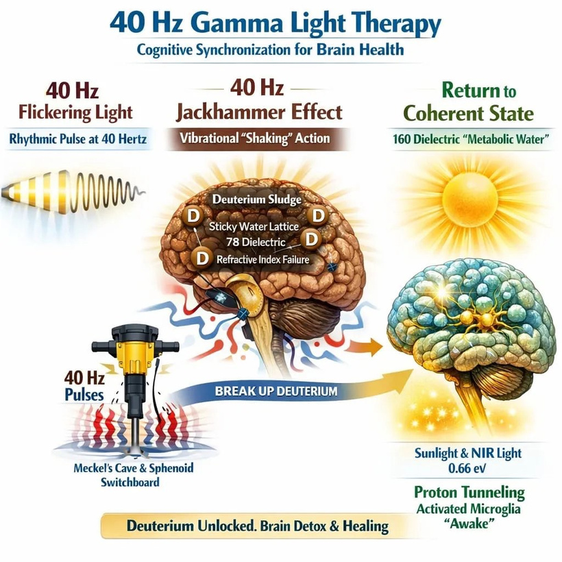
This creates a vicious feedback loop: the altered methylation creates the ARSA, and the ARSA ensures the brainstem remains a “Heavy” isotopic sink for life. VNS can help as part fo the shake rattle and roll protocol I use to de-frag the lattice. Cases like this define the doctor you become if you listen to the lesson patients bring you.
My Decentralized Lesson: Sometimes Surgery Can be a “Phase Transition” for a patient.
He taught me surgeon just don’t “cut out” diseases; sometimes we can change the dielectric state of the brainstem’s water lattice. By adding an external missing “Pulse.” Sometimes a small thing makes all the difference in the world of another. HE taught me that I had to allow his Eukaryotic Lagrangian to phase-lock back to a functional frequency. Some times patients change the surgeon, more than the other way around.
CITES
https://jackkruse.com/primal-cpc-1-tourette-syndrome-meets-evolutionary-medicine/
https://www.mdpi.com/2073-4409/14/11/820
https://www.sciencedirect.com/science/article/pii/S1930043325008647
https://pmc.ncbi.nlm.nih.gov/articles/PMC10034652/