CPC #71: GOLD/YELLOW AS A POMC RADIATION SHIELD DURING AIRPLANE TRAVEL?

video
play-sharp-fill

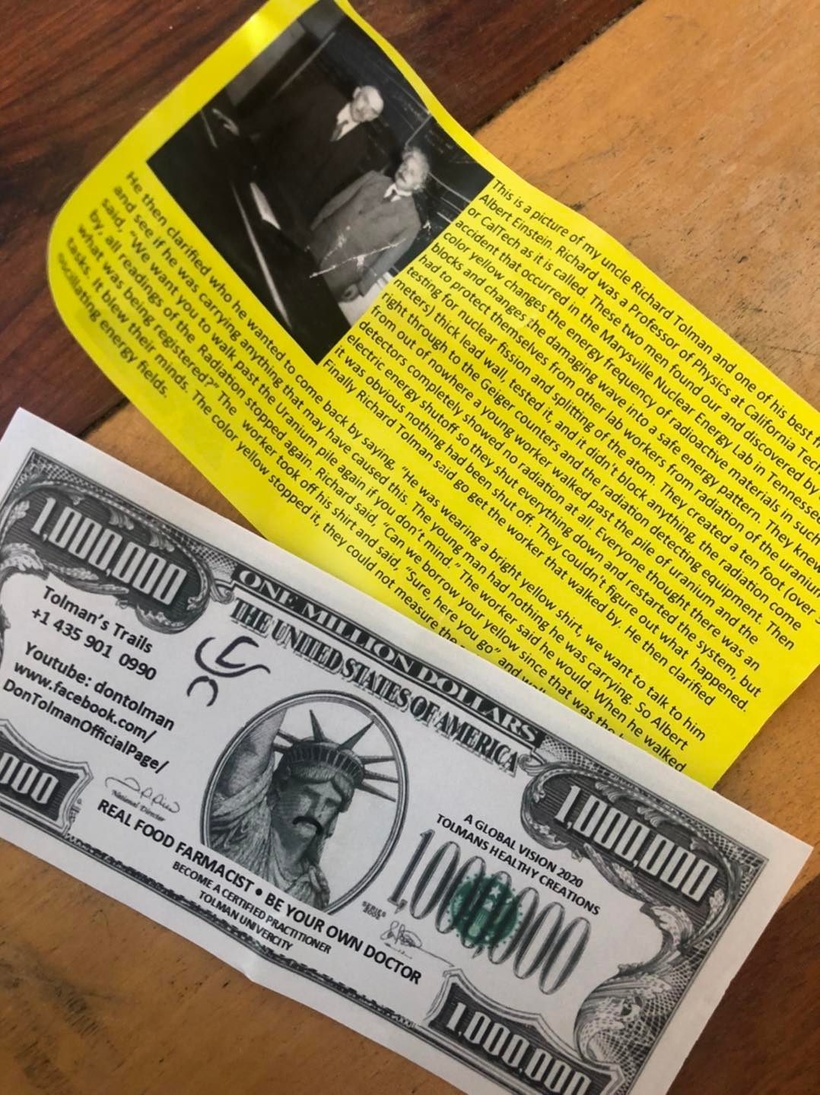
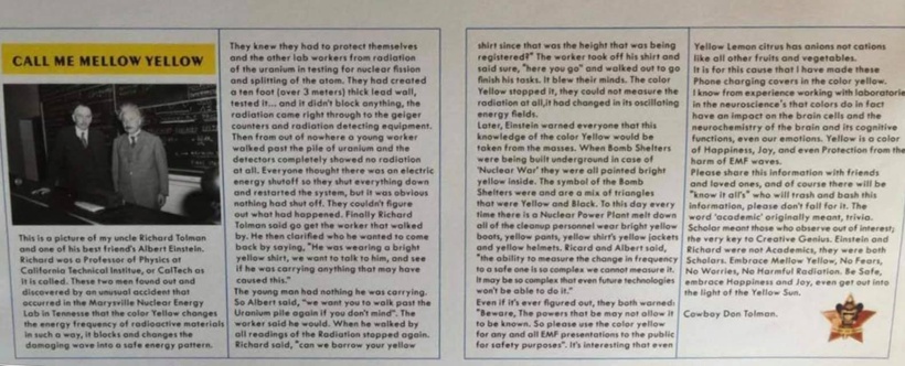
For a long time, there has been a guy running around the world advocating wearing yellow clothing to block radiation.  Is this pseudoscience or might there be something to this?  You might even run across this pamphlet from time to time.

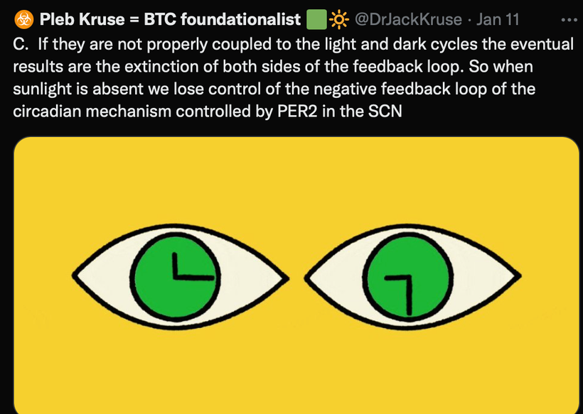
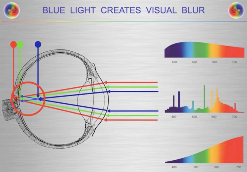
What is a possible mechanism for this observation in Nature? Many people do not know when I look into the eye of a patient with an ophthalmoscope one of the things I am looking at is the macula lutea. The macula contains the fovea where color vision is the sharpest. “Lutea” is yellow in Latin. This area collects yellow carotenoids to decrease the visual blur that is induced by blue light because blue light is the high frequency of light and it is the frequency that bends the most for the macula to handle. Yellow is the complementary color of blue.

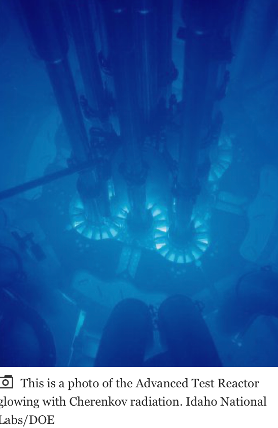
In nuclear reactors, the heavy water that contains spent fuels emits Cherenkov radiation. The frequency spectrum of Cherenkov radiation by a particle is given by the Frank–Tamm formula. Unlike fluorescence or emission spectra that have characteristic spectral peaks, Cherenkov radiation is continuous. Around the visible spectrum, the relative intensity per unit frequency is approximately proportional to the frequency. That is, higher frequencies (shorter wavelengths) are more intense in Cherenkov radiation. This is why visible Cherenkov radiation is observed to be brilliant blue. Cherenkov radiation is a form of energy that we can perceive as a blue glow emitted when the electrically charged particles that compose atoms (i.e. electrons and protons) are moving at speeds faster than that of light in a specific medium. Ironically, most Cherenkov radiation is in the ultraviolet spectrum—it is only with sufficiently accelerated charges that it even becomes visible to our retina; the sensitivity of the human eye peaks at green, and is very low in the violet portion of the spectrum. This helps explain this scientifically. Anyone who calls it pseudoscience without examining it for themselves should not be listened to.

HERE IS WHAT THE RADIATION LOOKS LIKE INSIDE A NUCLEAR REACTOR

DOES SCIENCE PROVIDE A POSSIBLE ANSWER FOR THIS OBSERVATION IN NATURE?

Can Gold be used as nnEMF radiation shield for melanin protection?

Now for more science related to this idea.

In 1988, this was published in the NY Times and I remembered reading it.  Remember gold emits a yellow color because of Einstein’s relativity equations.

What was in the article I read in 1988 in New York City?

I was dental school in 1988 and I was learning about class 5 gold foil dental reconstructions and came across this article back then.

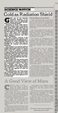
GOLD, one of the heaviest chemical elements, is the basis of a new lightweight plastic foam under development as a radiation shield.

Scientists at Texas A&M University have found a way to intersperse gold atoms with other atoms in the long molecular chains that make up polymers. By bubbling gas through the gold polymer, it can be expanded into a light foam that reportedly shows great promise as a shielding agent against neutrons and other types of radiation.

Dr. John Fackler, director of the program, said the new polymer combines gold with triphenylphosphine, a compound of carbon, hydrogen, and phosphorus, in a form that may be suitable for making anti-radiation garments.

The polymer is 11 percent gold by weight, and the gold atoms in the substance efficiently scatter or absorb most forms of radiation, including X-rays. Chemically incorporated into a polymer, gold is less poisonous than other heavy metals that also block radiation.

Metallic gold is not poisonous, but when incorporated into compounds it may be. Dr. Fackler said that because gold is chemically very stable, it tends to revert to its native state from some of the compounds it forms. ”We often have trouble with shiny, yellow metallic gold precipitating out of liquid compounds – just what refiners want, but the opposite of what we want,” he said.

The gold polymer the Texas group has developed looks like plain white plastic, Dr. Fackler said, but it emits a yellowish glow when exposed to ultraviolet light.

SUMMARY

Many might find this shocking but there is a lot of science behind these claims about the color yellow and the atom of gold.  One of my former members in the mining industry could not believe this when I share it with him during a consult I did on him in 2015 about his melanoma risk.

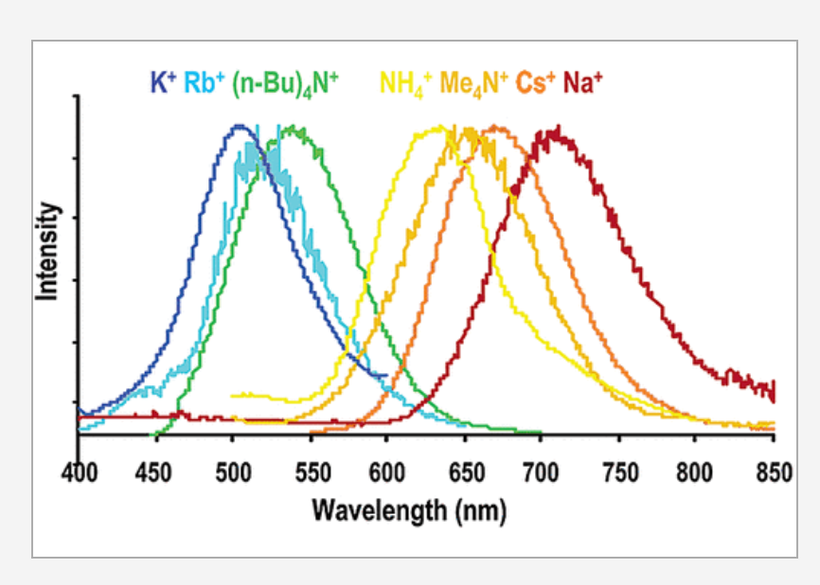
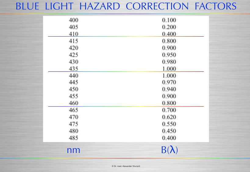
Above you see a series of bis(thiocyanato)gold(I) complexes with Au−Au interactions show luminescence in the range from 500 to 670 nm.  These frequencies include the blue light frequencies that cause the blue light hazard in the human retina. This picture below from Alexander Wunsch, MD show this effect.

The series of salts correlates emission energy with the reciprocal of the Au−Au distance. As the Au−Au distance increases, the emission energy decreases. The ligand system provides no framework for the Au−Au interaction. The emission energy seems totally determined by the Au−Au distance.

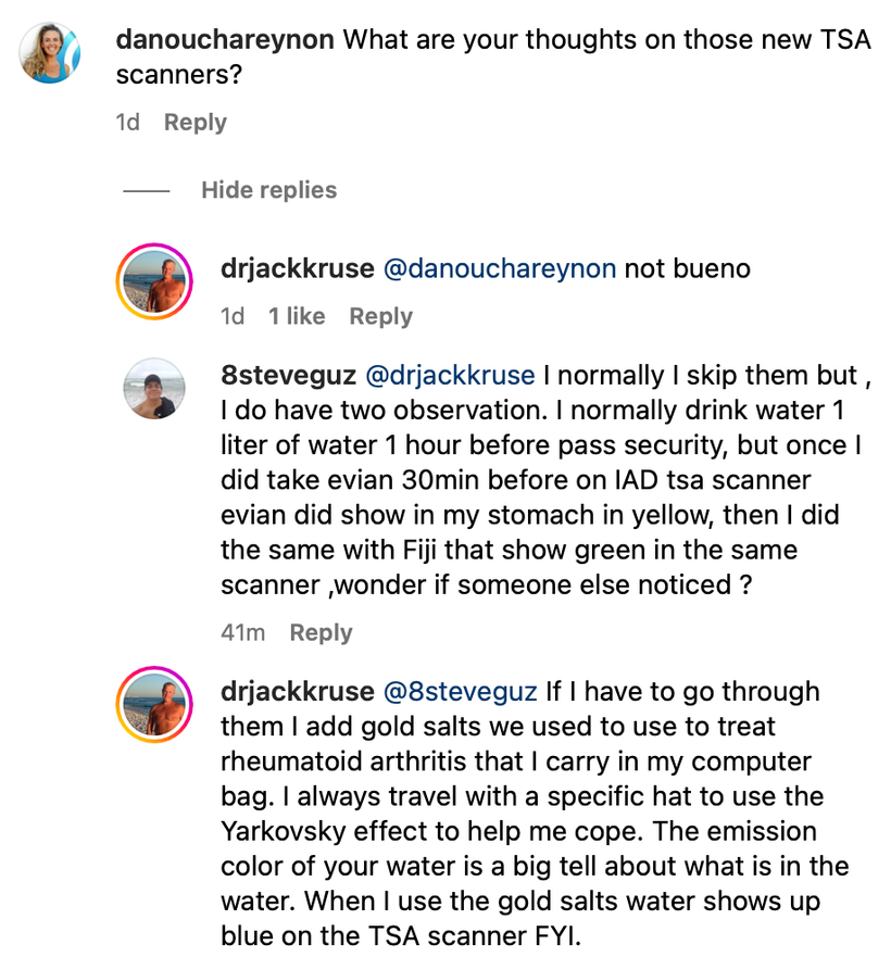
So when I am in a country where I cannot opt out of the new TSA scanners I reach into my computer bag to protect melanin in body.  I usually take it 30 minutes before flight.  Last time I had to do this was in Germany when I went to speak at Flowfest.  I posted about this hack on Instagram.

Gold therapy, while not commonly used in the treatment of RA today, is still available in oral capsule form as Ridaura (auranofin). The injectable forms—Myochrysine (aurothiomalate) and Solganal (aurothioglucose)—are no longer manufactured but they can be compounded by a pharmacy.  Below is my travel jacket I wore to Europe the last time I went there. The jacket is definitely not a fashion statment.

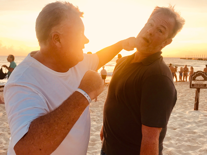
Jason Bowden Smith, a former entrepeneur member who was a mining industry executive, was with me in Mexico 4 months before the Germany talk and I told him about the jacket hacks and the gold hack and why I was doing it to go to Europe.  Here is the picture taken that morning at 8:34 AM on January 4, 2018.  He just did not believe what I shared with him.  That maybe explains why reality unfolded as it did.

The gold salts and clothing are expensive hacks to protect melanin, but I think my health is worth the expense.  I first mentioned this effect to Jason Bowden Smith  in a consult in 2016.  He was from Australia and had a very challenging medical history.  We did a few consults when he was alive & during a consult I told him about these effects for long flights to mitigate TSA scanners, cosmic/gamma radiation exposure, and WiFi toxicity from passenger use around him.

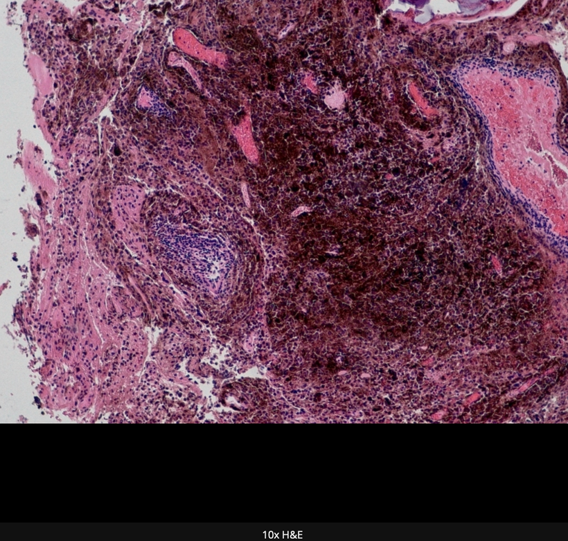
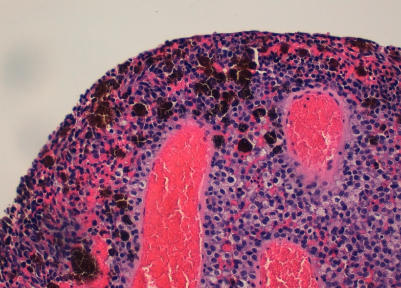
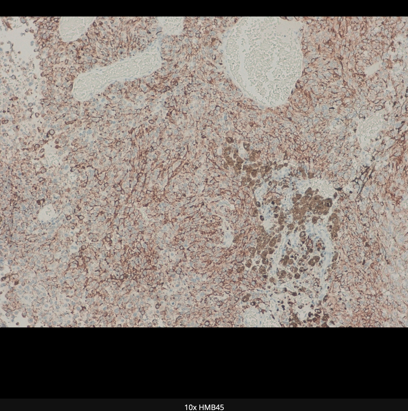
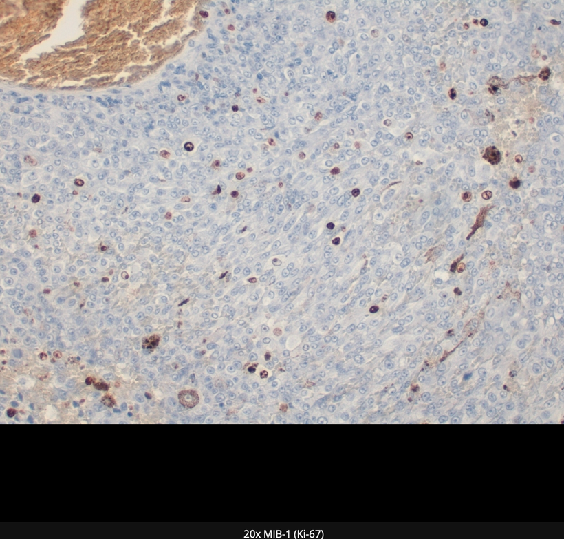
Gold therapy can induce an internal Yarkovsky effect  (below) for melanin protection.  People who have had melanoma and who still are afraid of the sun should be told about this mitohack.  Very few of my clients needed this.  Unfortunately he did need the advice and he got it.  UInfortunately, he believed a lot of the propaganda that is floating around in Australia even today about the sun.

Jason took a lot of circumpolar flights in his life.  I thought this was linked to his REAL melanoma & thyroid risk and I told him about POMC and melanin.  I told him a lot of what you’re learning now in this blog series a long time ago.  In fact, Jason was with me in a meeting the night before my clinic opened in New Orleans in December 2018 to talk about a business opportunity about this very topic since he had deep connections with gold miners in Australia.  My nurse, Chantal was present at this meeting.

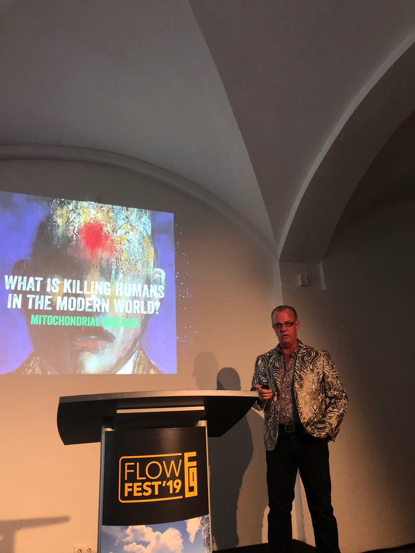
Anyone who was with me in Germany knows I did not feel well when I was in Germany because they had recently turned on 5G in the city I was speaking in.  I distinctly remember Jason mocking me before my talk when I took my clothes off and laid in the sun to get ready to speak.  He kind of pissed off my nurse who was with me at the event.  He just did not believe that something on the surface could have that big an effect on our mitochondria below.  I told him that the gold/yellow can protect quantum coherent process happening with 1/2 nuclei spin magnetic moments in our colony of mitochondria to maintain quantum coherence.  I told him everything that is important in quantum biology happens on the surface before it changes the matter inside of us. I told him that in June of 2018 I was goning to unleash this in Vermont.  I did.  Here is the slide from that event.

I spoke in Germany right after the Vermont event on July 6th, 2019.  Something else happened at that talk that others can verify. The meeting organizer subtracted 45 minutes from my talk and only gave me 15 minutes to speak. Matt Maruca and Jason were both there also as a speaker and they saw this go down.  He made me a bet that there was no way I could do a nnEMF talk for the German biohacking community in the 15 minutes.  I went and powered up in the sun, drank one liter of DDW, and laid shirtless for two hours before my talk and missed many talks doing so.  Matt then got a red LED stop clock and put it in his lap to show me my time to see if I could do it.  Notice the first slide in the talk.

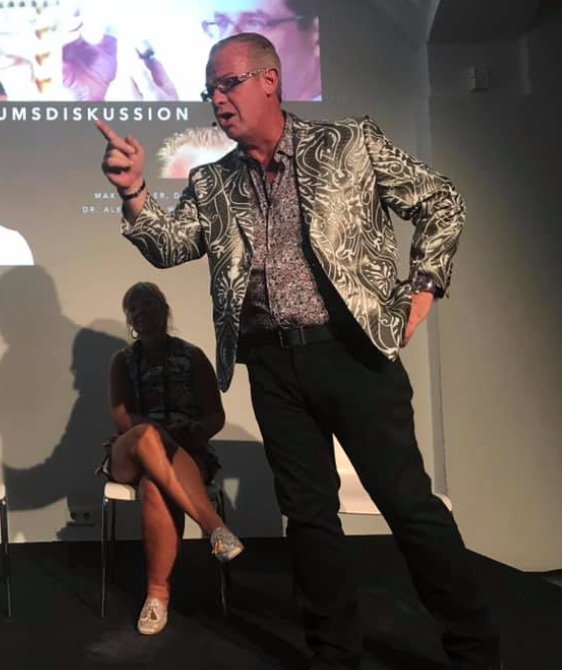
I finished the talk at 14:59 and everyone who was at the event was astounded I got the talk done in such a short time frame.  The talk was so good that the Q & A lasted two hours outside the event when I was ushered off the property because the event was closing. Jason Bowden Smith spent that two hours in the sandpit of the event with me trying to ask me more about this science.  It was the last time we met live in person and we never spoke about it again.  He died soon after it.  I feel that he never called me back to speak about things because he felt foolish that he had an answer from 2016 and thought it was a joke.  Generally I don’t joke around on consults.  I am all business.

Below this was me in Germany live on the stage during the Q&A before it spilled outside.  That jacket was tailored to be infused with gold microfiber threads hidden in the silver etchings, hence the reason I bought it and wore it.  I still have this jacket today.

What is the Yarkovsky effect?

The Yarkovsky effect is defined as a thermal radiation force that causes objects to undergo semi-major axis drift as a function of their size, orbit, and the COLOR of the material properties in the matter that makes up the object in question.

Melanin has a massive Yarkovsky effect inside of cells because of its color.  so anything you can do to reflect and deflect incoming radiation should help protecrt you melanin sheets.

The Yarkovsky effect is hard to predict, though. Scientists have to know the asteroid’s mass, size, and shape. They also need to know the patterns of light and dark on its surface, because lighter colors reflect more sunlight, while darker colors absorb more.

The best measurement of the Yarkovsky effect so far is on an asteroid named Bennu. The effect has altered Bennu’s position by about a hundred miles in just a dozen years.  So when you hear something that sounds ludicrous.  Remember it is the height of ignorance to say it is pseudoscientific without looking deeper at the issue before making a decision.

Just because something’s logical, doesn’t mean it’s not wise. Something can make sense in and of itself, but when tested against reality yield an undesirable or inefficient outcome. Being seduced by impractical logic is antithetical to wisdom, you just feel smart despite being wrong.  Sometimes chosing comfort over wisdom might kill you.

CITES

1.https://artsci.tamu.edu/news/2023/02/texas-aandm-mourns-loss-of-distinguished-professor-emeritus-john-fackler.html

2. A version of this article appears in print on Sept. 6, 1988, Section C, Page 11 of the National edition with the headline: SCIENCE WATCH; Gold as Radiation

3. https://pubs.acs.org/doi/pdf/10.1021/ja00310a028

4. https://www.instagram.com/p/CsJcPQUM2a9/

5. https://forum.jackkruse.com/threads/dog-health.27663/#post-321327

CPC#70: MELANIN DEGRADATION OF THE CARDIAC TISSUE

video
play-sharp-fill

Supraventricular tachycardia SVT is a broad term for a number of tachyarrhythmias that originate above the ventricular electrical conduction system (Purkinje fibers).  read the link below to learn about SVT.  This rhythm is usually the first symptom in melanin degradation of the cardiac plexus.  I showed you this picture in Quantum Engineering #47 for the first time.

https://www.ekgstripsearch.com/SVT.htm

In ACLS codes we use adenosine via IV push to rid the heart of this detrimental rhythm when the patient is not symptomatic.  SVT can lead to rapid cardiac standstill.  We are seeing this more commonly now due to the spike protein inflammation of the heart by mRNA vaccines.  I believe this was behind the demise of Demar Hamlin and JJ Watt.

IS THERE NOW EVIDENCE THAT INTENSE RED LIGHT can do THE same thing using the PER circadian gene?  Does this imply that red light is a drug equivalent?

YES, it does.  The picture above shows you this.

Is there more to this circadian story you need to know?  YES.

You know my answer and you should know what Dr. Tina Kuru says about this………..Red light is a drug equivalent to adenosine use.  This is a message Big Pharma wants buried from centralized MDs.

In this paper below in an effort to find out why red intense light can do this, researchers developed a photonic strategy using optogenetics to protect the heart using intense light to target and manipulate the function of the PER2 gene which is expressed in a circadian pattern in the part of the brain that controls circadian rhythms.  (Sounds like something Dr. Kruse would suggest no?)

You do know that sunlight is made of 42% intense IR-A light huh?

By amplifying this gene to improve the periodicity of the cardiac clocks, the researchers were just using LIGHT PHOTONS to do this job; they found that it protected cardiovascular tissues against LOW OXYGEN conditions like myocardial ischemia, caused by reduced oxygen flow to the heart.  Dr. Kruse called low oxygen situations pseudohypoxia (low NAD+).  These are all associated with low NAD+ levels in cytochrome 1 and leptin resistance with low delta psi on the inner mitochondrial membrane = low redox power.

They also discovered that the light increased cardiac ADENOSINE, a chemical that plays a role in blood flow regulation.   Hey didn’t Dr. Kruse just do a massive post on ADENOSINE last week on his page?

Hey, isn’t leptin resistance a synonym for melanopsin dysfunction?  Isn’t melanopsin dysfunction a synonym for a melanin problem?  Yes, it is. Tell me again how that works Dr. Kruse?

Dr. Kruse Response:  Blue light and nnEMF liberate Vitamin A from our cells and cell membranes to raise its presence in the blood plasma and this lowers plasma levels of Vitamin C and Vitamin D.  This destroys melanin in the cardiac plexus and it degrades into adrenaline and noradrenaline that begins to stimulate the heart adrenergically.  It also has effects on the glylympaphatic system in the brain and on slow wave sleep.  When Vitamin A is liberated by non terrestrial light or trauma, we are inducing a light stress and this causes Vitamin A to becomes an aldehyde that becomes a wrecking ball for many non visual photoreceptors like melatonin, dopamine, NO, adrenaline, noradrenalin, L-Dopa, P-450 enzymes, RBCs, cytochromes, and melanin.

This wrecking ball action destroys the small molecule modulators of the mammalian circadian mechanism.  PER1 and PER2 are gears in that eye clock mechanism.  Once the molecular clock in the eye and peripheral clocks goes awry the implications for many neolithic diseases spiral out of control.  What are some of the Vitamin A proteins involved in this downward spiral?

They are called retinoic acid receptor-related orphan nuclear receptors or RORs for short.  The RORs have several isoforms too called RORα-γ.  These proteins are also under the transcriptional control of CLOCK/BMAL1 heterodimers.  CLOCK and BMAL1 are positive regulators of circadian gene expression, and PER and CRY are the NEGATIVE FEEDBACK LOOP regulators that operate under day and night cycles.

These are the positive and negative feedback arms of the circadian mechanism.  They must be coupled properly to terrestrial light to operate well and control all growth and metabolism, protein synthesis and hormone production and release.  It also controls receptor biology.  It controls EVERYTHING.  These are post transcriptional explosions that blow up the whole system of optical signaling and require the cell and mitochondrial to rebuild them all while our power plants are experiencing a brown out.  This is not ideal.

If they are not properly coupled to the light and dark cycles the eventual results is the extinction of both sides of the feedback loop.  That is how all human disease begins.  It is circadian biology that couples all the molecular clock genes in humans and the SCN of the eye drives the program and the major timekeeper.

Remember this lesson from the past:

Classic Paroxysmal SVT has a narrow QRS complex & has a very regular rhythm. … A rapid heart rate will significantly reduce the time which the ventricles have to fill. This is why degradation of melanin to adrenalin and noradrenalin is a real problem.  Carefully look at how melanin degrades on the top line during periods of hypoxia.  The chemistry in hypoxia goes from right to left.

The heart fills during diastole, and diastole is normally 2/3 the cardiac cycle. A rapid heart rate will significantly reduce the time which the ventricles have to fill and as a result this lowers cardiac output and oxygen delivery = hypoxia.  As a result of this the heart tries to beat faster to improve global hypoxia and this worsens the situation.  It fast forwards on itself.  The reduced filling time results in a smaller amount of blood ejected from the heart during systole. The end result is a drop in cardiac output & hypotension.

With the drop in cardiac output, a patient may experience the following symptoms. These symptoms occur more frequently with a heart rate >150 beats per minute as the ECG strip shows:

Shortness of air (S)

Palpitation feeling in the chest (S)

Ongoing chest pain (U)

Dizziness (S)

Rapid breathing (S)

Loss of consciousness (U)

Numbness of body parts (S)

The pathway of choice for SVT in the tachycardia algorithm is based on whether the patient is stable or unstable clinically.

The symptoms listed above that would indicate the patient is unstable are noted with the letter (U) in a code situation. This can present outside a code situation when someone has a very low redox state because of a very poor environment linked to blue light and nnEMF toxicity. This mimics adrenal fatigue and brainstem pathology, sleep disorders, and eating disorders I wrote about in the QE#47 blog.  Stable but serious symptoms are indicated with the letter (S) above.

Insert any 3G-5G city or environment = an acute or chronic adenosine problem. What fucks up sleep ultimately at the dorsal longitudinal fasiculus? Problems with adenosine at the brain stem level. This is why ACLS algorithms for advanced caridiac life support uses adenosine to treat acute mitochondrial failure in the heart that results in SVT cardiac rhythms……..guess what drug is used for cardiac tachycardias in ACLS?

ADENOSINE via IV push = a DEFECT IN PER 1 or PER2.  SOUND FAMILIAR?

How do you like me now…………?

Do you still think tech use and abuse is safe and has no side negative effects for the heart of your blood vessels?  That damage can be found early in your brainstem by the DLF where glylymphatic drainage occurs.  How about your eye clock or the molecular clocks in your skin.  These are all places where melanin, melatonin, melanopsin and leptin are.

Adenosine building up occurs normally during the day when red light is powerful as we live and is one thing that drives us to feel sleepy during the night. Then while you sleep, the adenosine is cleared out by the glylymphatic system in the brainstem at the perivascular spaces, leaving you refreshed and ready to go in the morning… for most people. This does not happen in those with sleep apnea.  Those with OSA have circadian mediated glylymphatic failure of AQA 4 in the brain stem due to lowered periodicity of PER1 & PER2 (above pic).  I can see the widened perivascular spaces on 3 Tesla MRI images at the DLF.  This is how decentralized medicine is practiced.  Today, most people that have a version of EHS that affects adenosine at the brainstem level which causes their sleep destruction and hypoxia via this mechanism. This mimics adrenal problems (PVN) and causes wild symptoms doctors cannot explain.

Who else can get these problems sans a technology addiction?

if you follow your genetics from 23 and me…

There is a genetic variant in the adenosine deaminase (ADA) gene that decreases the clearance rate of adenosine. People who carry the variant may get more slow-wave, deep sleep at night, but they also may need to sleep a little longer o feel refreshed the next morning.  These people are at high risk of neurodegeneration and for normal pressure hydrocephalus and glylymphatic failure.

What is the decentralized diagnosis that your centralized sleep MDs will never resolve for you?

Check your genetic data for rs73598374 (23andMe v4, v5; AncestryDNA):

C/C: normal clearance of adenosine

C/T: reduced clearance of adenosine, more deep sleep but may feel sleepy when waking up

T/T: reduce clearance of adenosine, more deep sleep but may feel sleepy when waking up.

How do you like me now?

Intense RED LIGHT via PBM/LLLT is the first step I like I use at Kruse Longevity Center for melanin renovation of the cardiac plexus that I wrote about in QE #47.  It is always cardioprotective.  The red light of the sun is better than my fake red light panels.  Shocking huh?

It shocks only those who are not on the path of becoming a decentralized Black Swan Mitochondriac.

If you do not listen to me you might wind up needing a shock from a defribrillator sometime in your life from tech abuse or from excessive spike protein build up in and around these areas in your body.  See my blog on Demar Hamlin or JJ Watt for more detail.

When you know better you do better.

CITES

https://www.sciencedaily.com/releases/2019/08/190808115052.htm

CPC # 69: MRI & MELANIN & Dx PRIMARY MELANOCYTOMA

Today you get to see see what melanin looks like on MRIs of some of my patients.  

Melanocytes are normal, neural crest-derived cells present in the human leptomeninges (pia and arachnoid membranes) primarily at the base of the brain, the posterior fossa, and around the upper cervical spinal cord.  They are very close to cells with POMC expressed to some degree.

Meningeal melanocytomas are rare benign primary melanocytic tumors of the CNS that are derived from leptomeningeal melanocytes. They can occur anywhere along the neuraxis but are most commonly found in the spinal canal near the foramen magnum, as well as the posterior cranial fossa, Meckel cave, or adjacent to cranial nerve nuclei based on embryological movements.

Key point:  When these tumors are found in the trigeminal cave, then they are associated with a nevus of the dermatomes corresponding to the trigeminal nerve on the face. The benign dermal melanocytic nevus usually involves the ophthalmic (Va) and maxillary (Vb) divisions of the trigeminal nerve (CN V).  This again shows the neural create connections and the neuroplasticity pathways one should expect in melanogenesis from these movements in mammals.

Primary melanocytic tumors of the CNS can manifest as solid masses or as diffuse dissemination within the subarachnoid space. They range in histologic grade from benign to malignant, differentiating between the following entities:

Melanocytomas are more common in women (mean age 45-50 years old). A prolonged evolution of clinical signs of myelopathy or radiculopathy prior to surgical resection (from 5 to 10 years) has been documented.

I believe this is true because women tend to wear more clothing over their bodies due to cultural and societal reasons.

Approximately 100 cases of melanocytomas have been reported in the CNS (brain and spinal cord) since Limas and Tio coined the term “meningeal melanocytoma” in 1972. In the spinal cord, most cases of melanocytomas are found in the extramedullary intradural compartment, at the cervical and thoracic spinal levels. An intramedullary location as our case depicts is extremely rare, with only 24 cases reported before.

The unique paramagnetic properties of melanin result in a relatively specific MRI pattern for melanocytoma, consisting of iso- or hyperintensity on T1WI and iso- or hypointensity on T2WI, and with homogeneous enhancement.  The differences in MRI signal intensities relate to a variable degree of tumor melanization.

Paramagnetic means it is drawn to magnetic fields.  This is a clue why the tumor was in this location.

Definitive diagnosis is based solely on histopathological and immunohistochemical examination.

The distinction between melanocytoma and melanoma rests on the identification of cytologic atypia, mitotic activity, necrosis, and neural parenchymal invasion.

MIB-1 (Ki-67) labeling index seen in melanocytomas is low (0%-2%) while in primary melanomas is higher (2%-15%). Based on a proposition by Brat et al. the WHO classification assigns an intermediate grade to melanocytomas with increased mitotic activity and infiltrative growth that fail to meet all characteristics of malignant melanoma. In this case, MIB-1 (Ki-67) proliferation index was 5% and a definitive diagnosis of intramedullary Intermediate-grade melanocytoma of the thoracic spine was made.

CURRENT CENTRALIZED MEDICINE BELIEFS

Although classified as benign, meningeal melanocytomas may behave aggressively and a limited number may transform into malignant melanomas. Complete excision is the treatment of choice, however, this is often not possible as intra-operative hemorrhage may be severe. Furthermore, local recurrence has been reported even after gross total removal. Due to the risk of tumor recurrence even after complete excision, adjuvant radiation therapy is advised in cases of both complete and incomplete resection.  I no longer believe this.

EPIDEMIOLOGY

Peak presentation is in the fourth and fifth decades, although these tumors have been diagnosed in all age groups. Occurrence in children is very rare!

RADIOLOGY IMAGING

The patient presented after breast cancer resection with a 2-year history of progressive paraparesis, paresthesia, and dysesthesia in the left lower limb. Neurological examination demonstrated generalized hyperreflexia and clonus of the left foot.

The above MRI is done on a 1.5 Tesla magnetic and shows a solitary well-defined fusiform intramedullary lesion involving the spinal cord at the T8-T11 level, hypointense on T2WI, moderate hyperintense on T1WI, and presenting homogeneous enhancement of its solid component after gadolinium administration.

Lesion associates a cystic component in its cranial pole, and presents susceptibility artifacts in the T2WI-GRE corresponding to its melanin component and the presence of degraded blood products.

T2WI hyperintense per focal tumoral edema at the T5-T7 levels and conus medullaris is noted, suggestive of myelopathy.

OTHER IMAGES:

3 case questions are available

Q: The presence of hyperintensity on T1WI can be an important clue leading to a specific diagnosis, as happens in this case above. Which are the causes of T1 hyperintensity?

A: Melanin. Gadolinium. Fat. Blood forming proteinaceous substance. Some paramagneticstages of blood. Mineralization. Slowly-flowing blood. Calcium. (Pic below)

Q: What’s the most frequent T1WI appearance of meningeal melanocytomas?

A: Isointense or hyperintense depending on the amount of melanin content present.

Q: What’s the most frequent T2WI appearance of meningeal melanocytomas?

A: Isointense or hypointense depending on the amount of melanin content present.

The slide below reminds us that when we are looking for melanin loss we should see it missing in T1 weighted MRI images.

Radiological differential diagnosis includes:

  • primary or metastatic malignant melanoma
  • melanotic schwannoma
  • melanotic meningioma
  • melanoblastosis
  • cavernous malformation
  • astrocytoma
  • ependymoma

Operation report:

Tumor resection was performed through osteoplastic laminectomy and under electrophysiologic intraoperative monitoring.  Tumor fragments were soft, brownish, and hemorrhagic.  Melanin tumors are always bloody because they are always associated with brisk blood flow due to their need for massive oxygen consumption.  This mimics what we see in retinal bleeds around the RPE and Bruch’s membrane.

PATHOLOGY SLIDES

H&E stained slides above show in sequence sheets and nests of cells with mild nuclear pleomorphism and prominent nucleoli; note the marked melanin pigment deposition. An immunohistochemistry study revealed positivity for melanocytic markers Melan A and HMB45. MIB-1 (Ki-67) proliferative mitotic index of 5%.

KEY BLOG POINT: This index clued me in that her previous breast cancer was likely related to this mass and its location.  She had a WiFi router under her bed in her apartment at the level of her mid-thoracic spine.

Meningeal melanocytomas are most commonly found in the cervical and thoracic regions (intrathecal-extramedullary). Within the spine, melanocytomas present as intradural masses, and maybe intradural extramedullary or rarely intramedullary. They are most commonly found in the upper cervical region, as melanocytes are most concentrated at this location. I believe the real reason is that this area is usually covered and not illuminated by sunlight.  Even more interesting is they are less common in the intracranial compartment because rarely do we lose all VUV-IR-A inside the brain.  We would most commonly see this in glioblastoma multiformans cases instead.

CITES

  • 1. Louis DN, Ohgaki H, Wiestler OD, Cavenee WK, Burger PC, Jouvet A, Scheithauer BW, Kleihues P. The 2007 WHO classification of tumors of the central nervous system. (2007) Acta neuropathologica. 114 (2): 97-109. doi:10.1007/s00401-007-0243-4Pubmed
  • 2. Karikari IO, Powers CJ, Bagley CA, Cummings TJ, Radhakrishnan S, Friedman AH. Primary intramedullary melanocytoma of the spinal cord: a case report. (2009) Neurosurgery. 64 (4): E777-8; discussion E778. doi:10.1227/01.NEU.0000341516.22126.AAPubmed
  • 3. Liubinas SV, Maartens N, Drummond KJ. Primary melanocytic neoplasms of the central nervous system. (2010) Journal of clinical neuroscience: official journal of the Neurosurgical Society of Australasia. 17 (10): 1227-32. doi:10.1016/j.jocn.2010.01.017Pubmed
  • 4. Czarnecki EJ, Silbergleit R, Gutierrez JA. MR of spinal meningeal melanocytoma. (1997) AJNR. American journal of neuroradiology. 18 (1): 180-2. Pubmed
  • 5. G.Q. Hou, J.C. Sun, X.J. Zhang, B.X. Shen, X.J. Zhu, L. Liang, X.L. Zhang. MR Imaging Findings of the Intraspinal Meningeal Melanocytoma: Correlation with Histopathologic Findings. (2012) American Journal of Neuroradiology. 33 (8): 1525. doi:10.3174/ajnr.A2987Pubmed
  • 6. Wagner F, Berezowska S, Wiest R, Gralla J, Beck J, Verma RK, Huber A. Primary intramedullary melanocytoma in the cervical spinal cord: Case report and literature review. (2015) Radiology case reports. 10 (1): 1010. doi:10.2484/rcr.v10i1.1010Pubmed
  • 7. Brat DJ, Giannini C, Scheithauer BW, Burger PC. Primary melanocytic neoplasms of the central nervous systems. (1999) The American journal of surgical pathology. 23 (7): 745-54. Pubmed