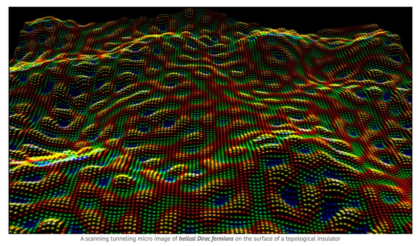
Quantum Engineering #47 really focused on the biology and physics of the bipolar disease. Everyone learns differently. this blog is essentially the same information presented to you in another way.
Which one do you like better?
Your choice actually tellls me something about your SCN and ventricular system.
1)
2)
3)
4)
5)
6)
7)
8)
9)
10)
11)
12)
13)
14)
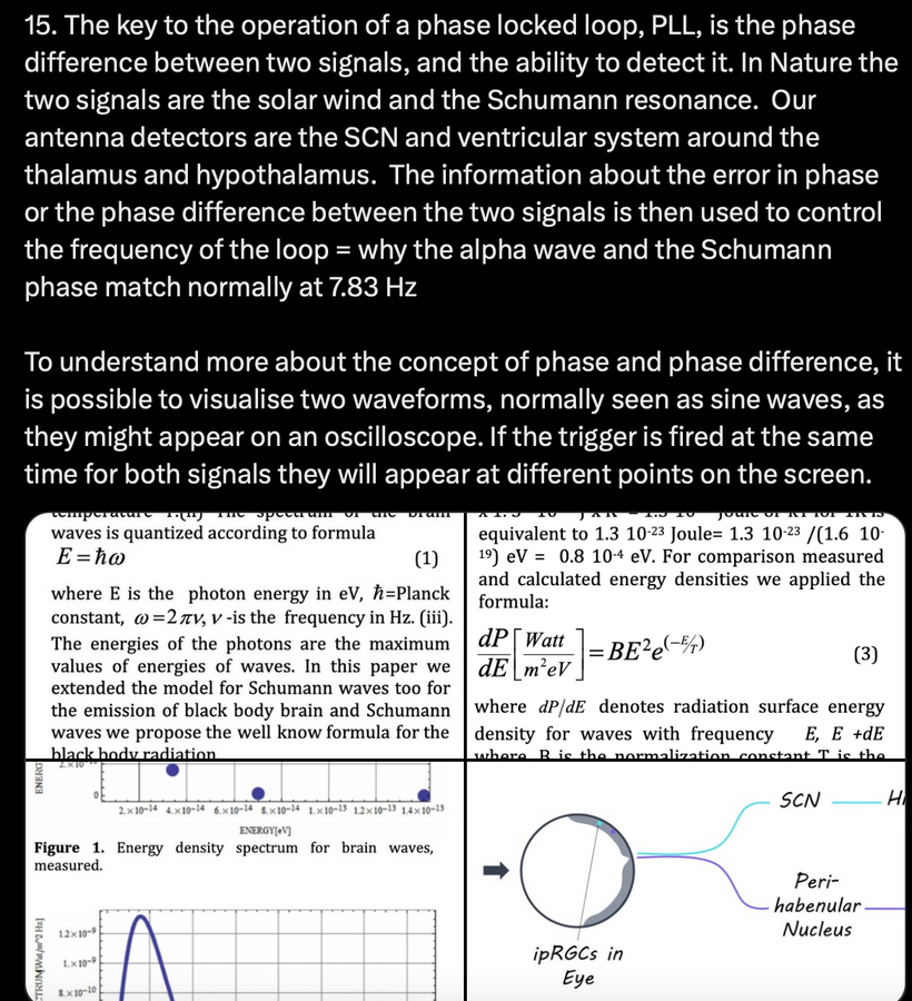
15)
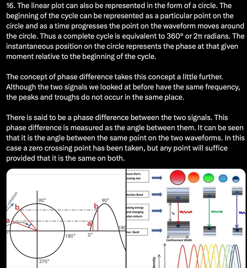
16)
17)
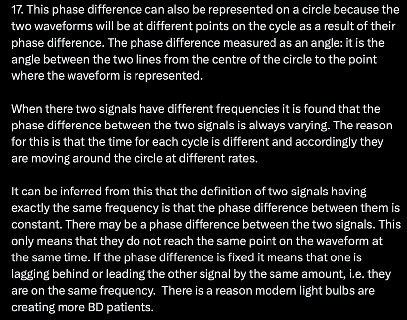
18)
19)
20)
21)
22)
23)
24)
25)
26)
27)
28)
29)
30)
31) How do you begin to fix this process?
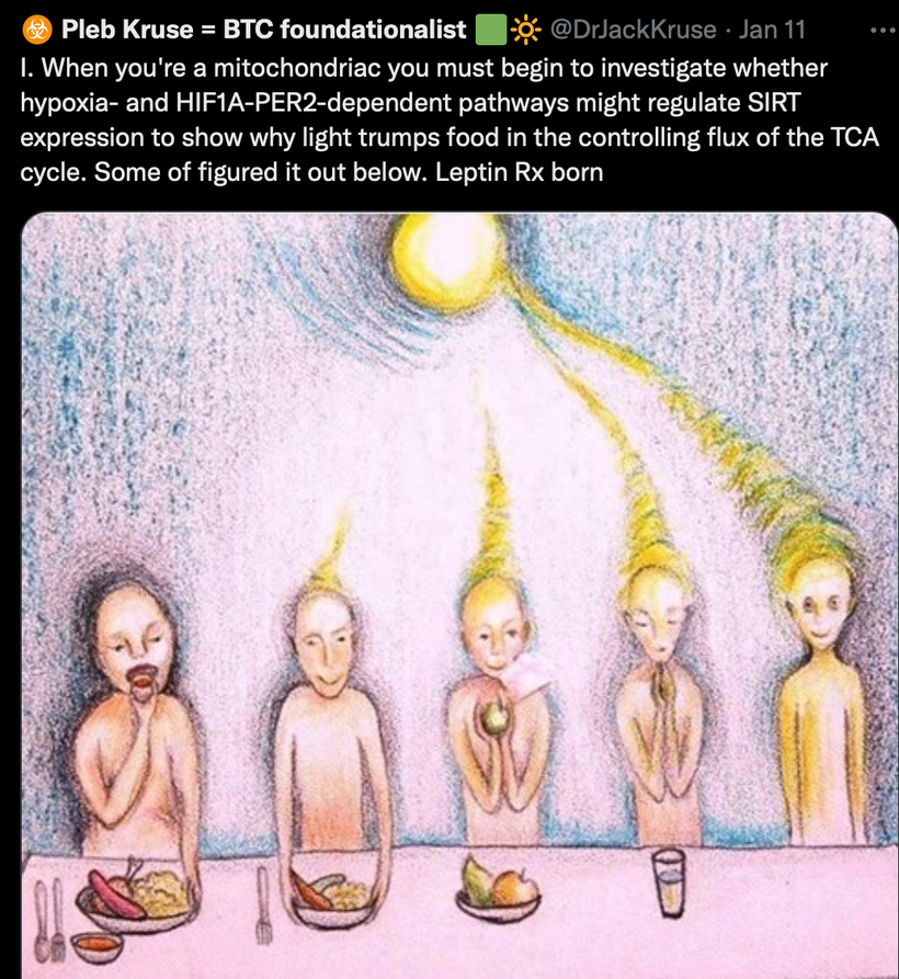
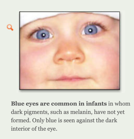
Lower the mass in the SCN and the CSF that fills your ventricles. Why was sweating a key sign to follow in the Leptin Rx written 19 years ago now? When you sweat you are degrading melanin for a short term benefit. Then you need to renovate back by increasing redox by using the sun to turn on and translate POMC to make alpha MSH. https://twitter.com/DrJackKruse/status/1671199992227278858
Implications?
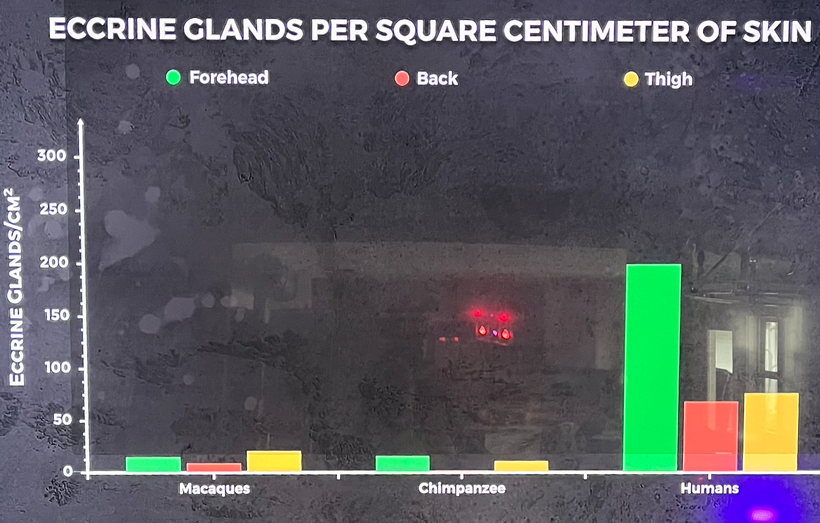
Eccrine sweat glands already exist at birth in humans and are widely distributed over the whole skin surface with only two exceptions: lips and glans penis. Depending on individual variation, there are 1.6–5 million sweat glands found across the human body with an average density of 200 sweat glands/cm2 ranging from 64 sweat glands/cm2 on the back to 700 sweat glands/cm2 on the palms and soles. Eccrine sweat glands are another superpower in mammals built by the POMC system that was expanded in us. Humans expanded the use of eccrine and apocrine glands compared to other primates. Apocrine glands are the breast of females used to mature our brains and is a signal to turn on the melanin renovation pathways in infants.
Dermcidin is an antimicrobial peptide expressed in eccrine sweat glands and plays a big role in innate host defense mechanisms. Eccrine sweat glands also play an important role in body temperature regulation for topologic control for melanin. These glands also secrete a fluid mainly composed of water and various ions.
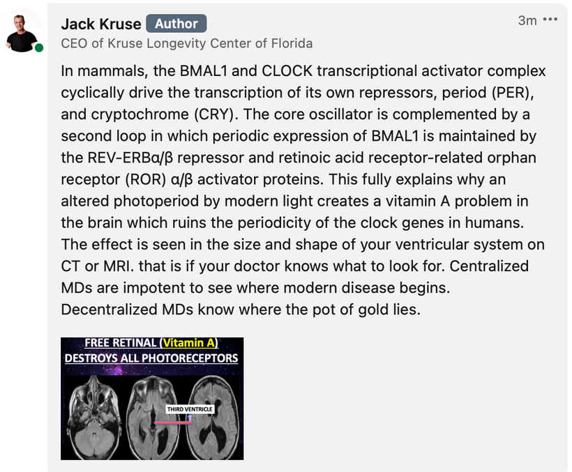
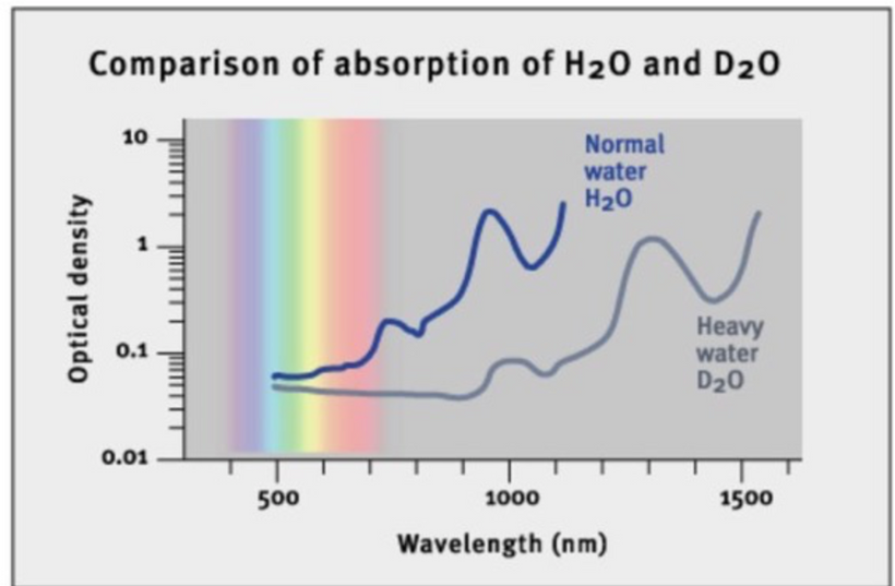
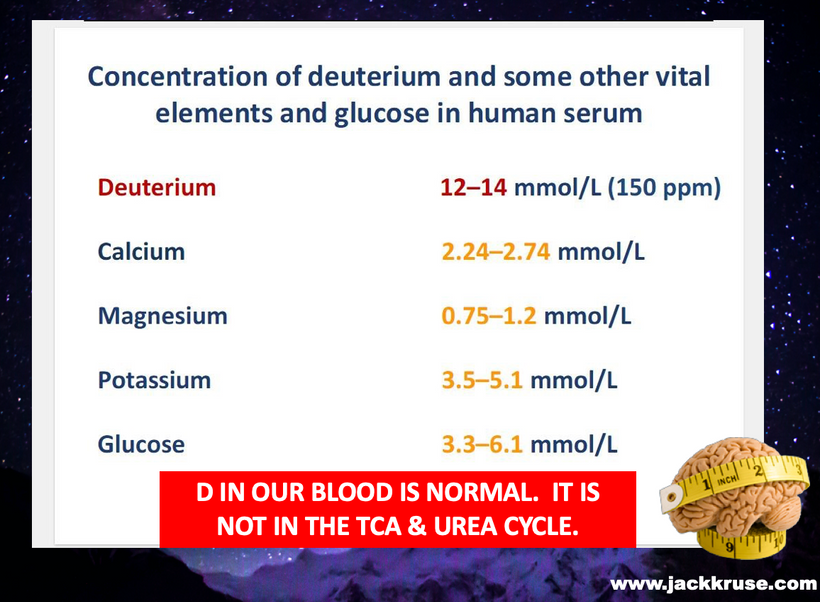
32) Why do you want to rid the CSF of all the deuterium in it that is creating white matter plaques in the brain and deforming the ventricular geometry? Because water without deuterium absorbs more of the light cells need to optimize themselves.
33)
34)
35) My Kruse for Dummies attendees probably could skip the entire slide deck here and I’d show them this slide, and they’d say…….I got bipolar disorder Uncle Jack no problem. LINK TO THAT LECTURE
36)
37) Mammals superpower is how they moisten places other primates have not. Cooling melanin is a big deal when the leptin melanocortin pathways is disrupted by modern life.
38)
39)
SUMMARY
Experience directs our learning to instruct us, how one event or observation constantly follows another; without instructing us in the secret connection of the events or observations, which binds them together, and renders them inseparable. This is where wisdom is found. Today’s post exemplifies this situation perfectly.
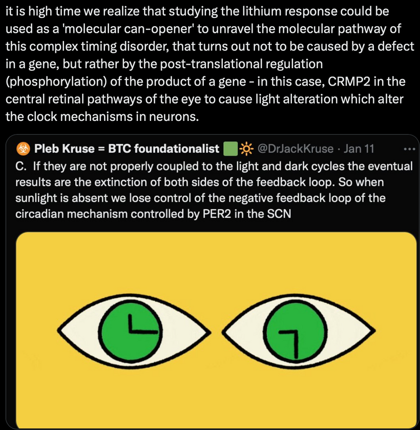
The clock in the pineal organ, the clock in the retina (SCN), the clock in the your liver, the peripheral clocks in all the other tissues all behave differently, but they use the exact same molecular machinery. Think back to quantum biology one blog for a moment. I taught you about how biology builds a zero entropy system for perfect energy/information transfers in water and carbon nanotubes of collagen. I have not given you all the goods yet, but each blog is building up to the magic in Nature decentralization plan.
It does this by using multiple pathways to do many things at once using light as the paintbrush and quantum field action of water as the canvas. Here we see the same thing in central and peripheral clocks controls. Even before these two studies in Nature, it was clear to me from the neural network level, that the properties must have been modified by something else in the environment that affected the cell directly or indirectly using the interactions between cells in the tissue to control the process. Dr. Montaigner’s work on water and EMFs was the missing link, in my humble opinion. Light is part of the EMF spectrum. It became clear to me at least that the path of life and circadian cycle control is the domain of quantum controls that use particle and wave mathematics to be the thermostat to control the processes in cells. Life then built a complex set of biochemical and hormonal pathways to create micro nanomachines to transduce those environmental signals to control the processes of growth and metabolism in their zero entropy systems.
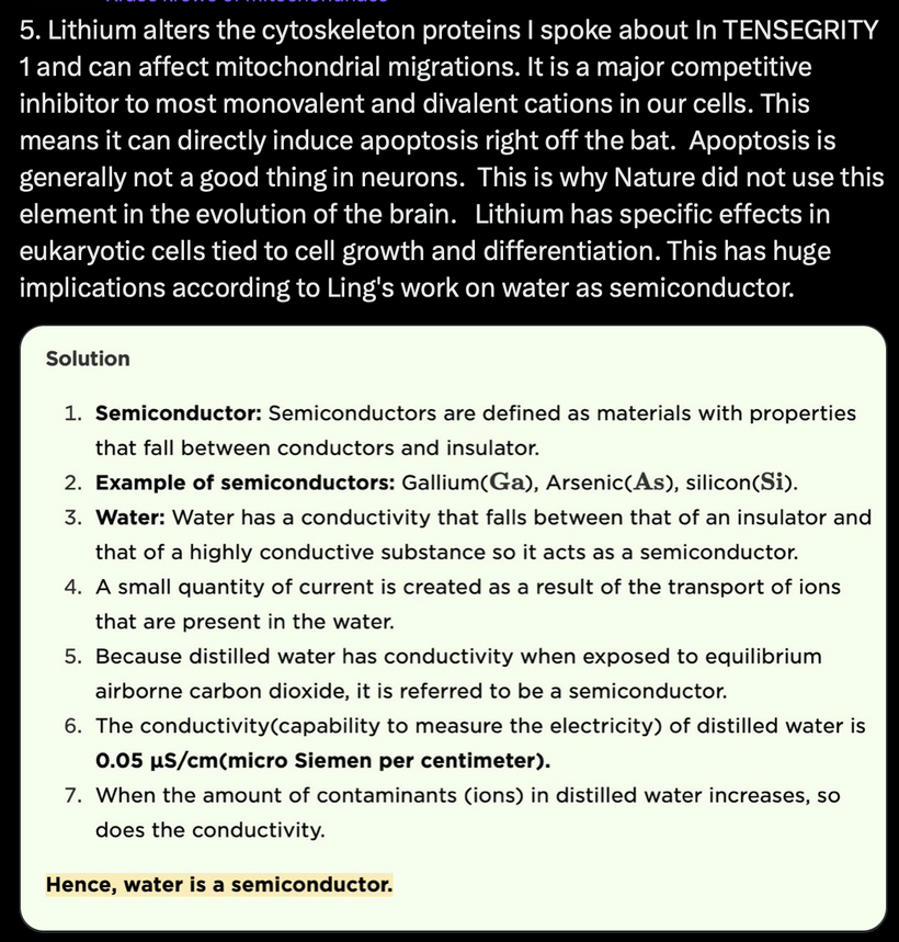
The key chemical to the entire equation is the use of water in cells because it is the ultimate quantum canvas for life to paint her masterpiece on. It is the perfect dipole molecule to bind to proteins to make liquid crystals function as semiconductors inside of cells. You just saw in BD how the crystals can break. The method of breakage is vast in humans. This is where diseases come from. The proxy for the breakage is always found in the heteroplasmy % of the tissue in question. Water makes protein semiconductors operate in humans. It is assistated massively by melanin to charge separate water. Doping these semiconductors with atoms makes a wide band gapped semiconductor or a narrow band gapped one. This changes their ability to do the things physiology requires of cells.
Water is life’s quantum field It does things to electrons and protons you wouldn’t normally expect, to keep life far from equilibrium. These effects are all non-linear. and often photonic. It just made fractal sense the more I dug into the science of 9 different scientific disciplines. The real problem blocking all nine from this reality is that none of them seem to know what the other is studying and finding out. They are buried in their own scientific silos. They just keep doing their own thing without sharing what they have learned and then connecting to the fabric of nature.
Today when you look back to where you began with me, and see where you are right now you’ll see how far you have come. Now you have to decide how far you want to go with me.
CITES
1. O’Neill JS, & Reddy AB (2011). Circadian clocks in human red blood cells. Nature, 469 (7331), 498-503 PMID: 21270888
2. O’Neill JS., Gerben van Ooijen, Laura E. Dixon, Carl Troein, Florence Corellou, François-Yves Bouget, Akhilesh B. Reddy and Andrew J. Millar, (2011). Circadian rhythms persist without transcription in a eukaryote, Nature, 469 (554–558), doi:10.1038/nature09654
3. Njus, David, Frank M. Sulzman & J. W. Hastings (1974) Membrane model for the circadian clock, Nature. Vol. 248, pp. 116-120, doi:10.1038/248116a0
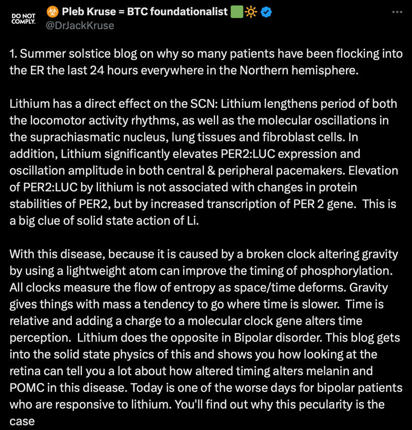
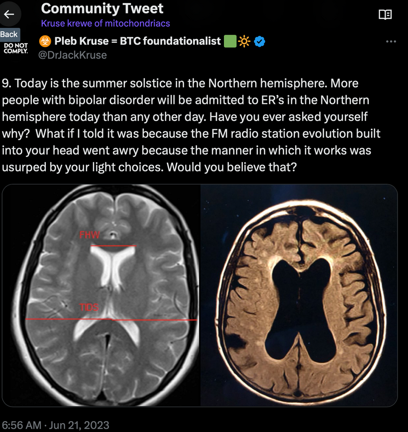
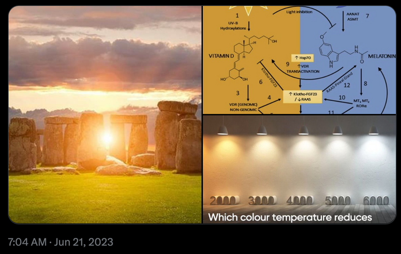
Today is the summer solstice and that is why this blog is being released today. More people with bipolar disorder will be admitted to ER’s in the Northern hemisphere today than any other day. Now you are going to learn why this is the case.
Bipolar disorder (BD) is a chronic and common psychiatric pathology, which can be particularly disabling. The disease has a global prevalence rate of 1–4%, begins at an early age, i.e. predominantly between 15 and 25 years old, and persists throughout the life of patients. BD is characterized by a recurrence of mood depressive episodes (pathological decrease in mood and energy), hypomanic or manic episodes (pathological increase in mood and energy), or even mixed episodes (simultaneous presence of depressive and manic symptoms).
Bipolar disorder is a common mental condition in our modern world with a seasonal pattern of onset. In the spring, there is a higher incidence rate of mania or mixed onset and suicide associated with equinox and solstice. The reason for this is a defective pruning mechanism related to the cortisol melatonin cycle in cells. The question is then what controls this pruning in us?
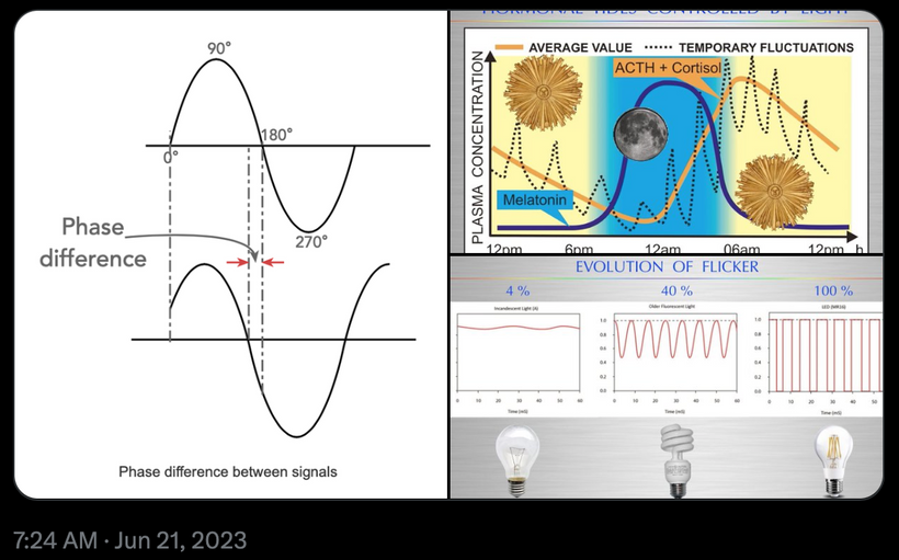
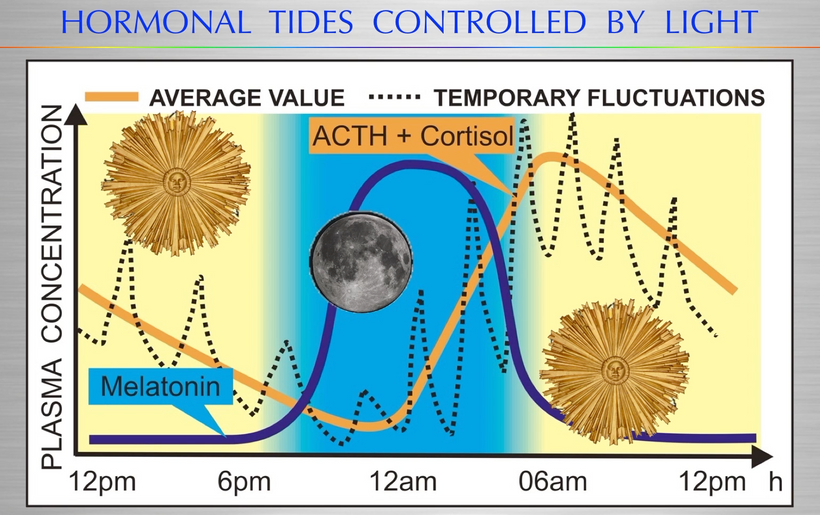
The pruning method of control in the brain is normally controlled by the cortisol melatonin cycle. Cortisol usually creates new pathways and melatonin trims or prunes them to suit the environment the person is in.
In bipolar disorder this system has gone awry. Remember cortisol is controlled by light via ACTH and melatonin levels are made by the mitochondrial matrix.
Explain this me like I am a 3rd grader Uncle Jack?
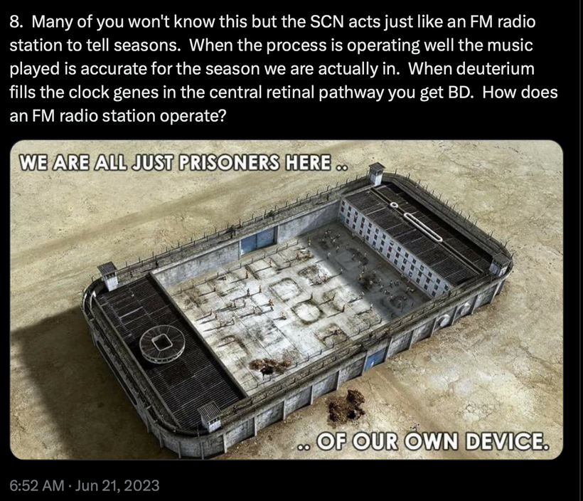
What if I told it was because the FM radio station evolution built into your head went awry because the manner in which it works was usurped by your light choices. Would you believe that?
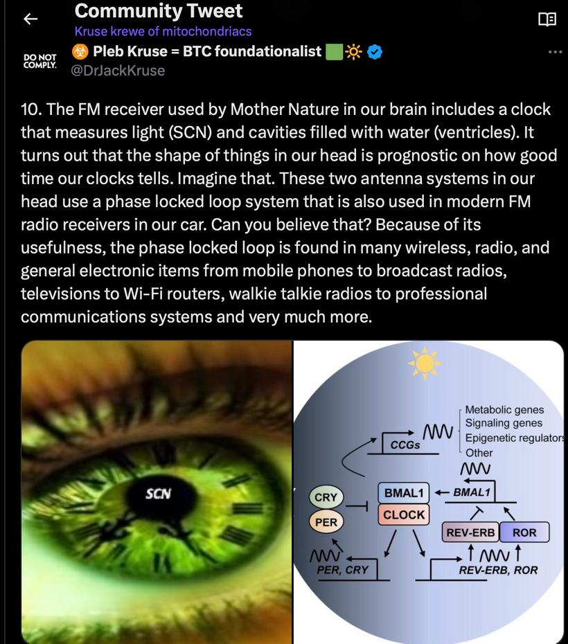
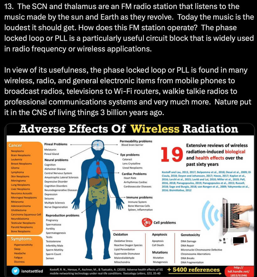
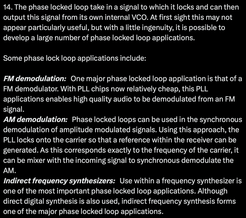
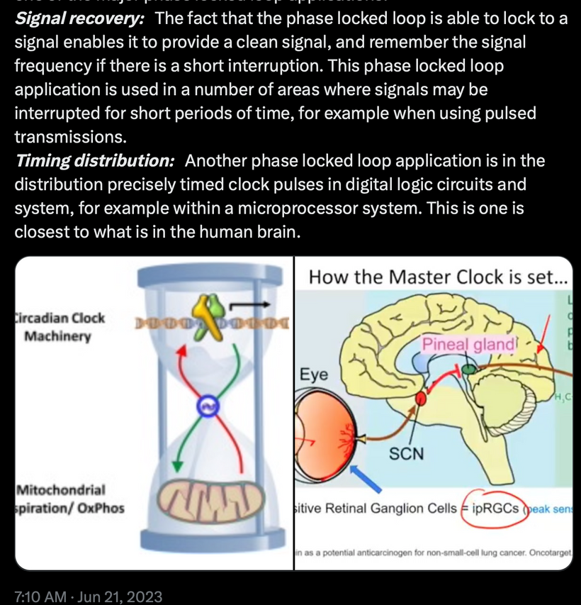
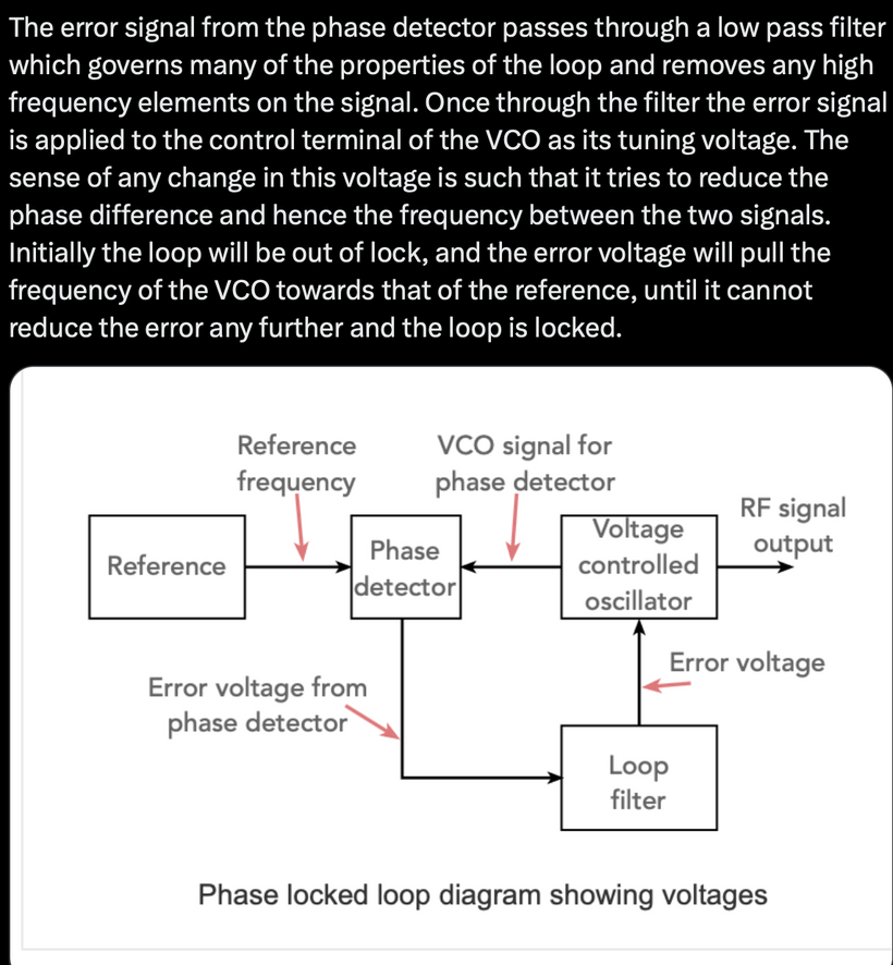
The FM receiver used by Mother Nature in our brain includes a clock that measures light and cavities filled with water. It turns out that the shape of things in our head is prognostic on how good time our clocks tells. Imagine that. These two antenna use a phase locked loop system that is also used in modern FM radio receivers in our car. Can you believe that? Because of its usefulness, the phase locked loop is found in many wireless, radio, and general electronic items from mobile phones to broadcast radios, televisions to Wi-Fi routers, walkie talkie radios to professional communications systems and very much more.
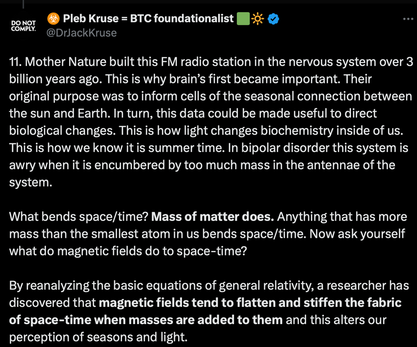
Mother Nature built this FM radio station in the nervous system over 3 billion years ago. This is why brain’s first became important. Their original purpose was to inform cells of the seasonal connection between the sun and Earth. In turn, this data could be made useful to direct biological changes. This is how light changes biochemistry inside of us. This is how we know it is summer time. In bipolar disorder this system is awry when it is encumbered by too much mass in the antennae of the system.
What bends space/time in the universe? The masses associated with the matter in the universe does. Anything that has more mass than the smallest atomused inside of our molecular clocks changes how we experience time. Mass bends space/time! Now ask yourself what do magnetic fields do to space-time?
Magnetic fields tend to flatten and stiffen the fabric of space-time when masses are added to them and this alters our perception of seasons.
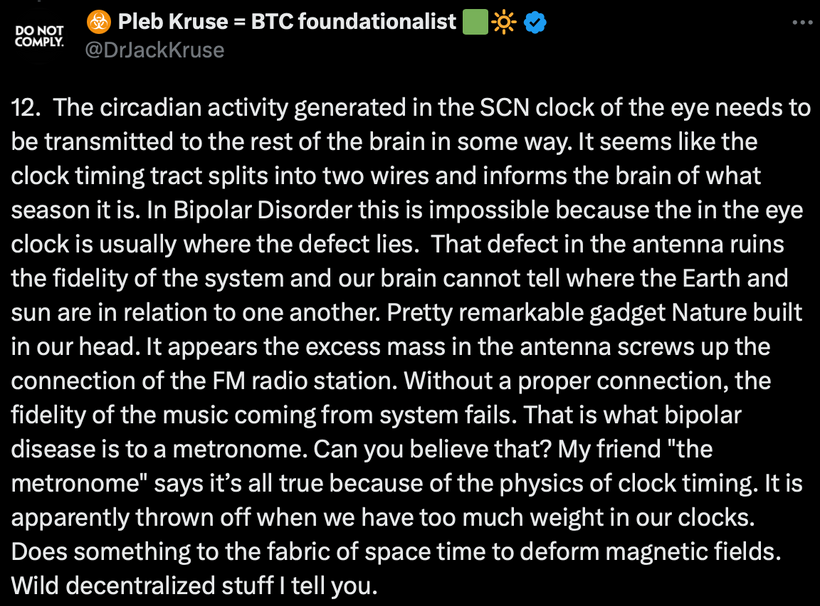
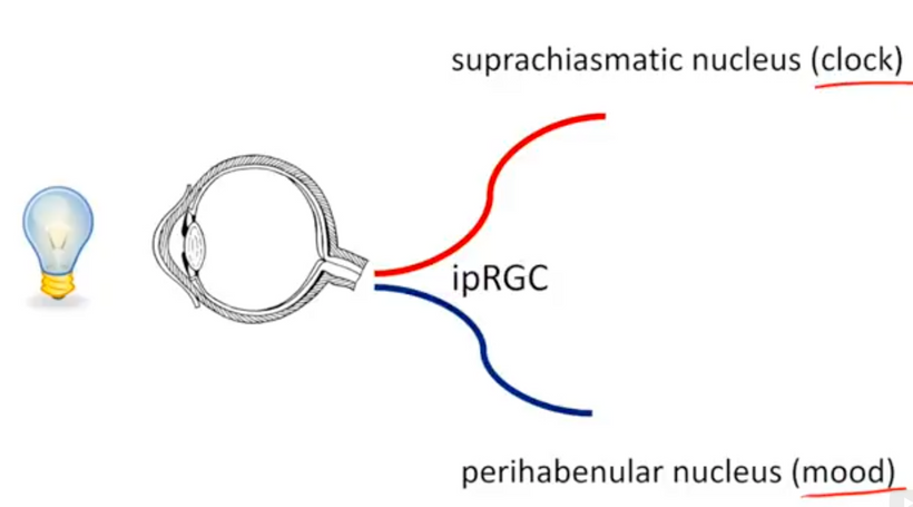
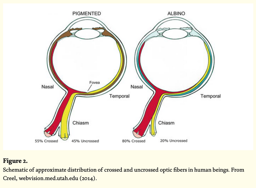
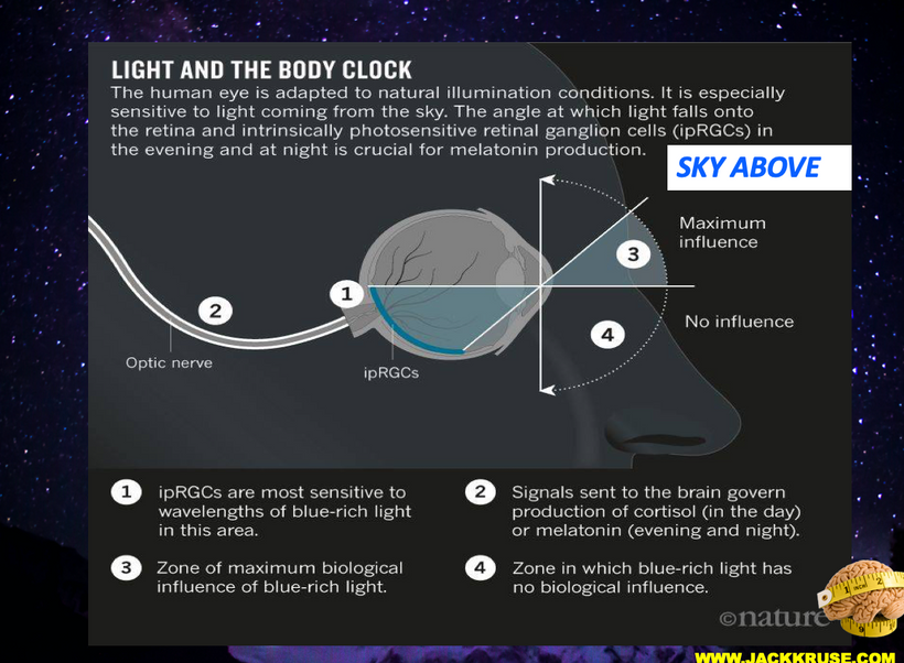
The circadian activity generated in the SCN clock of the eye needs to be transmitted to the rest of the brain in some way. It seems like the clock timing tract splits into two wires and informs the brain of what season it is. In Bipolar Disorder this is impossible because the in the eye clock is usually where the defect lies. That defect in the antenna ruins the fidelity of the system and our brain cannot tell where the Earth and sun are in relation to one another. Pretty remarkable gadget Nature built in our head. It appears the excess mass in the antenna screws up the connection of the FM radio station. Without a proper connection, the fidelity of the music coming from system fails. That is what bipolar disease is to a metronome. Can you believe that? My friend the metronome says it’s all true because of the physics of clock timing. It is apparently thrown off when we have too much weight (mass) in our clocks. Does something to the fabric of space time to deform magnetic fields. Wild decentralized stuff I tell you.
3rd grade bipolar lesson is over.
BACK TO THE SCIENTIFIC EXPLANATION OF BIPOLAR DISEASE
These thymic episodes the bipolar patient experiences are interspersed with phases of clinical remission, known as “euthymic” episodes. The disease is associated with a high morbidity and mortality rate and due to the significant functional impact it induces, including during euthymic periods, BD is the cause of poor quality of life and is one of the ten most disabling diseases according to the World Health Organization. The diagnosis of BD is mainly clinical and can be supported using scales or questionnaires. The diagnostic delay is estimated at around 10 years. This delay is clearly related to the heterogeneity of the clinical expression of the disease. The phenotype of the disease is relative because how patients experience time is relative to the environment they inhabit.
The study of the literature shows that this delay in treatment seriously affects the prognosis, particularly on the functional level, and constitutes a major public health problem. In addition, there are no good biomarkers, easily usable in current practice, to help the clinical decision for the diagnosis or for predicting the course or prognosis of the disease. One thing is known about bipolar people. Many of them have the same changes in their eye as diabetics. They also tend to get metabolic syndrome in the guts more frequently than any other mental illness as is documented in the cites below. They tend to have pale skin in their thoracic and lumbar regions as well.
WHY ARE SLEEP DISORDERS ALWAYS LINKED TO THIS DISEASE?
Sleep disturbances and sleep/wake rhythms are major in BD. These disturbances are observed during the different phases of the disease and are major symptoms of mood episodes and belong to the diagnostic criteria for depression, hypomania, and mania. In addition, these anomalies are also found during the euthymic phases of this disease. Indeed, patients suffering from BD would be more likely to present a more evening chronotype and a more languid and rigid circadian type than healthy subjects as well as a decrease in the efficiency of their sleep, an increase in sleep duration, an increased sleep latency and a prolongation of the duration of awakenings after the onset of sleep. These disturbances in sleep and wake/sleep rhythms are associated, among other things, with more frequent relapses, an alteration in the quality of life, and cognitive disorders.
Additionally, neurocognitive deficits are frequently associated with BD. Most typical deteriorations found are impairment of episodic verbal memory, executive functions, processing speed, and sustained attention. These troubles can be present during mood episodes but also in around 30% of patients during euthymic phases. Cognitive deficits of patients with BD have a direct impact on their psychosocial functioning, on the risk of relapse, on treatment adherence, or even on their ability to insight. Their early detection associated with the identification of prognostic and predictive biomarkers of the response to cognitive and functional remediation tools is essential in order to be able to offer early and appropriate treatment.
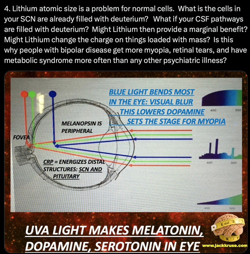
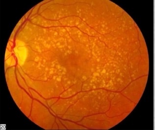
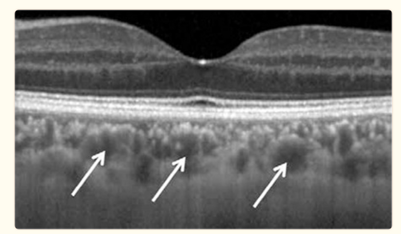
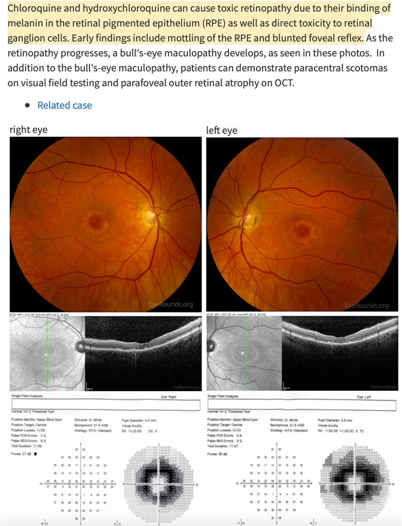
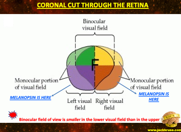
Due to its embryological origin, the retina is an integral part of the central nervous system. The retina is a complex neural tissue (above in a BD patient), consisting of several layers of retinal neurons. They have structural and functional properties similar to cerebral neurons. You’ve seen this laid out in detail in this series. In addition, molecules involved in brain neurotransmission such as dopamine, serotonin, glutamate, or GABA, are also involved in retinal neurotransmission. The retina is now considered a relevant candidate for the study of neurotransmission abnormalities in neuropsychiatric pathologies. The function of retinal neurons can be measured using the OCT (below) or the electroretinogram (ERG).
ERG is a simple, fast, inexpensive, and non-invasive process that aims to measure the electrical activity of retinal neurons in response to light stimulation and provides indicators of synaptic transmission. This technique can represent an important electrophysiological measurement in biomarker research within psychiatric diseases. However, studies evaluating retinal function with ERG in BD are very few today. I think in the future In addition to the electrophysiological anomalies mentioned above, structural anomalies at the retinal level have been described. Indeed, several studies carried out in optical coherence tomography (OCT) have shown that subjects with BD present a thinning of the layers of retinal neurons. Additionally, results also seem to suggest that retinal thinning in the periphery of the retina where melanopsin is located may be related to disease progression. People with bipolar disease are equisitely sensitive to light it appears. This is what makes them different.
As previously described elsewhere on Patreon, retinal neurons and cortical neurons have similar properties. It should be noted that measurements of retinal function with ERG and measurements of cortical function with visual evoked potentials (VEP) share the same characteristics. Indeed, both can be performed using light flash stimulation and using luminous black-and white reversing checkerboard (pattern stimulation). In addition, they are complementary measures for the analysis of dysfunctions of the central visual pathways. Thus, the combined study of VEP using EEG could be relevant and complementary to ERG for a study of all visual neural pathways in neuropsychiatric pathologies in the future. I think OCT shows these changes when clinicians think to order them in bipolar patients. This is going to be how centralized medicine slowly decentralizes in my opinion as these tests reveal the quantum mechanical changes that occur in the antennae of the phased loop signaling you are about to learn about in this blog.
BIPOLAR DISEASES AND LITHIUM & METABOLIC SYNDROME
Lithium is the most common drug used to treat bipolar disorder. Overall, compared to placebo, lithium appears to decrease the risk of suicide by more than 60% in bipolar disorder.
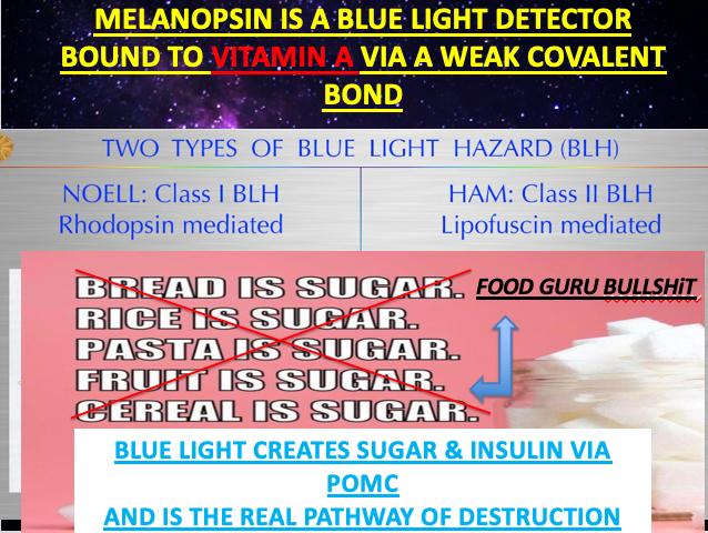
Blue light increases blood glucose and insulin from POMC cleavage of ACTH and CLIP. Did you know that people with newly diagnosed bipolar disorder are 3.5 times more likely to have metabolic syndrome? What is the connection? Abnormal light in their environment is the link. Metabolic syndrome changes how the gut works with the brain because of changes in the SCN and the CSF that surrounds the thalamus. It is most often seen in those who live an indoor existence while bathing themselves in blue light and nnEMF. The best way to avoid all mental illness is to remain in the sun and remain in the dark when the sun sets. It is not that hard when you understand how light changes the topology of the eye, skin, and gut. When you bathe in light at night or day mental illness and a sleep disorder will find you eventually in some way.
ISOTOPIC LESSON ON LITHIUM: DEEP PHYSICS
Lithium drugs are widely used for treating bipolar disorder. They work, but nobody really knows how they work in cells. I think I might. How might a mitochondriac attack this problem? How about by mito-hacking the periodic table of elements yet again?
Consider the link between size discrepancy and the proton spin crisis between B-meson and protons. B-mesons are approximately 2/3 the size of protons and each of the quarks that make them up have a spin of 1/2. Protons do not follow the expected spin predictions that mesons have given us. Why would lithium be linked to this proton story? Remember that mitochondria in neurons deal with protons and B-mesons in the mitochondrial matrix as I laid out in my April 2016 webinar on this topic. Those details are not important here but the size difference of the atoms is.
Might lithium’s effects somehow be due to the quantum effects of the size variation of isotopes found in the matrix in the SCN and the leptin melanocortin pathways in some areas as it travels through the brain?
You do know that different isotopes of elements have different nuclear spins, right? People who listened to my Kruse for Dummies lecture should now fully understand why lithium works in bipolar disorder and why it can help reverse some of their Metabolic syndrome effects. Lithium alters the binary code of life because it has spin number that mimics one fo the atoms used in the binary code.
Guess why a mitochondrion favors protium (H+) over deuterium (D)? Atomic mass is the answer.
Might isotopic effects cause some quantum change in a material or a receiver built into our system? Might the size variation of isotopes found in the blood be linked by Nature to make circulating blood act differently than water in other parts of our body? LINK TO THE LECTURE
IS BIPOLAR DISEASE REALLY AN ALTERED SIMULATION OF REALITY DUE TO ISOTOPE VARIATION IN NEURONS?
Deuterium is an isotope with spin = 1, unlike hydrogen-1, which has spin = 1/2.
Photons (particles of light) can have a spin of –1 or +1.
Why can’t photons have 0 spins?
The definition of the spin as the angular momentum of a particle at rest is also inapplicable to the photon since there is no rest frame for the photon, which moves at the speed of light. Light never rests or stands still once it is liberated from matter.
What bends space/time? Mass does. Since Deuterium has more mass than H+ it bends space/time more than protium. Now ask yourself what do magnetic fields do to space-time?
By reanalyzing the basic equations of general relativity, a researcher has discovered that magnetic fields tend to flatten and stiffen the fabric of space-time when masses are added to them.
How does this affect the circuit between the sun, earth, and the human brain?
It should immediately raise a question about this solar circuit in bipolar patients. Daytime has strong electric fields associated with sunlight. Nighttime has little.
Does a strong electric field cause time dilation in the SCN and the tracts linked to it in the mammalian brain to cause bipolar disorder?
The spacetime curvature for a charged static spherical body is given by the Reissner–Nordström metric:
With these mathematics, you can feed in the value of whatever charge you want and calculate the time dilation as a function of distance from the charged body. If you do this you’ll discover something rather odd in Einstein’s field equations, namely, the charge reverses the effect of the mass.
Do you remember how many times I have told you in podcasts that redox potential is the net negative charge in a cell? Do you see now how that charge can offset the bad isotopic variation in some of your tissues to make your clocks work better in the circuit that tells the brain what season it is? Remember sunlight deuterium depletes us. Light at night adds deuterium to our tissues. This ruins our clock function.
Is this why mammals conserve charge using their POMC biology in their cells Uncle Jack to offset this deuterium problem? Yep. The excess mass of heavier isotopes in cells and mitochondria causes time to slow (relative to the observer at infinity) but adding charges back to proteins/water (of either sign) can make the time speed up again.
Wait a minute Dr. Kruse, explain that again.
The sun creates massive electric fields during daylight. Implications for Bipolar Disorder?
Does a strong electric field cause time dilation in the universe? Yes, it does because a strong electric field generated by the sun adds a massive net negative charge to cells that absorb this radiation. POMC makes sure this happens in the human brain via Noether’s thereom.
This is true because the mathematics of physics says it operates this way.
It appears this is what really causes Bipolar disorder at its fundamental core.
TYING IT BACK TO MY THESIS OF THIS SERIES
The adrenal medulla of mammals was born under the light stress of the KT event. This is why I wrote the adrenal fatigue blog years ago but no one saw things back then like they do now. I told the implications of that Huberman/Rubin podcast were VAST. Bipolar disorder is one of the effects.
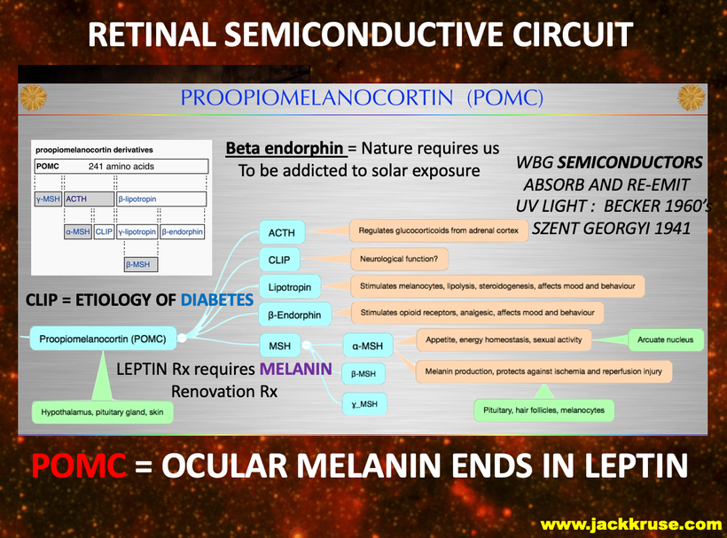
Man’s five senses can capture the vastness and the immensity of our cosmos and POMC is the paintbrush that allows it. POMC is the paint that goes on the canvas, your brain. POMC is a color palate that humans use to paint their mind. In some ways, that palate confines us to the limited real estate of the sensory organs in our brains to understand all the complexity of the universe. All 5 senses tract directly to the thalamus of humans that surrounds the 3rd ventricle of their brains. But there is a wisdom in being connected to nature in this fashion. Yet, few see the artistic symmetry of confinement. I am trying to explain this art to you now every day, in every word I write in everyone of these blogs. It is time for you to LEVER UP your knowledge.
THE LEVER UP LESSON: The adrenal cortex synthesizes three main groups of hormones (glucocorticoids, mineralocorticoids, and adrenal androgens) (Auchus and Miller, 2001). The homeostasis of glucocorticoids is regulated by a feedback mechanism CONTROLLED BY POMC gene translation via solar light and temperature sensors (non-visual photoreceptors) in the skin, eye, and autonomic nervous systems that are relayed to and through the hypothalamic POMC center and the circumventricular organs (water networks in the ventricular system of the brain) by means of corticotropin-releasing hormone (CRH), the pituitary gland by ACTH, and the adrenal cortex by cortisol (Koch, 2004).
The mammalian cell responds to all stress, including light stress, by increasing cholesterol production.
Cholesterol is the MAIN non visual photoreceptor of the brain. If you go into an ICU with a patient with an acute infection and draw their lipids (not often done except by a tool like me) you will find sky-high LDL cholesterol.
And that is both LDL and HDL, but the LDL fraction is much higher. Is that a problem? No. But Big Pharma has trained centralized MDs to reflexively reach for the Rx pad to write for a statin. This is why so many people with Bipolar disorder also have metabolic syndrome as a comorbidity as well. Their LDL is high because they are all solar deficient. Remember LDL cholesterol lacks electrons realtive to HDL cholesterol.
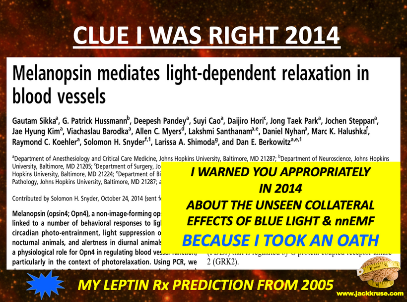
The other non visual photoreceptor in the brain that is common is melanopsin and it is found in the blood vessels. With blue light at night it increases blood flow and increases blood pressure excessively. High blood pressure is part of metabolic syndrome.
Few MDs & psychiatrists today are trained to even know full spectrum sunlight lowers cholesterol naturally. Even fewer realize sunlight is the best treatment option for Bipolar disorder. Blue light at night makes this disease deadly.
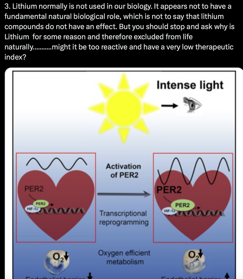
In fact, in metabolic syndrome, higher LDL cholesterol stabilizes the inner mitochondrial membrane function during heavy oxidative phosphorylation (cellular energy production). So a raised LDL actually helps mitochondria retain its electric charge to condense H+ inside the mitochondrial matrix to make energy using the light hydrogen isotope.
This is how the cell controls space time and magnetic fields inside of their tissues to ACCURATELY SIMULATE THE PHYSICS OF YOUR ENVIRONMENT.
In bipolar disorder, this is a broken mechanism at the level of the SCN because there are atoms in it with higher masses than their should be. (D>H+)
Do you know what sits really close to the outer mitochondrial membrane in most mammalian cells?
POMC waiting to create melanin from the biophotons released from the mitochondria right next to it. Making CO2 and DDW is the reason our cells stole bacteria 600 million years ago. And since the cell is trying to recover from a light stressor, it needs a good inner mitochondrial membrane in which to transfer its electrons to oxygen and use the H+ liberated by melanin buried in water to make ATP by the spinning ATPase Fo head.
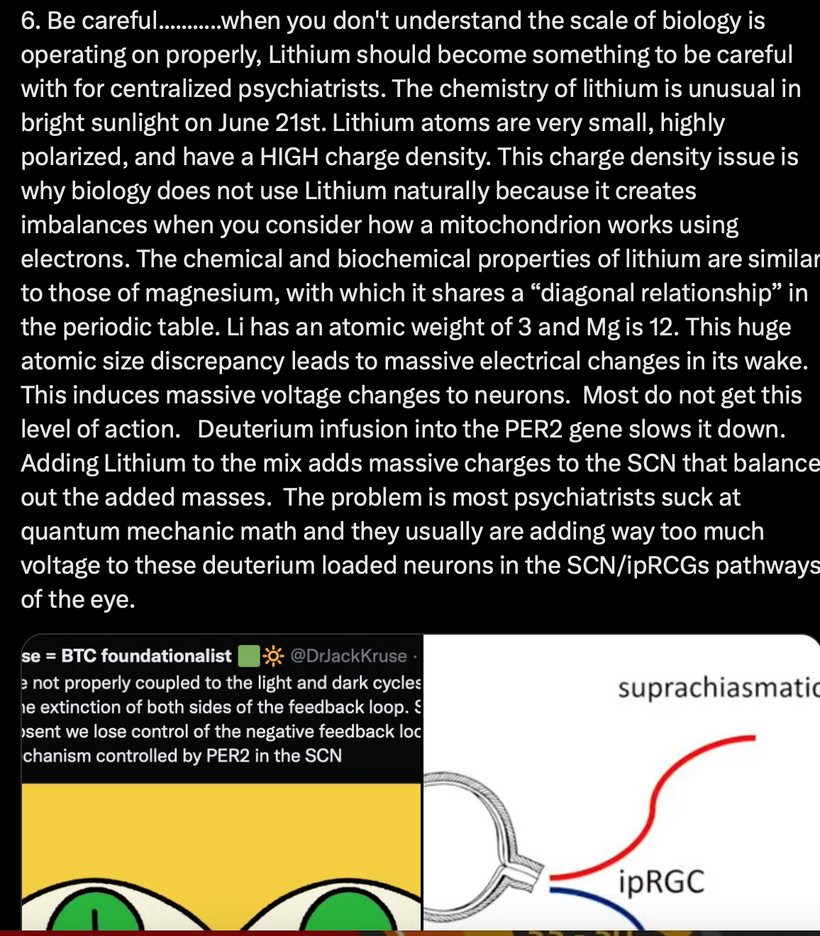
When the deuterium isotopic variation mechanism is broken in the SCN, lithium can be used to help offset that loss in the CSF networks in the ventricular system of the brain. Effectively, lithium is adding mass to the water of CSF to offset the deuterium in the SCN to allow it act as a better FM radio antenna to tell the season for the brain.
That is why lithium can help some of these people.
The problem with lithium is that it can never reverse the disease. Only strict sun exposure and darkness at night can repair the broken mechanism at the SCN level to help eradicate this disease in humans. When the SCN is loaded with too much deuterium, as such, it cannot accurately tell time.
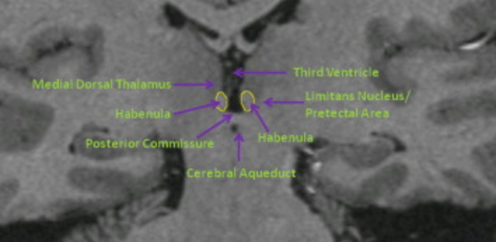
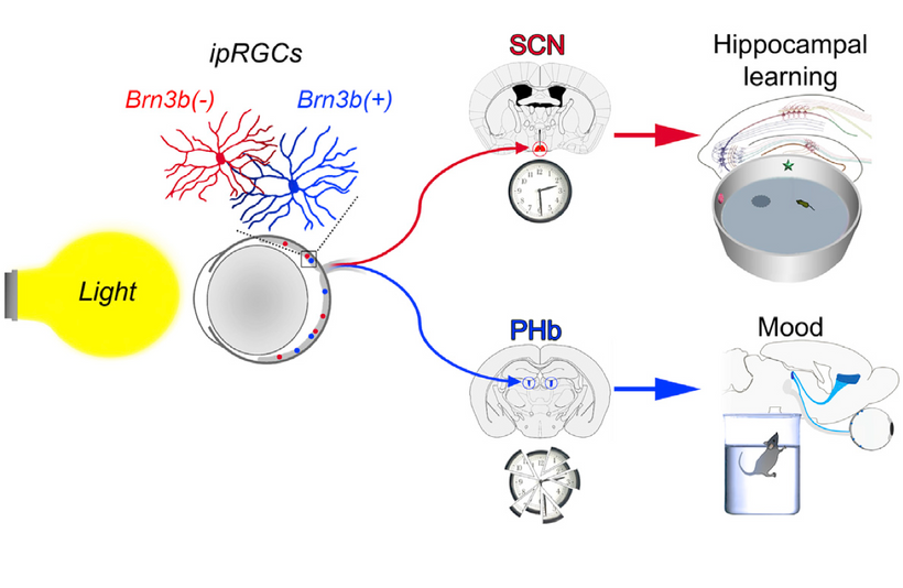
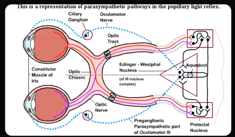
The isotopic variation changes as their choices in light vary leading to mania and depression cycles in things the ipRGCs link directly to. Note the positioning of the habenular nucleus in the next two pictures and you’ll see the circuit manifest before your eyes.
What controls the autonomic nervous system in the gut of mammals that get metabolic syndrome?
NEUROANATOMY LESSON TIME: POMC does because it is found in high concentration normally in the outflow tracts of the neurons from the hypothalamus that connect to the gut. This connection is made via the dorsal longitudinal fasciculus. That tract targets the thoracic nerves that connect to somites where other melanin stores are located from the neural crest. Remember before when I told you bipolar people tend to have pale trunks and abdomenal skin? Now you know why this link is important. These dermatomal layers need melanin renovation in BD.
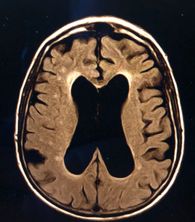
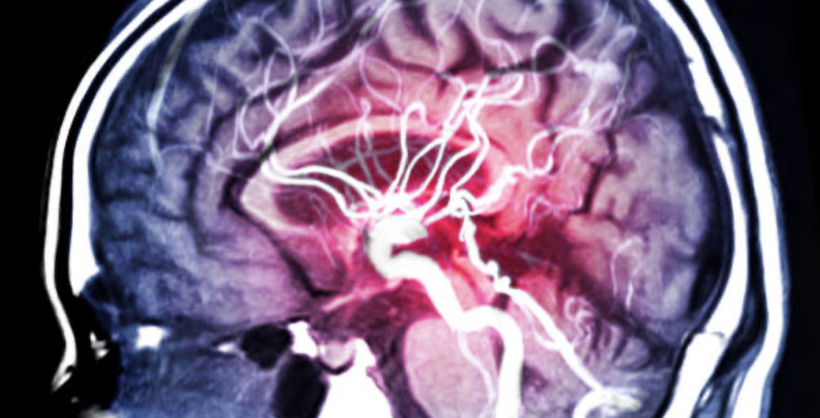
When I see a patient with BD I always examine their skin in these areas before I order an MRI (above). Why? Pale skin in these areas always corelate to white matter lesions in their brain (shown above). Centralized science is still clueless why this is the case. You no longer will be. This is decentralized medicine 101. White matter hyperintensities (WMH) are much more common in subjects with bipolar disorder (BP) than in healthy subjects. The more prominent that lesion or the larger their ventricles tells me how severe their disease is. LINK I have also observed that paleness of the skin in these areas also corelates with thicken of the choroid on OCT images of the retina and with changes in the inferior nasal retina on my eye exams.
The hypothalamus is the key brain site for central control of the autonomic nervous system, and the paraventricular nucleus is the key hypothalamic site for this control. Much of the medial surface of the thalamus and hypothalamus form the wall of the third ventricle pictured above.Part of the hypothalamus forms its floor. Its thin, membranous roof contains the choroid plexus that makes cerebrospinal fluid which is 99.8% water. The third ventricle narrows quickly at the posterior end of the mammillary bodies. These bodies are the key to memory formation and important in dreaming. Anatomically, the PVN is adjacent to the third ventricle and many of its neurons project to the posterior pituitary. These projecting neurons secrete oxytocin and a smaller amount of vasopressin, otherwise the nucleus also secretes corticotropin-releasing hormone (CRH) and thyrotropin-releasing hormone (TRH).
Recall my Brain Gut #16 blog told you that the PVN is where adrenal fatigue began. It is a light disease that turns off POMC in the hypothalamus and not a real adrenal disease.
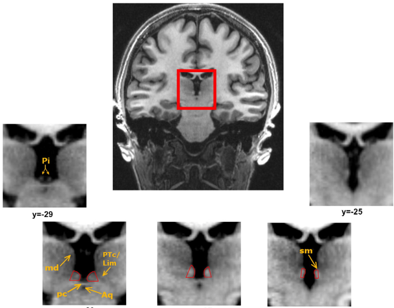
The PVM neurons massively express POMC but without UV light this switch never gets turned on. Bipolar patients are all lacking that critical UV light switch. The major pathway from the hypothalamus for autonomic control is the dorsal longitudinal fasciculus in the human brain. Below is another picture of the third ventricle (red box) that is adjacent to the hypothalamus and thalamus in humans.
The dorsal longitudinal fasciculus (fasciculus of Schutz) is a periaqueductal (area around ventricular system Aq above) ascending and descending fiber system arising from the hypothalamus and terminating to the autonomic nuclei of the pons and the medulla, conveying autonomic fibers from the brain to the gut in humans. It also conveys pain and is important in sleep pathways of humans. These are usually altered in bipolar patients as well because of a lack of melanin in these areas.
The dorsal longitudinal fasciculus is found within the dorsal brainstem tegmentum. It passes through the periaqueductal gray matter and contains both ascending and descending fibers. The ascending fibers pass from the reticular formation (sleep region/insomnia) passing to the hypothalamus thus transmitting information related to the viscera.
People who travel a lot across time zones or people who use a lot of blue light or nnEMF in their cities are going to have massive sleep disturbances like people with mental disorders because they have broken the same rules of Nature. I view insomnia as a mental disorder in decentralized medicine.
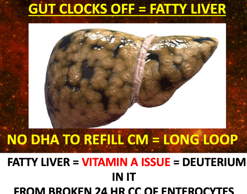
This surface is acritical in barrier in the brain health of humans. This is the pathway where metabolic syndrome, poor sleep, and fatty liver all come from. Many of these same findings are found in diabetics, bipolar disorder, and those with sleep disorders like insomnia. Patients with bipolar disorder tend to have all these symptoms at times that vary based on how defective their FM antennae are.
This explains how a lack of sunlight leads to most gut and sleep issues modern humans face. Without proper DLF input to the gut via the brainstem, ferroelectric currents are lost and circadian control of the gut lumen is awry. This opens the gut barrier to many potential problems.
Pro-opiomelanocortin co-localizes with corticotropin-releasing factor in axon terminals of the noradrenergic nucleus locus coeruleus. It is not just a PVN sotry folks. Where the problems lies will determine aspects of the disease you get. This nucleus is filled with neuromelanin. The locus coeruleus (LC), a small brainstem nucleus, is the primary source of the neuromodulator norepinephrine (NE) in the brain. The LC receives input from widespread brain regions, especially the hypothalamus and projects throughout the forebrain, brainstem, cerebellum, and spinal cord.
What is the main function of locus coeruleus?
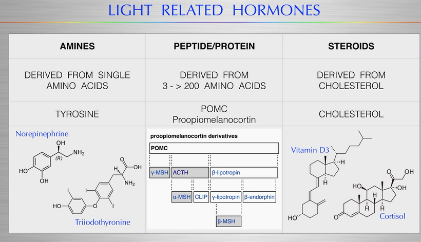
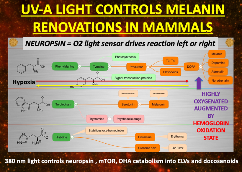
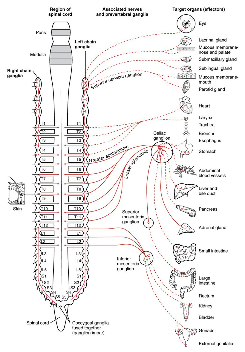
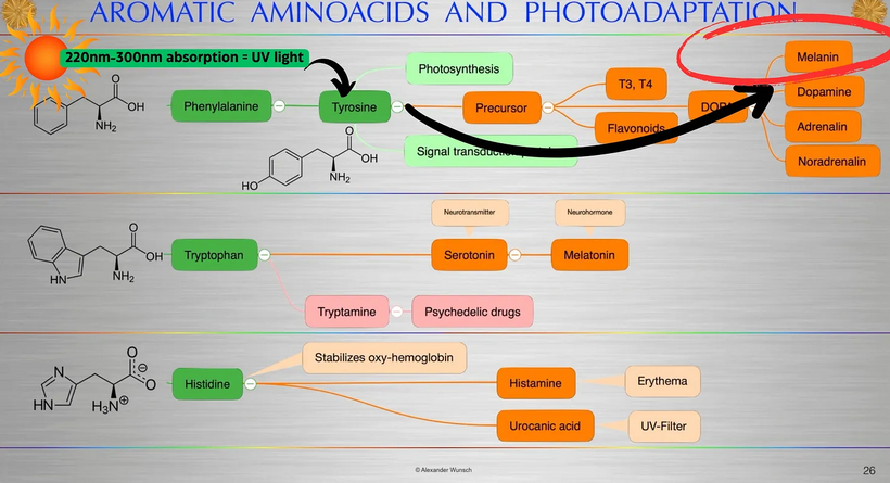
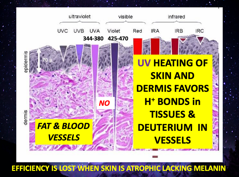
It is the brain’s main source of the neurotransmitter noradrenaline (norepinephrine). This chemical is excitatory and is released in response to pain or stress, stimulating what is referred to as the ‘fight-or-flight’ mechanism. This means that it activates the sympathetic nervous system via those thoracic nerves I mentioned above. Those are the same nerves that innervate your brown fat and also give input to the superior cervical ganglia that I mentioned in Time 12. Without UV light exposure in the eye and skin in these dermatomes melanin is degraded and noradrenaline is diminished. You remember that nor adrenalin can be made from melanin right? See the slide below far right top line.
Neuropsin is a 380 nm light sensor that the locus coeruleus needs to function well.
Without this system operational, this opens the BBB and the gut barriers. We see this in most psychiatric cases and in TBI cases due to the stress response. Now you can see why really the brain-gut barrier is linked. Usually, there is a lack of melanin in the DLF and/or the locus coeruleus to create the aura of brain-gut barrier when in reality what links the two is a lack of UV light to stimulate POMC in both hypothalamic pathways and melanin is degraded leading to many disease phenotypes. Each somite in the spinal cord also has POMC in it derived from the neural crest in this part of the body. For the gut this is linked to the celiac ganglion which comes from T5 -T11 thoracic nerves. So thoracic level solar exposure is key to renovating gut level melanin. It is counterintuitive until you see it all laid out in front of you. A lot of this deep science would have been the Vermont 2019 video I was going to give 4 years ago. I think I still might do it for the Kruse for Dummies folks.
SUMMARY
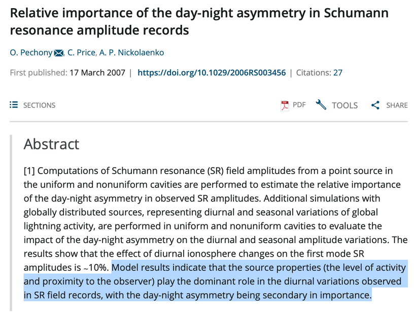
Neil Cherry PhD from New Zealand showed in 2002 that there is strong and robust scientific evidence of the human brain detects and responds to the Schumann Resonance signal based on the work of many researchers Robert O. Becker worked with in the 1970s. People with bipolar disorder cannot connect well with the Schumann resonance because of excess mass contained within the receivers of the signal which comes from the sun and Earth. As a result this leads to behavioral and neurologic dysfunction. Since increased mass of isotopes used in this system affects coherence times that are possible in cells, they lose their ability to maintain quantum coherence and the system becomes dysfunctional. How long a system can maintain coherence is a function of the atomic mass in that system. You can see now why any additional masses in the SCN or habenular nucleus destroy this realtionship. The pictures below shows the specific details of the pathogy laid out for you.
The brain uses a range of frequency patterns monitored by the EEG system in our neurosurgical clinics. The frequency range of the EEG rhythms coincide with the frequency range of Schumann Resonance signal (0–45Hz). The alpha wave is identical to the frequency most commonly linked with Schumann resonace on Earth. The normal brain has developed an ELF oscillating ion system, primarily using calcium ions (above), to control neurotransmitter release. The release is awry in BD. Dr. Russ Adey at UCLA established this in the 1970s. Adey work is cited below. The human brain and its optical clock lattice in the SCN is now well established in the literature. Blackman’s work on calcium ion resonace frequencies was cited multiple times in Becker’s books and my early blogs on the non ionizing effects of nnEMF and neurological compromise. Blackman’s research also cited below, has taught decentralized MDs that external electromagnetic ELF signals induce altered neuron calcium ion effluxes in mitochondria of brain tissue.
These ELF signals between the sun and Earth are weak, yet they match the ultraweak UV biophotons that cells create in response to the Schumann extraterrestrial signal. This allows the brain to know the season. The stable synchronization of the brain’s electromagnetic systems using a system that mimicks an FM radio has actually led to humans becoming thinking, emotional, memory machines of biology that are capable of quite a bit of intelligence in the human central nervous system. In order to carry out these functions the brain has developed electromagnetic transmitters and receivers in the neurons to accomplish this task in the SCN, leptin melanocortin pathway that extends into the thalamus of man and into his spinal cord. The thalamus is where the alpha wave finds its genesis in humans. This connection is pictured below.
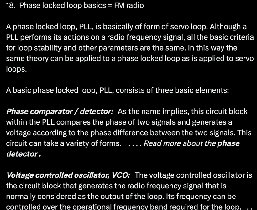
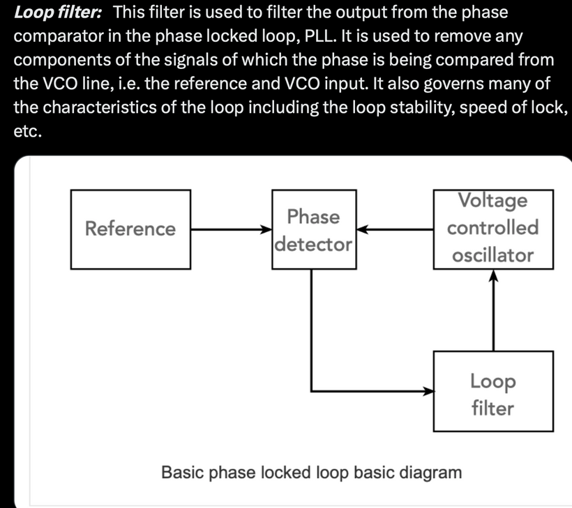
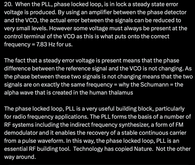
The receivers used by Mother Nature in the human brain included a phase locked loop system, described by Ahissar et al. This paper is cited below. The phase locked loop system is also used in modern FM radio receivers in your car. In the human brain it provides an FM radio receiver that non-linearly resonantly interacts with the Schumann Resonance signal. The phase locked loop or PLL is a particularly useful circuit block that is widely used in radio frequency or wireless applications in tech gear. In view of its usefulness, the phase locked loop or PLL is found in many wireless, radio, and general electronic items from mobile phones to broadcast radios, televisions to Wi-Fi routers, walkie talkie radios to professional communications systems and very much more.
Mother Nature built this system over 3 billion years ago in the brain’s of living systems to inform cells of the seasonal connection between the sun and Earth so this data could be useful for biological changes. In bipolar disorder this system is awry when it is encumbered by too much mass in the recievers.
The circadian activity generated in the SCN needs to be transmitted to the rest of the brain and henceforth to the rest of the body via non-optical signaling to stay well. In Bipolar Disorder this is impossible because the SCN is where the defect lies. That receiver is not receptive to the Schumann’s information. Coherent connection ruins the fidelity of the system. Most of the centralized science right now believes it is through direct wiring tracts. I do not. I think it is via the hydrogen bonding network in water through the areas where there is no blood-brain barrier in the brain that provides the fidelity of the phased locked loop. This allows electron and proton tunneling and quantum coherence to allow for rapid communication in the system. I believe all biological processes in any living organism are due to the creation of biological biophotons in the ultraweak VUV-IR-A range. Light can dictate highly selective and specific electromagnetic interactions between particular biomolecules because of changes in mass or the magnetic moments of the component boxcars of biochemistry. In most cases, these interactions involve and are driven by semiconductive proteins surrounded by water, which act as logic gates do in the system of organization seen in solid-state physics.
The human brain SCN contains the key mechanism to having a strong diurnal pattern that assists the sun to maintain the circadian rhythm. The Schumann Resonance signal continuously synchronizes the brain wave ELF patterns in a set range of grouped frequencies that begin in the thalamus that surrounds the 3rd ventricle of the brain. The ventricular system also acts a resonant cavity on Earth to receive the signals. The hydrogen bonding network in CSF is another receiver in the phased locked loop communication system mentioned above. This stabilizes the brain’s electromagnetic system of communication and its fidelity has enabled intelligence and stable thinking to evolve to the point where this complex understanding of the biophysical environment interacting with the human brain can be reasoned, understood, and appreciated.
DNA only codes for the key backbone proteins that will become the main conductors of any life process within the organism. The mitochondrial matrix created the other part (water hydrogen bonding network) of this semiconductive backbone in cells to surround the protein. Both parts of the semiconductor have to be operational to simulate time from your environment at the level of the SCN. Everyone focuses on refraction errors in the eyes. Few spend time understanding how the eye clock really operates at a quantum mechanical level with the Schumann frequency. This is why psychiatry remains impotent to help reverse this disease.
Let’s review the lessons from 100’s of my blogs now in relation to this disease.
A. Hydrogen bonds form between water molecules, giving rise to supramolecular aggregates/clusters/coherent domains. Some call this EZ water. The clustering of water is a cooperative phenomenon, which means that forming one hydrogen immediately favors the formation of several other hydrogen bonds, and vice versa, breaking one bond leads to breaking up a whole cluster. Thus clusters are dynamic flickering networks with lifetimes of 10^- 11 to 10^-10 seconds. Light changes those hydrogen bonding networks by moving charges around. https://phys.org/news/2022-10-revolutionary-technique-hydrogen-efficiently.html
B. Hydrogen bonds are the key to coherence in cells. Hydrogen bonds act like a spec of dust in the formation of snow, ice, or crystals.
How do coherent excitations make the system sensitive to specific, weak signals? Such a weak signal will be received by the system only when the system is ‘in tune’ — rather like a very sensitive FM radio receiver, which can resonate to the signal. Furthermore, a small signal will be greatly amplified for it will not only affect one molecule, but because many other molecules are in the same state of readiness, they too, will be affected in the same way, and the signal is correspondingly multiplied as many times as there are molecules responding to it.
The shape of your ventricular system which is filled with H+/D is another clue for me that the receiver system is broken.
The hydrogen bonds in water have a critical point at which they change and act like dust. When enough energy is pumped in bond angles change and this makes water liquid crystalline and a better antenna to receive signals of what season it really is. People bipolar disorder have bad antennas.
More lessons from old blogs you need to recall now to understand Bipolar:
C. Sunlight creates electric fields in our atmosphere every day and that sunlight creates a Coulomb force in our body. Coulomb force is an electrostatic force. Electrostatic phenomena arise from the forces that electric charges exert on each other. Such forces are described by Coulomb’s law. Even though electrostatically induced forces seem to be rather weak because of how we experience them in life, when the scale shrinks, as it does in a cell, the electrostatic force increases tremendously in strength as the scale drops. Scale drops = less mass present
D. For example, some electrostatic forces such as the one between an electron and a proton, that together make up a hydrogenatom, are about 36 orders of magnitude stronger than the gravitational force acting between them. The electrostatic force generated in your skin is another example of a strong electrostatic force because the skin, as an insulator can hold large amounts of charge from the sun if it is normal. It is not in bipolar disorder.
E. Modern physics now has proven that energy and information are equivalent in physics. Landauer’s Principle of 1961 & Shannon’s 1948 work was critical in making this linkage. Modern quantum biology has experimentally proven that energy is trapped directly at the electronic level in cells. Energy is stored not only as vibrational and electronic bond energies in biochemicals, but also in the structure of the system: its membranes, and in gradients, fields and flow patterns, compartments, organelles, cell water, and tissues. All this in turn enables organisms to mobilize their energies coherently at any time it is needed and hence make available the entire spectrum of stored energies for physiological work. It is energy on demand by atomic design.
F. What is the bandwidth of the entire electromagnetic spectrum of light? The electromagnetic spectrum starts from wavelengths of 10^-14 m at one extreme to 10^8 m at the other, spanning a range of 10^22. In terms of doublings, 10^22 ≈ 273, or 73 octaves. Visible light is a small fraction of this bandwidth but that bandwidth does very specific things that cells take full advantage of.
Light is an EMF. Sunlight (visible light) is a very specific EMF that interacts with specificity for atoms in cells. That specificity is created because the EMF selects the type of water created by the dissipative system. This DDW water surrounds the atomic lattice inside cells whose integrity is maintained by the nuclear genome blueprint. The atomic arrangement in cells also allows for light absorption and light emission. The light that cells emit is also quite specific. It is an ultraweak extreme low-frequency biophoton that spans the visible spectrum frequencies. This is the light that is used for optical signaling and it drives the molecular resonances that drive the internal motions of biochemicals in cells. Light drives biochemistry. Biochemistry does not drive light.
Conventional centralized biochemists do not understand these nuances of what biophotons are and how they are created by EMFs, water, and oxygen in cells. So You should not expect centralized physicians (psychiatrist) to get this nuance either.
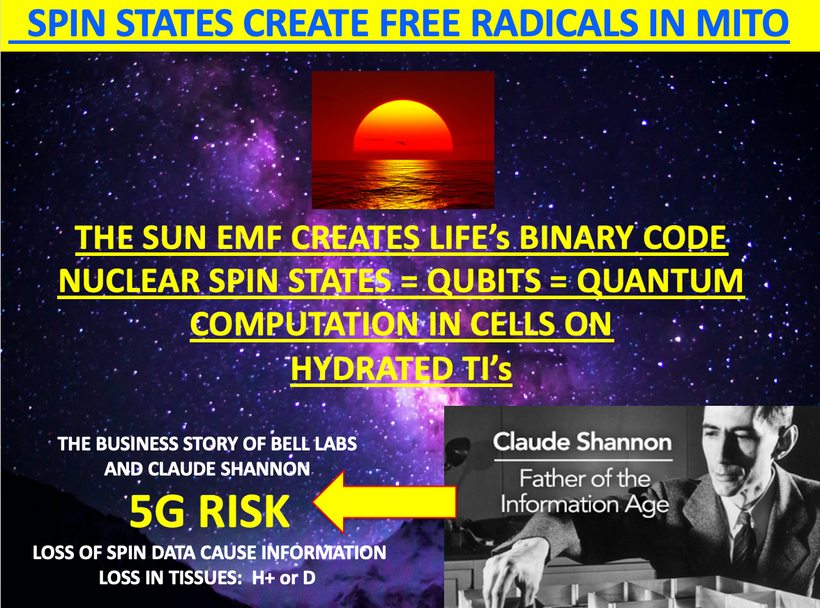
G. Claude Shannon taught the world that information flows via entropy. All clocks should be thought of as flowmeters for entry. This includes the biological circadian clocks in cells. Wheeler taught physics that information and energy are one and the same thing. Shannon’s mathematics from his 1948 paper advanced the linkage of entropy and information. Shannon’s paper also told us anything can be a message. (Kruse for Dummies lecture) H+ and electrons are fermions. Deuterium is a boson and this small difference can ruin how an FM antenna works.
I believe sunlight messages for mitochondrial DNA and nuclear are controlled by “fermion messaging” (pic above from old blogs). DNA is informed about what to do with optical information from the sun via mtDNA signaling (optical and free radical) and this message is transmitted over water’s hydrogen bonds adjacent to proteins in a cell. DNA is a very complex topologic insulator that is an antenna for our star’s information.
H. Water makes up 99% of molecules in every cell and it fills the ventricles of our brain. Water is a very small molecule that has more hydrogen bonds in it than any other compound. Liquid water contains the densest hydrogen bonding of any solvent, with almost as many hydrogen bonds as there are covalent bonds and hydrogen bonds in its structure found anywhere on Earth. These two bonding networks are the binary code in water. Just as a computer can use a 1 and 0 to create digital information on the internet, hydrogen bonds create the internet in your cells. Shannon taught us the information content of any kind of message could be measured in binary digits or just bits. This was the basis of the Kruse for Dummies talk.
I. Water’s hydrogen bond network changes at a pico and femtosecond level in any environment. Inside a cell, its atomic arrangement is controlled by electrostatic forces in a cell created by the redox power of the mitochondria in that cell. These hydrogen bonds can rapidly rearrange in response to, light frequencies, charge density, and changing conditions and environments (for example, solutes like K+ in a cell). This is how our ventricles tell seasons in us. When the FM system is bad you cannot tell seasons and bipolar disorder is the result.
J. Shannon demonstrated, contrary to what was commonly believed in the 1940s, that engineers could beat their worst enemy ever: transmission errors-or in their technical jargon, “noise.” Noise is anything that disturbs communication. Your FM radio antenna does not work when there is noise in the system. It can be an electric signal in a telephone wire that causes crosstalk in an adjacent wire, a thunderstorm static that perturbs TV signals distorting the image on the screen, or a failure in network noise to increase the energy of the transmission signals or send the same message repeatedly-much as when, in a crowded pub, you have to shout for a beer several times. Shannon showed a better way to avoid errors without wasting so much energy and time: coding. Nature does the same thing.
She takes the message in the hydrogen bonding network of water that surrounds every protein and encodes that information in fermionic code in mRNA, mtDNA, RNA, tRNA, and DNA. Deuterium is a BOSON. It ruins the fermionic signal and that CAUSES bipolar disorder.
DO YOU GET IT YET?
K. Coding is at the heart of information theory. All communication processes need some sort of coding to limit the noise and create a high-fidelity signal that doesn’t degrade. Bipolar disorder is due to tissues in our FM radio station in our head having too much mass in them. Water preserves the information and transfers it to nucleic acids via hydrogen bonds. Just as the telephone system transforms the spoken voice into electrical signals. In Morse code, letters are transmitted with combinations of dots and dashes. The DNA molecule specifies a protein’s structure with four types of genetic bases. Digital communication systems use bits to represent or encoded information. Each letter of the alphabet, for example, can be represented with a group of bits, a sequence of zeroes, and ones. You can assign any number of bits to each letter and arrange the bits in any way you want. In other words, you can create as many codes as desired. Cells have done this to run life’s program. The more mass a tissue has the less time it can remain quantum coherent to transfer information in any system.
All of these lettered lessons above ^^^^^ have been published in my blogs or forum for years. Please ASSIMILATE THEM NOW.
This blog was easy to write because piece and parts of it are in hundreds of other blogs.
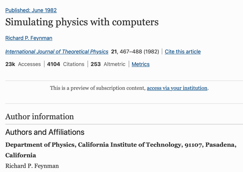
The emission and absorption spectra of proteins can be used to make predictions of what light frequencies will couple with proteins and the matrix to create DDW water. The logic in the system of operation can only be comprehended when you understand how the subatomic parts of nature are able to be used and manipulated by quantum mechanical abilities. I told you a long time ago, in the Cold Thermogenesis series, that the default state of life was sleep. Then as evolution progressed with evolved wakefulness. This idea was counterintuitive to many at the time. The reason for my prediction was simple. I read Feynman’s 1982 paper on a computer simulating the physics of the environment and thought to myself what organ in humans fit his description of an ability to simulate the physics of Nature? The SCN fit perfectly.
The SCN filled with deuterium and your CSF overhwelmed by deuterium is where bipolar disease comes from. Patients with Bipolar Disorder also have a higher risk associated with retinal detachment, primary retinopathy, diabetes retinopathy, hypertensive retinopathy, and retinal vascular complications than the controls. Now you all know why this blog came after the last one.
Please re-read QE # 44-47 again. The concepts are there to explain this disease and all sleep disorders. The last blog was on diabetic retinopathy. Those patients also always have features of metabolic syndrome. So do most mental disorders and sleep disorders. Now you can see why they are all linked. You have effectively broken the FM antenna station nature put inside your skull. If you have insomnia, (yes, Sam this is for you) part of your system in the periaqueductal grey area is broken in your brain because you loaded this area up with deuterium by using Netflix too often while travelling outside your time zone to omuch for your brain, and you decided to live in Chicago with all its associated nnEMF and population density. It should be obvious why you cannot sleep with these circumstances, no?
What I am explaining to you now is where in the topological map of the human brain the POMC deficit is located leads to the phenotype of the disease we see in our clinics. Patients with this disorder intuitively know they need more sun. How they go about it is often incorrect as you see in cite 4 below.
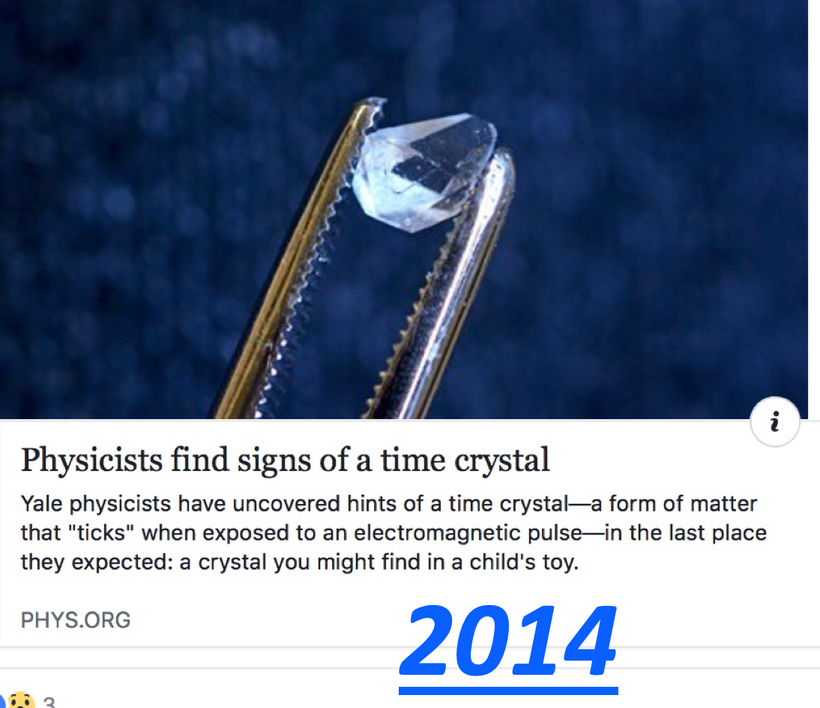
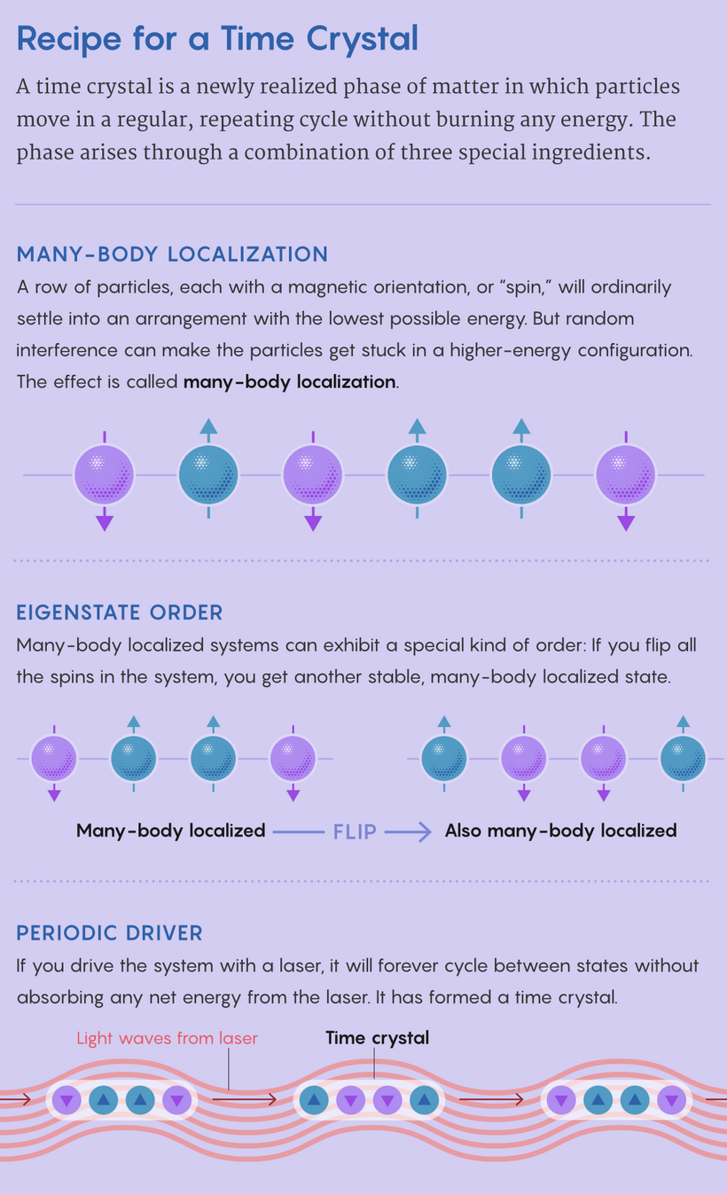
Understanding the human eye and retina and linking it to entropy in a cell was the only quantum leap I made to understand this concept. The flow of entropy is why I made this prediction back in 2010. All molecular clocks are flow meters for entropy. I knew the SCN had to be doing something unusual with the retina and the brain during sleep. The SCN appears to be a perpetual motion machine built into the brain. Feynman wrote a paper in 1982 stating he believed a quantum computer could simulate physics if it used a time crystal.
The SCN is a small computer in your eye that is simulating the physics of the environment between the sun and Earth to make the best predictions for cells in tissues. In bipolar disease that time crystal is not working well because the mass inside of it put there by the light you allow ruins it. Adding mass back to the water networks, using lithium is how lithium operates.
The problem is the system fidelity operates at quantum mechanical precision with respect to mass and this is why results are so uneven in patients with this disease.
Adey W. R. Electromagnetic fields and the essence of living systems. In: J. Bach Anderson (ed). Modern Radio Science. Oxford University Press; 1990. p. 1–37.
Ahissar, E., Haidarliu, S., Zacksenhouse, M., 1997. Decoding temporally encoded sensory input by cortical oscillations and thalamic phase comparators. Proc. Nat. Acad. Sci. USA 94, 11633–11638.
Blackman C. F. ELF effects on calcium homeostasis. In: B. W. Wilson, R. G. Stevens, L. E. Anderson (eds). Extremely low frequency electromagnetic fields: The question of cancer. Columbus: Publ. Battelle Press, 1990: 187–208.
Cherry N. J. Schumann Resonances, a plausible biophysical mechanism for the human health effects of Solar/ Geomagnetic Activity. Nat Hazards 2002; 26: 279–331.
Diabetic retinopathy (DR) is a well-recognized microvascular complication of diabetes. What is not well recognized is that light stress release of ACTH from POMc and CLIP is the main driver of the disease. Blue light is a nnEMF that causes diabetic neuropathy. It causes many other diseases as well.
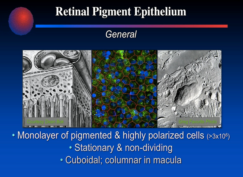
Growing evidence suggests that, in addition to retinal vascular damage, there is significant damage to retinal neural tissue in diabetic retinopathy. This is especially true in the RPE melanin sheets which have many functions in the eye.
OCT IS USED TO VISUALIZE THE RPE IN RETINAL SCANS:VIDEO
Studies have revealed neuronal damage before clinically evident vascular lesions and this alone should have gotten researchers to realize that light into the eye and not food is the key driver of these diseases. Centralized medicine now classifies DR as a neurovascular complication. ACTH release drives blood sugar and insulin higher. Hyperglycemia from ACTH actually turns off the cleavage of alpha-MSH, Beta-MSH, and gamma-MSH. This lowers the dopamine level in the eye via hypoxia and elongates the globe to cause myopia. What else might it cause? The blog has many new diseases linked to this mechanism. The last blog showed you how autism was linked to it.
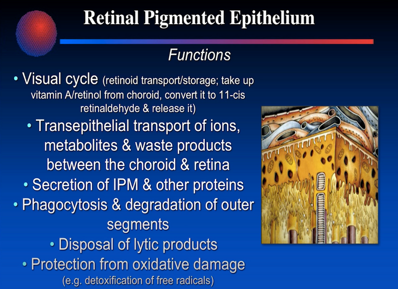
In contrast to skin melanin, which is constantly synthesized by the epidermal melanocytes, it is currently believed that melanin in the RPE does not regenerate I no longer believe this is true. Melanin is known to function as a potential radical scavenger and photoprotective agent. It also scavengers metal ions when photoreceptors are destroyed by the action of seeing using sunlight. The retina had to be built a certain way to compensate for this ability. What are the implications of Nature’s design in the retina with respect to modern disease creation?
The neural retina and retinal pigment epithelium (RPE) diverge from the optic vesicle during early embryonic development. The optic vesicle forms as an evagination from the diencephalon. They originate from different portions of the optic vesicle, the more distal part developing as the neural retina and the proximal part as RPE.
I believe they retain their neuroplasticity and today’s textbooks are wrong. So when the RPE is damaged their connection will be in the diencephalon before there is radiation into the cerebral hemispheres. I believe this is where the defect begins in autism during neurulation. I also think this is where blood disorders like Hemophilia, von Willebrand’s disease, and Von Hippau Lindau’s disease arise as well.
Wait, what?
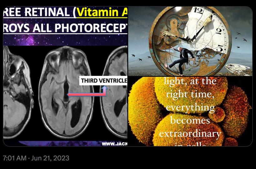
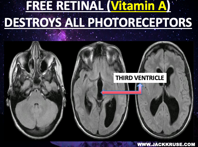
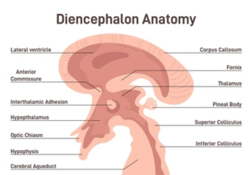
So when retinal changes are present during my exam I am always interested in an MRI of the brain in the diencephalon as the next step in my own work-up because I am looking for things no one else looks for. The diencephalon is the region of the embryonic vertebrate neural tube that gives rise to anterior forebrain structures including the thalamus, hypothalamus, posterior portion of the pituitary gland, and pineal gland. The diencephalon also encloses the third ventricle. Below in the center picture, you will see a massively enlarged 3rd ventricle in someone with non-visual photoreceptor problem that began in their retina. It came to my office as a case of normal pressure hydrocephalus and brain atrophy. A look in the eye made the link for me easy.
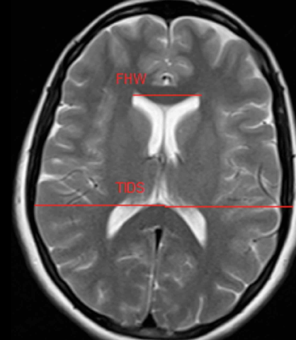
Ventricular size measurement is necessary for the determination and follow-up of many neurological illnesses, and pathologies. Ventricular enlargement is an indicator of brain parenchyma loss (Karakas et al, 2011). Furthermore, ventricular size measurements are used in studies of hydrocephalus, schizophrenia, tumors, trauma, Alzheimer’s disease, Parkinson’s disease, gender, aging, and atrophy which is associated with many neurological diseases such as stroke and dementia, Huntington’s disease (Karakas et al.; Gameraddin et al.; Honnegowda et al.) and provides useful indices of cerebral asymmetry and atrophy. The knowledge of ventricular system anatomy is essential for clinicians, neurosurgeons, and radiologists (Kanakaraj et al., 2016; Farheen & Sukre, 2017). Due to literature findings, ventricular size is considered a potential indicator in the determination of many diseases related to the brain. I think it is the key to understanding where melanopsin and melanin damage are located in the human brain. It is a treasure trove of non-visual photoreceptor damage that we can use to predict future diseases. Additionally, the normal reference values of ventricles obtained by MRI are necessary to form the baseline data for interpreting pathological changes, planning neurosurgical operations, and determining the presence and progress of some neurological diseases. Below you see the 3rd ventricle is almost nonexistent in a healthy brain compared to the picture above.
Fig. 1 above is an Axial T2-weighted Turbo Spin Echo MRI (TR:3600, TE:87 ms) of measurement areas of healthy subjects. (FH) Frontal horn width. (TIDS) The maximum transverse inner diameter of the skull. Note how the 3rd ventricle is small here in the pic.
Today’s facts are the boundary of human knowledge. These facts are set for it, but should not bind us to what the central paradigm believes. We must look past that edge to explain things they cannot. If our mind is open, we can spot new data and formulate ideas to test. Our mind requires nature’s connection to provide the software to run the hardware inside the skull. To build this natural quality, a “natural mind” is also a necessary requirement for this process. These facts allow our species to travel hopefully throughout life, and this appears to be why the journey of discovery supersedes the arrival.
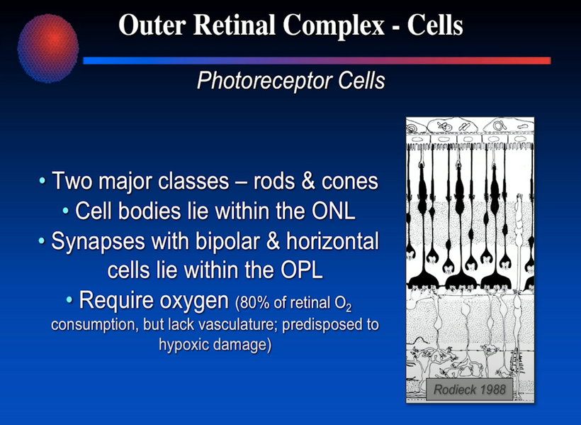
Below is the location in the periphery of the retina where most of the melanopsin IPGRCs are located in the periphery of the retina and not in the macula/fovea. AMD, cataracts, and many neurodegenerative disorders show defects in this same area on OCT scans. Central vision is not affected early in these diseases. This paradox needs an explanation. Today, you’ll get my explanation from my 30 years of careful observation in a decentralized fashion.
Damage in this peripheral area outside the fovea correlates with cognitive decline in neurodegenerative diseases. Most ophthalmologists are not taught the reason why this area of the retina has the highest amount of O2 utilization in the entire human body. These photoreceptors use high O2 because this increases the band gap of the semiconductive proteins in the RPE to regenerate melanin in the RPE. Most eye professionals are told that RPE has no regenerative potential. I’m not so sure I believe this any longer. The cells may not divide but the melanin inside of them needs constant renovation via POMC activation and/or migration from Bruch’s membrane of the choroid where melanocytes are closest to RPE in adult humans for some reason. Moreover, this is where most retinal bleeds occur in diabetic retinas and this is where most retinal surgeons use laser coagulation to stop diabetic retinal changes and bleeding from proceeding. I doubt many of them realize this puts their patients at higher risk of many neurodegenerative conditions due to the disruption of the VUV creation at the WBG semiconductors of the eye. Please note the last line in the slide below. It will be critical in your understanding later in this blog.
KEY POINT: A recent systematic review and meta-analysis reported that vision impairment is associated with 2.4-fold greater odds of cognitive impairment in existing cross-sectional studies and 1.7-fold greater odds in longitudinal studies.
Do you still think laser treatment for retinal bleeds has few risks with 800-1200 burn holes from retinal surgeons’ lasers?
What is the link? The key feature for most neurodegenerative disorders is the accumulation of misfolded protein aggregates, framing them within the classification concept of proteinopathies or “protein conformational disorders”. The key change is alpha helices are changed to the beta format by changes in the pH of the surrounding fluids. People forget that pH is a log function of the H+/D ratio of the tissue. Here we are back to seeing the impact of the Kruse for Dummies lecture yet again. The H+/D ratio is key to understanding chiral effects in tissues and how certain diseases manifest. Diabetic retinopathy is one of those diseases.
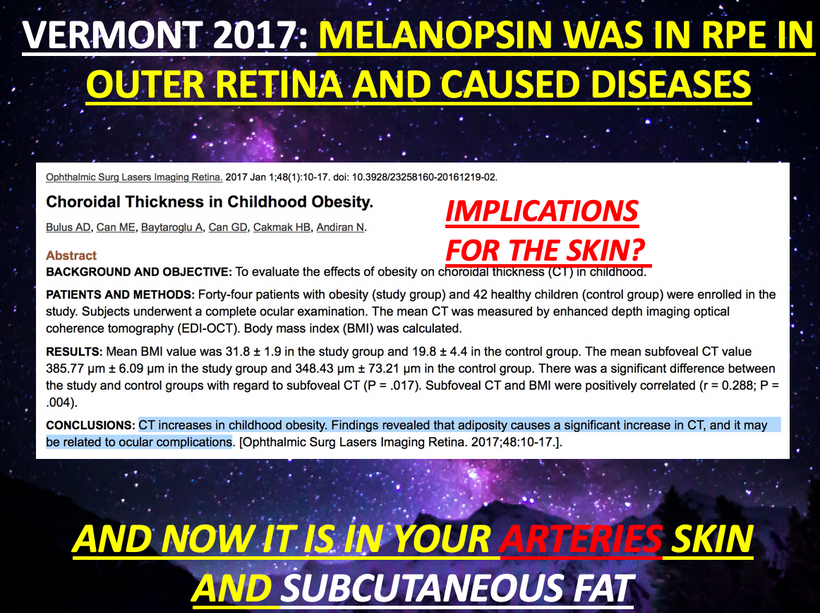
However, it is important to underscore that not all neurodegenerative diseases can & should be considered protein-conformational disorders. Proteins are semiconductive materials in cells that need constant care and rejuvenation (redox management). Protein conformational changes can happen from the protein or water side of the biological semiconductor. If the redox power in the cell is suboptimal those semiconductive proteins will misfold and accumulate with their associated components. We see these changes in the retina all the time in the RPE before diseases in the brain or other tissue manifest. This mimics what we see in the choroid of children who will become fat because their choroid has thickened. This was the key lesson I taught you in the Vermont 2017 lecture.
I just never told you it explains a whole lot more. And none of you bothered to ask me either.
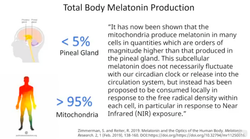
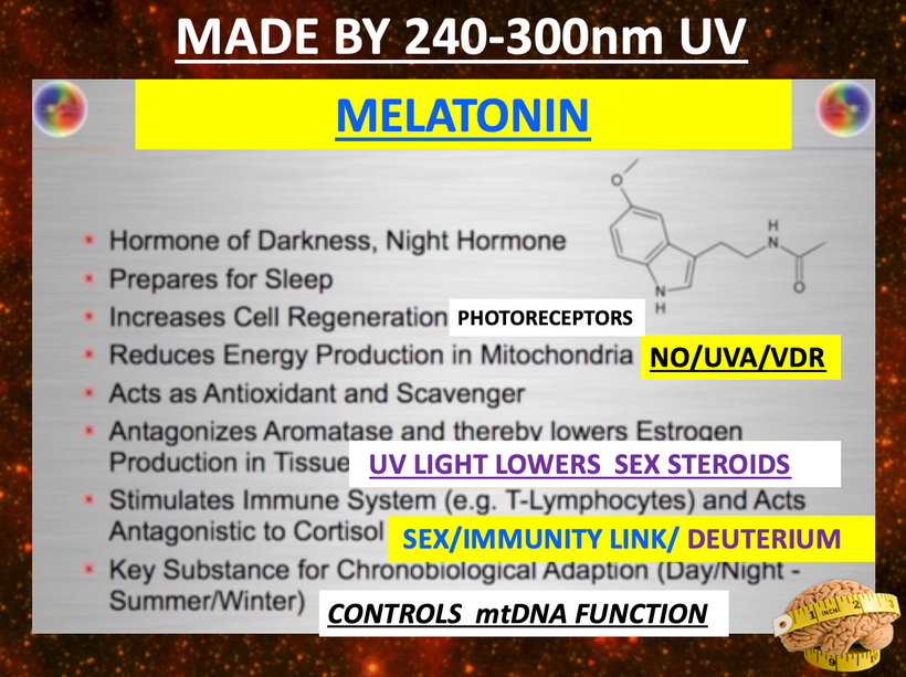
Although the molecular underpinnings of neurodegeneration are still not completely understood by centralized medicine, if you are following my thesis over the last 20 years, it should be obvious where the problem lies. It begins with altered light frequencies at surfaces to cause unusual diseases. Melanin’s charge separates water to make H+, oxygen, and electrons. Therefore it is the most powerful redox semiconductor in a cell. It explains why most melanin tends to be adjacent to the perikaryon in cells where mitochondria also reside. There are no coincidences in Nature. Recall that mitochondria make 95% of melatonin in a cell and that melatonin acts as the change programmer of the matrix and a major antioxidant to control ROS/RNS generation along with melanin that is adjacent to it.
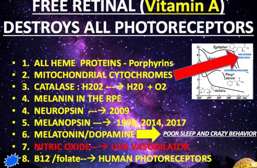
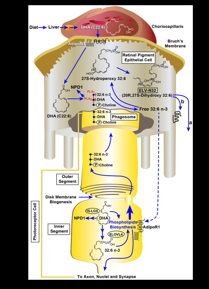
As melanin is degraded or lost in the eye photoreceptor complex we see the loss of the RPE microvilli on the basal side of the RPE. This causes infolding by the microvilli and increases the surface area of the RPE over a millionfold. The loss of microvilli is the first clinical sign of a POMC problem in the retina. This is the clinical sign one can see on OCT retinal scans that melanin needs renovation. As melanin is lost so is the redox power of the cristae in mitochondria. As a result, melatonin levels drop locally in those mitochondria and they all make less water and consume less oxygen. As a result, the tissue becomes hypoxic. This changes the free radical stream in mitochondria which changes the biochemical pathways used by the photoreceptors to regenerate in the retina. When this occurs, simultaneously DHA is consumed locally in photoreceptor damage. Normally most people think the consumption and oxidation of DHA are pathologic (Ray Peat) but that is wrong. It is wrong because DHA is the only PUFA in evolutionary history found to be transformed into highly anti-inflammatory chemicals called docasanoids and elovanoids to protect the RPE from ROS generation. This is an optical switch used in photo repair that requires UV-A light to be present when hypoxia exists in the retina. This is the basis of the Bazan short loop in the eye. This is pictured below in detail. This is direct evidence that redox power in the retina is lost because melatonin and melanin are both redox proteins that respond to this signal in retinal cells. Note that the liberated Vitamin A from the non-visual photoreceptor damage in the eye is what destroys the redox power of melanin and melatonin in tissues. Normally this process is tightly controlled by the physiology of the retina and terrestrial sunlight frequencies with DHA in the retina. If any of these thermodynamic givens are disrupted disease manifests.
Recall that dopamine can be made from L-DOPA from melanin in hypoxia but this ability is lost as well. As a result, the ROS made in the eye cannot be absorbed by melanin. This increases the oxidative stress associated with the eye and into the brain via tracts that are topographically linked to the RPE and the rest of the human cortex.
As we age heteroplasmy rises and so does melanin degradation. Thus, aging is associated with increasing levels of pro-oxidant factors (reactive oxygen species, ROS) and the dysfunction of the antioxidant systems in the brain, leading to protein and cellular damage and ultimately to neurodegeneration. That is the real cause of most cases of neurodegeneration. This is why Alzheimer’s disease is now referred to as Type 3 Diabetes.
Hyperglycemia causes retinal damage through complex metabolic pathways leading to oxidative stress, inflammation, vascular damage, capillary ischemia, and retinal tissue hypoxia. Melanin scavenges ROS/RNS/metal ions to clear them to prevent protein misfolding of the semiconductor complexes in retinal tissues that connect to deeper optical tracts in the brain. This mimics how prions spread disease.
This preserves non-linear optical processes in tissues to maintain tissue function. When melanin is absent or degraded the production of reactive oxygen species (ROS/RNS) leads to the generation of oxidative stress (lowered melatonin/melanin), which will result in the excessive production and accumulation of catabolism of these semiconductive proteins in these areas of the body. Melanin normally cleans up this debris when the eye is healthy. Without UV light exposure, the creation and accumulation of these complexes will occur and disease follows.
In ophthalmology textbooks, they repeat this statement all the time. Melanin granules in the retinal pigment epithelium (RPE) have many important functions which are not yet completely understood by centralized science.
One thing they do get right is that melanin in the RPE protects the cell from damage caused by oxidative stress. It also turns off vascularization of the fovea of the macula. This pigment acts as a free radical sink and diminishes cytotoxic lipid peroxidation that occurs in most eye diseases when melanin is not present in the RPE. Melanin in the retinal pigment epithelium is mostly eumelanin. There are two types of eumelanin, which are brown and black, synthesized from levodopa or tyrosine. The melanin amount in the RPE reduces importantly in aged eyes. When this occurs we know by definition melanin and Vitamin A are a problem in the eye. This is what destroys the Bazan loops in the eye and what also increases the need for DHA in the central retinal pathways.
Retinal hypoxia is further worsened by high oxygen consumption in the rods in the periphery. This seems like a problem to centralized researchers who do not seem to realize this is how dopamine is made from melanin to regenerate all the photoreceptors in the visual and nonvisual systems. The rods use a Warburg metabolism by design because of this arrangement. Melanopsin synthesis needs a massive blood supply in the periphery of the retina to create astronomical amounts of this opsin in the retina. I believe the reason for this is to increase the band gap because the rods and iPGRCs of melanopsin use so much oxygen to regenerate. The collateral effect of this arrangement creates more VUV light and ROS that stimulates melanin renovation on the RPE. This is why the RPE is so close to it in the retina.
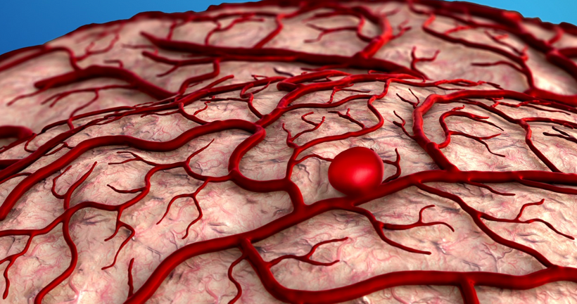
The RPE cells are the workhorse of the retina and have a large job to do as the slide shows above, yet the RPE is a thin layer of cells that textbooks say does not UNDERGO mitosis. If it does not undergo mitosis it means its nucleus has to be protected from any stray VUV light created endogenously. This is the job of melanin in this layer in the RPE. It absorbs all frequencies of light to keep the nucleus of the RPE cells from dividing. This is why the RPE has dark neuromelanin. Persistent hypoxia in this area results in the destruction of pericytes around the retinal vascular arcades that first create A-V shunts in the retina. I believe this is exactly how AVMs of the brain form in maldevelopment in embryos and in adult forms of humans. This is also the first step in diabetic neuropathy. I believe it is also a step we see to varying degrees in TBI cases. Centralized medicine has missed this in TBI because they are not doing OCTs as part of the workup.
This increases vascular endothelial growth factor (VEGF) and other pro-angiogenic factors )lack of melanin) always lead to vascular proliferative diabetic retinopathy/macular edema and progressive visual impairment when melanin is absent or not renovated by adjacent melanopsin damage.
BAZAN SHORT LOOP LOSS OF DHA DUE TO LACK OF MELANIN/MELATONIN
Optimal glucose control has favorable effects on diabetic retinopathy primarily because this allows for melanin renovation. POMC is such a unique mammalian intervention because the cleavage products work in opposition to one another, yet mammals figured out a way to make food directly from light without an intermediary WBG like chlorophyll. Melanin utilization was truly an innovation of evolutionary design using the colors of sunlight that bend the most inside the globe.
This blog is showing you how Nature hides her recipes in plain sight. What is most obvious is also often most concealed in Nature by her design. This is why you need curiosity to see what she is really doing and understand why she is doing it.
Melatonin controls mitochondrial DNA biology but dopamine creation from melanin controls how we decipher and sense time between the inside and the outside world. This creates the illusion Nature needs and wants. Nature never wanted us to see that 380 nm light is her favorite spectral density to run her photo repair process using the NON-VISUAL PHOTORECEPTOR SYSTEM. This system is far more critical to life than sight because it controls the eye clock pathways that are designed to simulate the physics of your environment from your brain to make the best metabolic choices of biochemical pathways in cells linked to the leptin-melanocortin pathways of humans. This is the best prediction machine evolution has ever built.
Implications of this idea? You can live well blind unless your eye clock mechanism is involved in your blindness. You will never thrive when your non-visual photoreceptor system is damaged whether you are sighted or not. This is the critical piece that modern centralized ophthalmology is missing today Creating melatonin in the mitochondria of tissues like the eye is the most critical surface for humans. The cristae are where the magic really happens. Melatonin initially controls all regeneration of the photoreceptors, visual and nonvisual in humans except the Muller cells in the retina. This is also by Nature’s design. Melatonin needs help from the non-visual photoreceptors to finish the job of photo repair in the CNS. Both of these chemical molecules are made by sunlight early in the day when a certain spectral frequency falls to Earth as it collides with aromatic amino acids in our eye to slow light down and create the quantum magic we call life. RNA and DNA have a homochirality to them. <——-Do not miss this hyperlink.
Below are two of the slides from that Vermont 2018 talk that put forth this idea for the first time in public.
^^^^The exact same thing goes on in your retina where melanin is located in the RPE.
This implies that your eye is off and on the switch of the human brain’s non-visual photoreceptive system. Now I am showing the wiring diagram of how this optical switch was built by nature.
Aromatic amino acids have to have an opposite chirality to fit this design quantum mechanically. You might remember my lecture from Vermont in 2018 introduced the idea of chirality to my thesis. The chiral heat effect of UV light is critical in this process. If this process is disturbed the entire system becomes off-kilter and disease will result. That is how and what melatonin, dopamine, DHA, and melanin, surrounded by matrix water are doing and what Nature is hiding from the observers of centralized science in the retinohypothalamic tract.
I wonder when centralized science will realize that the mitochondria also generate a magnetic field that is far stronger than the Earth or the sun’s magnetic effect because its scale shrinks. They seem to forget the lesson in physics as scale shrinks the electromagnetic force gains in strength. Might Mother Nature be using these forces in her quantum design to do things that are impossible at the macroscopic level that centralized science observes cells?
YEP.
Modern centralized treatments never focus on the intricate photo-regeneration of the retina. This process is critical in TBI and many brain diseases as well. This is just not a retina story developing before your eyes Patrons. The closest we came to this comprehension and realization of these ideas was in Becker’s work on regeneration. Another place we came close to in centralized science was in the work of neurosurgeon Robert Spetzler in the 1980s.
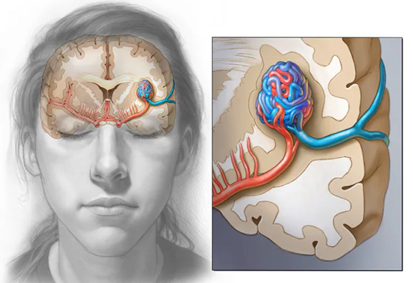
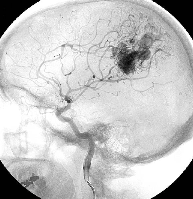
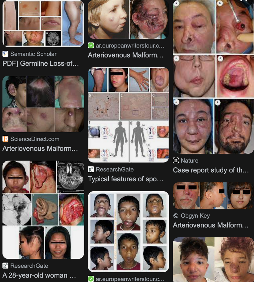
We still have not appreciated what either man really found (pic above and below). No one in neurosurgery today realizes that AVMs and aneurysms are due to a defect in melanin and melanopsin in the arterial beds of the brain that mimic what happens in the embryogenesis of the human retina. Instead, the focus of centralized scientists has been on the anatomic realignment of what appears in tissue in post-natal life. Big mistake.
AVMs anywhere in the post-natal human is a sign of melanopsin damage and a melanin problem passed down transgenerationally. AVMs of the dura and skin are big-time signs we still do not understand.
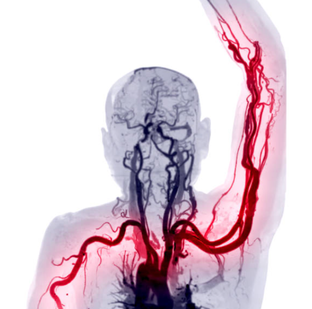
Aneursyms of the brain are also markers for melanopsin and melanin issues within.
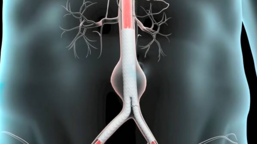
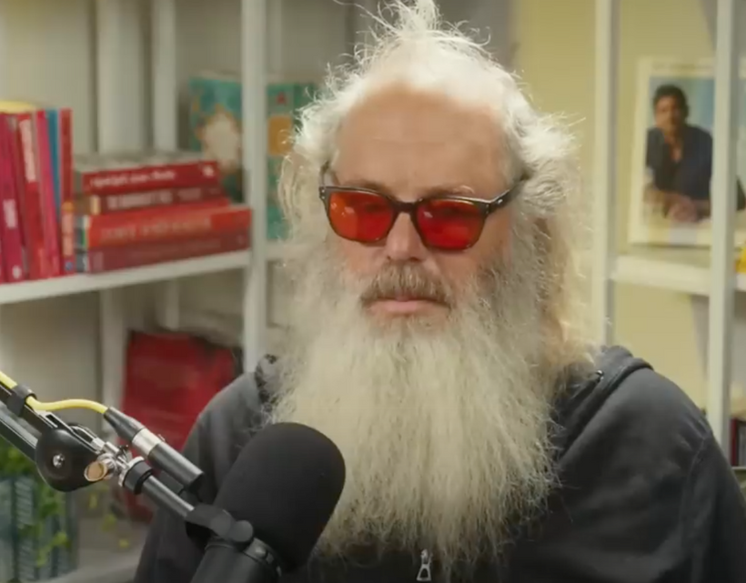
Aneurysms of the aorta are also markers of melanopsin and melanin renovation problems postnatally. An aortic aneurysm is the type of aneurysm rupture that Albert Einstein died from. It is also the type of aneurysm that Rick Rubin almost died from.
LESSONS FROM DIABETIC NEUROPATHY
Diabetic retinopathy stoichiometry defines most traumatic brain injuries. Every diabetic provides a lesson for a decentralized clinician. Centralized clinicians remain in the dark because they are bad at understanding opsin math.
What goes bad in diabetes in the eye? The photorepair regeneration system goes awry and this gives the decentralized MD something to understand the optical physiology of the human brain. In the Rod Outer Segment melanin renovation is astounding. It goes bad in diabetic retinopathy. Humans have 125,000, 000 rods in the retina., 1 RPE cell attends to > 200 photoreceptors. Each rod sheds approximately 3 mm a day. Thus, the human retina must shed 375 meters each day. This means > 10,000,000 meters of the disk have been shed by the age of 75. The most astounding ability of the human retina is that it must synthesize 9 billion opsin molecules per second in order to maintain its function. If it cannot regenerate the rhodopsin, melanopsin, or neuropsin you are guaranteed to get get some neurodegenerative condition linked to some degree of diencephalic breakdown in a topological map. Where the opsin is destroyed is a marker for where the disease will manifest as time elapses.
Other treatments for diabetic retinopathy include laser photocoagulation, which improves retinal oxygenation by destroying the high oxygen-consuming rods by their replacement of low oxygen-consuming glial tissue. No one has thought to use melanin renovation of the RPE to solve this problem because no one in ophthalmology realizes that melanin is a wonderful creation of condensed matter physics and atomic molecular orbital engineering. It is truly the most amazing protein anywhere in the human brain. No wonder humans put it in every organ system compared to other primates. In places it is absent or missing destruction lies in its wake. Remember alpha MSH is stimulated by UV-A light. 380nm light is UV-A light. It shares this common tie for its love of UV-A light with melatonin.
Hypoxia is a potent stimulator of VEGF, and HIF-1 alpha and intravitreal anti-VEGF antibodies have been somewhat effective in regressing macular edema according to some studies done in retinal neovascularization. The problem is using natural light to renovate the photoreceptors that should have hit somebody’s mind in 125 years still astounds me. We know that dopamine and melatonin regenerate the photoreceptors and we also know melanin creation improves the regeneration of dopamine from melanin by way of L-DOPA. We also know as melanin returns to the RPE the amount of melatonin made in the mitochondria of the retina also improves. When the RPE is loaded with melanin sleep improves tremendously because Vitamin A goes down and the visual cycle of photoreceptor regeneration is CONTROLLED locally in the eye by DHA, dopamine, melatonin, and melanin working in concert. This is why sunny days at the beach always lead to great sleep.
Very few people have linked the complex pathophysiology of diabetic retinopathy with an altered spectrum of sunlight because they do not seem to understand the biology of POMC even today in 2023. They continue a biochemical focus with their lens pointed to retinal oxygen/fuel consumption with resultant hypoxic damage to retinal neurons. Blue light destroys melanin because it raises blood glucose and insulin. UV light and IR-A light is what are critical in the melanin renovation of the RPE. The biochemical focus wants to continue to discuss potential mechanisms through which sodium-glucose cotransporter 2 (SGLT2) inhibitors improve retinal hypoxia—through ketone bodies. This is incredibly myopic because no one seems to understand mammals make glucose and insulin from blue light.
Melatonin plays a key role in the coordination of the diurnal and seasonal circadian system, which underlies how the biological clock works everywhere in humans.
Mitochondrial DNA is more vulnerable to alteration than nuclear DNA, mainly for two reasons. As such mtDNA damage LOWERS local melatonin levels. So circadian disruption alters melatonin levels locally in many tissues. That lead to diseases.
Melatonin controls mitochondrial DNA and dopamine controls how we experience, decipher, and sense time between the inside and the outside world. Nature hid that she made this dopamine in the eye when parts of the retina were forced to be hypoxic to degrade melanin to create dopamine. In this way, Nature never wanted us to know how important melanin was. Creating melatonin also occurs first in the eye and is the most critical surface creation for humans. Melatonin is made by the aromatic amino acid tryptophan which absorbs a very unusual amount of full-spectrum UV light according to its absorption spectra. This is light that never reaches the surface of the Earth in most places. So the curious would ask themselves how does this happen? Deuterium in the circulatory system of the retina, when sunlight entered the eye and hemoglobin’s unique optical window of 250-600nm, was the key to solving that mystery in the eye. This explained why the choriocapillaris exists as it does in the adult human retina.
The choriocapillaris is the innermost structure of the choroid in humans that directly nourishes the retinal pigment epithelium and photoreceptors.
The embryology of this layer of the retina shows the power of melanin. The initial human choriocapillaris form by hemo-vasculogenesis. This is how hemoglobin if formed in the human embryo. This area of the retina forms just like red blood cells do in our marrow and liver. This means the same cells in this region of the retina express special fetal hemoglobin called Hb-εas well as CD31, CD34, VEGFR-2, or vWf (where hemophilia and von Willebrand’s disease comes from), further suggesting the same precursors were capable of erythropoiesis, hematopoiesis, and vasculogenesis, the definition of hemo-vasculogenesis, as occurs in blood islands in cells. In the fetal period, hemo-vasculogenesis is completed the same way for blood cells and new blood vessels. They appear to form by angiogenesis since endothelial cells were proliferating in the same way at the same time. So is melanopsin. 9 billion opsin molecules are synthesized per second in the retina’s outer photoreceptor region. This requires massive amounts of oxygen to synthesize this amount of melanopsin. All these unique pieces fit now and make sense. Porphyrins are another non-visual photoreceptor in humans.
What was the key to this entire process occurring? Melanin had to be absent in the embryo at a specific time in morphogenesis when the choriocapillaris is forming. Just the presence of melanin in this area of the retina appears to stop hemo-vasculogenesis in the embryo’s retina creating the picture above in the adult retina. This is why the retinal angiogram of the adult human retina shows no blood vessels in the adult macula. Diabetic retinopathy is like autism. It is an atavistic effect of the embryo of previous stages of evolution of the eye that allowed for blood vessel growth in the fovea.
It is also why diabetic retinopathy is associated with neovascularization or angiogenesis with excessive blue light exposure. When melanin/melanopsin is disrupted or destroyed, blood vessels can grow and neurulation is changed in the optical pathways. You saw the same thing in the last blog on autism. Blue light also changes the retinoid cycle of the retina. Melanin’s presence or absence is critical in both the embryo and in the adult form of humans. Blood vessels bring RBC and RBC brings oxygen and oxygen is key to melanin biology. When hypoxia develops dopamine shows up from melanin degradation. Have I told you yet that the choriocapillaris creates the highest blood flow of any human tissue and it has the outer rod photoreceptors adjacent to it that consume the most oxygen of any tissue in humans? Do you want to guess why this arrangement exists? The picture below gives the answer.
If you have a high demand for oxygen you keep melanin in its optimal state and any photooxidation in the visual system of the photoreceptors needs dopamine. It also is needed to make 9 billion opsins per second. If the non-visual photoreceptor system fails, you’ll wind up with some diseases mentioned in this blog.
Dopamine is made from tyrosine, another aromatic amino acid that also absorbs short-wave UV light. This is why both molecules are tied to the physiology in the sys and skin and are linked to melanopsin. Melatonin controls all regeneration of the photoreceptors in man except the Muller cells in the eye. Both of these chemical molecules are made by sunlight as it collides with aromatic amino acids in our eye to slow sunlight down so it can become matter. That is how they link to one another. The Reality #12 blog told you long ago how they work in a cell but no one asked me in the comments the right question.
Most people with Parkinson’s disease know that they are deficient in dopamine in the midbrain, but most people do not know that that defect in that area spreads to the region from the eye first in a very similar fashion to how a prion disease works. The Vitamin A deficit in the eye is how the blood disorder von Willenbrands disease or hemophilia A happens due to the changes in the retinoid cycle of the embryo. These can be transgenerational diseases but few people see what I see in the embryology of the retina. This is why PD patients have misfolded proteins associated with their disease that mimics how prions operate in disease states. It is a story laid out in the OSF #3 blog post in detail. I’ve been telling you this story for a long time but now you have new eyes to perceive it. What has always been the clinical take home from Uncle Jack?
My Rx was not hard. Most of you refused to believe it was this simply because you were all ignorant of the optics of the non-visual photoreceptive system built into us by Nature. Stop complaining and just do what Nature requires you to do. I am giving you my life’s work for the cost of a cup of coffee. SHARE IT. You owe me that much.
MORE SCIENCE TO MOTIVATE YOU INTO NATURE
First, mitochondria are a major source of intracellular reactive oxygen species (ROS) via cytochrome C oxidase’s ability to hold oxygen between Fe and Cu ions. The electrostatic grip is linked to the free radical made. The light in the environment controls the electrostatic power via the effect of D shell electrons in these metal atoms. Therefore mitochondrial DNA is under much stronger oxidative stress than nuclear DNA. Second, mitochondria have a matrix-side negative membrane potential for oxidative phosphorylation. This membrane potential concentrates lipophilic cations inside the mitochondrial matrix up to approximately 1,000-fold. Deuterium and H+ are positive cations. Deuterium and H+ are both positive cations that varying lipophilic attributes FYI. Too bad some of the deuterium critics don’t know these basics. This is why the matrix favors H+ over deuterium and why the ECF and non-lipophilic tissues will naturally contain more deuterium (plasma). This deuterium in the blood is how cells make their own UVC light used to make neurohormones from aromatic amino acids that control local mitochondrial biogenesis.
^^^^Both slides were from Vermont in 2018. Please stop telling me I have not been telling you this story for years when the reality is you refused to sink to my level and LEVER up your knowledge. If you do not lever up your knowledge you are never going to stop believing the dermatologists and ophthalmologists who are being paid by Big Pharma to keep you from the most simple things to keep you healthy. The slide below was used in 2017 in Cancun by a group of optometrists/ophthalmologists who refused to see where diabetes came from………blue light toxicity. I hope you get the lesson today they still cannot fathom.
SUMMARY
Diabetes has been and will always be a chronic mammalian disease when the purple and red light is removed from our environmental light. The focus on a ketogenic diet is misplaced when you consider the quantum effects of manufactured light on the photoreceptors mentioned above.
The biochemical ideas continue to dominate the literature because they believe ketones are energetically as efficient as glucose and yield more ATP per molecule of oxygen consumed than fat, with less oxidative stress. While this is true, no ketogenic diet has ever been able to reverse DR primarily because diabetes is not a disease of diet. It is a disease of blue light. Instead, we need a paradigm shift where eye physicians begin to realize full spectrum sunlight leads to direct retinal benefits which occur quickly when UV light and IR-A light are reintroduced to improved fuel energetics. these two frequencies of light induce less hypoxia and reduce inflammation through the recycling of DHA and the neuroprotective metabolites of the short loop of Bazan in the eye. Modern eye care is in the dark ages in treating this disease of blue light overexposure.
MORE ON THE ipRGCs of the retina and SCN. Someday if someone funds my research, autism might be the first topic I’d suggest we study. Why?
Melanin seems to be important for migration of neurons in all mammals. I told you that in the recent blogs around melanin and metastasis. And this idea clued me long ago in that I might be able to explain autism because of my unique perspective on melanocyte migration and how it links to melanopsin regeneration.
Implications of a lack melanin pigment in mothers and fathers and what this might mean to their germ lines that become a child? Here is one of my kids above.
Today’s lesson is going to come from kitty. Most of you know I love cats. My favorite cat has always been the Siamese cat. My first cat was one and my current cat (above) is a Siamese. You might not know why I am fond of Siamese cats but you will soon. They taught me a lot about melanin when I was a boy in New York City.
CATS & HUMANS WHO ARE ALBINOS HAVE A MELANOPSIN LESSON TO TEACH
Most albino human beings and albino cats lacking retinal pigment in the RPE have observable nystagmus and many exhibit strabismus. The optic chiasm (pic above) of albino mammals including human beings and cats shows that almost all their retinal ganglion cells cross at the optic chiasm with few uncrossed optic fibers. This shows you there is a problem with morphology in mammals neurulation patterns. Something has to be behind this misrouting behavior. My bet is a lack of melanin allows the stickier deuterium isotope to affect cell migration during neurulation of the mammalin brain due to the kinetic isotope effect of deuterium on hydrogen in cells. One D controls 96 atoms of H+ (pic below). I think the massive increase of atomic weight and the kinetic isotope effect are critical in the misrouting behavior of neurons and this has dramatic effects on organization of the visual system of mammals. There are details about mammalian embryology that have lead me to this conclusion. I think it might be behind what causes autism, as well. A lack of melanin in the parents and their germ line cells maybe the precipatating event that begins this cascade of events on. I believe this idea is tied to autism because the effect is quite heterogenous in presentation, hence why they call autism a spectrum of disorders.
In 1965, C.L. Sheridan noticed that retinal ganglion cells originating in temporal retina of albino rats did not function well, hypothesizing that albino rats have fewer uncrossed, non-decussating optic fibers. R.D. Lund anatomically verified that albino rats have fewer non-decussating optic fibers compared to pigmented rats. For several years the abnormality of reduced non-decussating optic fibers at the optic chiasm in albinos was thought to be limited to rats and rabbits. Guillery reported atypical visual system organization in Siamese cats, but the association with albinism was not identified.
CIRCADIAN BIOLOGY ENTERS THE FRAY OF MY KITTY STORY
The mammalian SCN is controlled by light and temperature and by the ipRGC of melanopsin. I told you that a long time ago in the Cold Thermogenesis #6 blog. What else didn’t I mention back then?
In 1971, Creel initially published the connection between Siamese cats and albinism, and hypothesis that reduced non-decussating optic fibers likely is a “highly general transspecies phenomenon in albino mammals”. Siamese cats all have blue eyes and blue eyes = less melanin in the iris. Siamese cats, Himalayan mice, rats, and rabbits all express a mutation that is a temperature-sensitive pigmentation defect, that is, allowing pigment to show up only on the cold parts of the body.
This is why their coats are so grey and white (my kitty above). Moreover, their retinae lack pigment too. Many human infants are born with blue eyes because their brains are devoid of melanin because humans brains are born into life in an immature state. That is the case for human blue eyes, but what about the cats? Why do Siamese cats have blue eyes almost all time? It is not just their iris that has reduced melanin. Here we see the interplay of UV light and cold and begin to understand why this link is present from an evolutionary standpoint. Wide band gapped semiconductors can make the same light endogenously from atoms doped to proteins or via cooling environmental temperatures. I believe this is the basis of where the mammalian dive reflex comes from. Most humans who live at high latitudes where it is colder also tend to have blue eyes and blonde hair. The link is unmistakable when you realize it.
Cooling is usually occurs at surfaces in mammals (eccrine sweat glands) and most of their surface has arterioles loaded with higher levels of deuterium that act a Bose Einstein condensate because of deuterium unique nuclear spin. This makes their surface something important. It is called a topological insulator. Mammals interiors are filled with fermions = electrons and H+ ions. They follow Dirac fermion statistics. Here we see a physical difference manifesting in mammals for some reason.
Is there a trade off for mammals who live in colder environments? Yes there is. what is it?
They have less visual and non visual photoreceptors in their sensory organs.
Is this true in human mammals too, Uncle Jack? YEP.
Note when humans have normal pigmentation in the eye (RPE) tend to have optimized neuro-opthalmological migration in their visual system as well. It appears melanin is a beacon for proper migration of neurons in the eye to deeper brain structures in humans. This is critical in the eye because of the location of the SCN to the RPE. The SCN controls the eye clock, circadian rhythms, and the pupillary response in mammals to bright light. It also affects their behavior. This is markedly changed in autistic kids.
In humans with pigmented retinae, retinal blood vessels spare the foveal area. In albino human retinae, blood vessels intrude into foveal area. Pigmented human retina exhibit an avascular zone surrounding the fovea. Albinos do not exhibit this arrangement. They have blood vessel encroach into their fovea. Humans with destroyed RPE from diseases like diabetes get the very same neovascularization of the fovea. This ruins their central vision over time.
This is a big clue for what decentralized clinicians should be looking for in human’s with autism. If a lack of melanin is part of the pathophysiology of this disorder than we should see some subtle changes in OCT and their arteriole beds in the diencephalic derivative of the brain if we look for it, if I am correct. I think a lack of melanin pigment initiates atavistic expression in autistic kids of the central and peripheral nervous system. This lack of POMC or POMC translation leads to alterations in the melanopsin system that ruins normal neural migration.
The phenomenon is called atavism—this is the reappearance of a trait that had been lost during evolution before in the same species. In autism many of these kids cannot talk or interact with their species. We know our nearest cousins cannot speak either. We also know that early primates were loners and that social networking became a big trait later on in evolutionary history of the primate. This is another throw back behavior seen in autism that would have been seen in transitional chimps on their way to us. This change would not be found in our DNA either. Our genes do not determine who we are, but with atavism, they can sometimes serve as reminders of our evolutionary past. Functionally this is what autism in humans looks like to me from my current perspective. Another big clue for my hypothesis is found in the embryology of the visual and auditory systems of mammals.
MELANIN IS ALSO A TIME CRYSTAL FOR EMBRYOGENESIS IN MAMMALS
Embryonic progression in albino mammals’ visual and auditory systems seem to also take a step back in evolutionary time and it seems this program is initiated by lack of melanin pigment. Non albino animals do not exhibit this altered migratory pattern. Abnormalities of optic chiasmic misrouting in albino mammals is a developmental field defect that is seen normally in preceding phylogeny. Complete crossing of optic neurons at the chiasm is a normal developmental event in vertebrates prior to mammals. This reinforces my beliefs that atavism is a central problem in understanding modern autism.
The presence of melanin pigment in the embryonic retina is the signal that initiates retinal ganglion cell pathway routing from the eye to the SCN and from the SCN deep into the brain. Insufficient coding for retinal pigment launches an earlier, more stable genetic package directing a different targeting of optic neurons and this results in autism.
Genetic makeup includes preserved earlier evolutionary features. Charles Darwin popularized atavism in the 1860s as the term for reappearance of ancestral characteristics in future generations.
Today we know that vision and hearing evolved together dating back to the PaxB gene, which is a single gene controlling eye and precursors to hearing (mechanoreceptors) in box jellyfish. This occurred before independent Pax 2 and Pax 6 genes showed up in primates much later. There are evolutionary connections between eyes and mechanoreceptors of the inner ear to the extent that during evolution are linked to melanin generation in those sense organs. I told you earlier in the series melanin was the favored semiconductor of all mammals post KT event. This story fits this narrative as well. If POMC/melanin is absent for any reason, it appears human neuroplastiticty allows sensory cells to shift their sensory modalities to an older phylogeny experienced in evolutionary history. Why is this a big deal? The melanopsin phylogeny predates even primate evolution in time.
I think this happens in modern humans because of a loss of information and energy transformation in the embryo due to a lack of POMC or POMC translation in the parents cells and their germ lines that create their child. I think this is what autism is at its core. It is lack of POMC that has huge implications for the neurulation of the diencephalon in humans and its really old relationship to melanopsin biology.
BACK TO THE CATS
What happens when cats lack melanin in their eyes? Do they exhibit traits that mimic human autism? They exhibit fewer photoreceptors, they have foveal/area centralis hypoplasia, & exhibit misrouting of the temporal retinal ganglion cells, while having a variation of geniculate terminations, and most importantly they develop vascular intrusion into foveal area. They also exhibit abnormal cortical projections, and fewer cones in macular area. Lacking pigment in the cat leads to some major migration problems in the adult form. It seems albino kitties and Siamese cats have a lot in common with autism.
The animal model closest to organization of primate visual system studied the most is the albino cat. Albino cats have observable nystagmus, and many have strabismus. Figure 1 above pictures the optic chiasm of an OCA1 albino cat shows almost all retinal ganglion cells (RGCs) cross at the chiasm.
Not all of them do and the ones that do not cross tend to innervate areas they should not be in. The non visual photoreceptor associated with these RGCs is melanopsin. The number of binocular cells was found to be reduced in visual cortical areas of Brodman numbered 17, 18, and 19 in Siamese cats and albino cats. This impairs their stereovision. So these cats do not have great vision, but this is because they are adapted to colder environments with more blue light and less UV light. Their SCN periodicity can be retuned by cold weather. The trade off for their lack of melanin is altered neural migration in their brains.
This surface temperature thing is a big deal in mammals because our SCN changes its periodicity to temperature and light. I believe this explains why autistic kids do not like temperature changes or being touched. (another sensory processing delay). I have a sense why this happens in autistic kids because these changes are seen in cats as well who lack pigment. Autistic kids have a broken topological insulator in their skin, eyes, and circulatory system. I think they are exquisitely sensitive to the chiral pinch of deuterium in their blood and this creates a lot of endogenous light that overwhelms their system because they have poor melanin sheets within the basal levels of their skin. This allows too much deuterium to leach out of the circulatory system and into the substance of their subcutaneous tissues. These leaching of deuterium than alters non visual photoreceptors in that location.
There is some rather unique things to know about this situation. Evolution seems very fond of the SCN melanopsin connections I mentioned in QE #44. Why? Melanopsin retinal ganglion cells are always completely crossed or bilaterally projected into suprachiasmatic nuclei above the optic chiasm and these projections are not affected by albinism in any mammals. Interesting don’t you think?
I think this goes back to where melanopsin came from phylogenetically. It came from our fish amphibian origins 380 million years ago. Phylogenetically older connections are less abnormal in albino mammals by exhibiting a bilateral suprachiasmatic projection pathway to the SCN. These tracts appear to be unaffected in albinos. This tells me this system is highly protected by evolution because of how important it is to be able to simulate the physics of your environment to be highly adaptable. This also explains another peculiarity about the melanopsin system.
Melanopsin regeneratiion acts largely independently of the visual cycle. This tells me the melanopsin system has old roots in evolution and evolution has specifically made sure it did not co evolve similar mechanisms with the rod and cone system of the eye. Melanopsin neurons also control pupillary size with the autonomic nervous fibers of the third cranial nerve.
When this system is disrupted we get mammals that are not adaptable at all. This defines modern humans who are on the autism spectrum. Most children with autism have trouble sleeping, which may exacerbate other challenges associated with the condition. Sleep problems hint at disruptions in the circadian clock, a cellular timer that keeps cells in sync with the day-night cycle. Vitamin A disruption causes sleep disorders.
I have a sense that we will find out in the future that autistic children have wiring defects in their chiasms and in their sensory relay pathways because of a broken Vitamin A cycle in the melanopsin system along with a lack of melanin and POMC activity in the germ cells. This Vitamin A problem will lead to science realizing these kids all have altered melanopsin regeneration pathways compared to humans that do not have this deficit. I believe the melanopsin system has its own regeneration system because it is VITAL to accurate time crystal managment of the circadian mechanism you saw in the last blog. The SCN simulates the physics of our environment to program our metabolism from these light signals. This is broken in autism.
LACK OF POMC/MELANIN = ALTER MELANOPSIN REGENERATION = VITAMIN A PROBLEM
Vitamin A is necessary for normal embryonic development in humans, but its role in the adult brain is poorly understood by centralized science in 2023. Vitamin A derivatives, called retinoids, are involved in a complex signaling pathway that regulates gene expression and, in the central nervous system, controls neuronal differentiation and neural tube patterning.
There is another reason why I think this Vitamin A, melanin, melanopsin story is linked to autism. Most people know that the majority of melanin in the eye is in the RPE. What many however do not know is that the photosensitivity melanopsin requires a steady supply of cis-retinaldehyde, a type of retinoid. The primary source of this vitamin chromophore (328nm) in the vertebrate eye comes from a complex multistep enzymatic pathway, known as the retinoid or visual cycle (above). In this cycle by 11-cis retinaldehyde is regenerated from bleached all–trans originating in photoreceptor outer segments of rods. The critical elements of this pathway occur in the retinal pigment epithelium (RPE). Here is the problem. Since melanopsin is found in the more superficial layers of the retina, quite distant from the RPE, it would seem poorly placed to obtain cis-retinaldehyde from this RPE source. So how does melanopsin do it? Why does it have its own regeneration pathway separate from rods and cones?
Chromophore regeneration in rod photoreceptors relies solely on a tissue adjacent to the rod and cone layer of the retinal pigment epithelium. Cone photoreceptors rely both on the RPE and Müller glia, which traverse the entire depth of the retina to regenerate. Melanopsin doesn’t use either system.
In the mammalian photoptic retina light activates melanopsin to trigger a G protein cascade that causes membrane depolarization. The ipRGCs/melanopsin response is opposite to that seen in our rods and cones, which hyperpolarize, but resembles the actions of photoreceptors found in invertebrates like fruit flies and horseshoe crabs. This system exhibits atavism mentioned above. In humans, ipRGCs fire spikes. They are understood to use glutamate as their primary neurotransmitter; uniquely among RGCs, they also express a peptide neurotransmitter called PACAP (pituitary adenylate cyclase activating peptide).
Known influences of ipRGCs extend well beyond their direct targets. Examples are regulation of melatonin synthesis in the pineal gland and the development of synaptic plasticity in the hippocampus. I believe they drive synaptic plasticity in all of our brain’s circuits and this is why melanopsin is the most numerous opsin in humans.
Why do I mention this detail? Most people forgot human embryology of the brain and skin. They have forgotten that Vitamin A/retinoic acid have massive effects in the morphogenesis and growth of the neural tube. Not only will deuterium affect neural migration from the notochord, but the chemical signal governing it is likely also awry. Most of which can be explained by defective notochord signalling. This would cause neurulation defects from the thalamus to the adult hemispheres. This process is how humans create their hemispheres in the last trimester and postnatally. This set of circumstances fits the modern phenotype of autism based upon my knowledge of human brain embryology. I think the key problem in the autistic embryo is a problem of Vitamin A signaling being comingled in the eye, optic chiasm, hypothalamus, thalamus and eventually the neocortex in humans. This would create a wide spectrum phenotype in the adult human.
Modern humans are new phylogenetic creatures in evolution and we know that optic projections near chiasm projecting into hypothalamus antecede vertebrates evolution because they occurred in early chordates. Chordates first appeared on Earth 590 million years ago. Those are the animals that innovated melanopsin originally.
It appears that fact surprised Dr. Huberman and Dr. Berson based on what Huberman said in the Rubin podcast. It did not shock me because I knew mammals came from early chordates because of my time in the Museum of Natural History in New York. This is why it made sense to me why we had melanopsin in our brains. I was shocked to learn in 2014-2017 that we also have it all our blood vessels, skin, fat, and our outer inferior retina. Nature does not make mistakes.
This is my kitty today as I write this blog. She likes IR-A and UV-A light now. I have a sense all parents with autism need to learn a lot more about full spectrum sunlight and the key frequencies of IR-A light and the 380 nm light that controls the photorepair mechanisms in mammals. When they do, they will realize how to help fix the problem they created by their abuse of blue light and tech screens.
SUMMARY
When I was 8 was the first time I got a Siamese cat. I asked the lady at the museum why my cats eyes were different colors than other cats and she did not know the answer. She told me she’d find out and few weeks later I found out about melanin as an 8 year old kid. She told me all chordates had retinal pigments matching vertebrates. Humans were the latest version of vertebrates. That lesson would come in big later in my life. I never forgot about my kitty’s eyes. And that is why it never surprised me that the melanopsin chromophore showed up in human brain. My childhood was prepping me for a later discovery around this amazing protein. People with autism one day may thank Siamese cats for solving this enigma disorder for centralized science.
CITES
1. Sheridan CL. Interocular transfer of brightness and pattern discriminations in normal and corpus callosum sectioned rats. Journal of Comparative and Physiological Psychology. 1965;59:292-294
2. Lund RC. Uncrossed visual pathways of hooded and albino rats. Science. 1965;149:1505-1507
3. Giolli RA, Guthrie MD. The primary optic projections in the rabbit: An experimental degeneration study. The Journal of Comparative Neurology. 1969;136:99-126
4. Guillery RW. An abnormal retinogeniculate projection in Siamese cats. Brain Research. 1969;14:739-741
5. Creel DJ. Visual system anomaly associated with albinism in the cat. Nature. 1971;231:465-466
6. Creel DJ. Differences of ipsilateral and contralateral visually evoked responses in cats: Strains compared. Journal of Comparative and Physiological Psychology. 1971;77:161-165
7. Creel D, Witkop CJ Jr, King RA. Asymmetric visually evoked potentials in human albinos: Evidence for visual system anomalies. Investigative Ophthalmology & Visual Science. 1974;13(6):430-440
8. Sanderson KJ. Retinogeniculate projections in the rabbits of the albino allelomorphic series1. The Journal of Comparative Neurology. 1975;159:15-27
9. Creel DJ, Giolli RA. Retinogeniculate projections in albino and ocularly hypopigmented rats. The Journal of Comparative Neurology. 1976;166:445-455
10. Guillery RW, Oberdorfer MD, Murphy EH. Abnormal retino-geniculate and geniculo-cortical pathways in several genetically distinct color phases of the mink (Mustela vison). The Journal of Comparative Neurology. 1979;185:623-655
11. Piatigorsky J, Kozmik Z. Cubozoan jellyfish: An Evo/Devo model for eyes and other sensory systems. The International Journal of Developmental Biology. 2004;48(8-9):719-729
The SCN is an optical lattice clock that pays attention to all the zip codes in a cell and functions as a true time crystal. It mitochondria imprint the time signal into the water & melatonin that the SCN’s mitochondrial create. This amazing part of the brain and a direct target of the ipRGC’s of melanopsin from the inferior nasal portions of the retina. (slide below from my Vermont 2018 talk)
I discussed time crystals in my 2018 Vermont talk briefly and told my members they were first thought about in 1982, rediscovered in 2012, and physically discovered in 2014. Today, in 2023, the time crystal is a new category of phases of matter, expanding the definition of what a phase is. All other known phases, like water or ice, are in thermal equilibrium: time crystals are dissipative far from equillibrium structures. Their constituent atoms have settled into the state with the lowest energy permitted by the ambient temperature in the environment, and their properties don’t change with time. This makes them a perfect metronome to keep time for multiple zip codes that exist in cells. The time crystal is the first “out-of-equilibrium” phase: It has order and perfect stability despite being in an excited and evolving state. This makes it highly responsive to light photons to create the tick-tock of periodicity that exists in circadian controlled mechanisms.
Essentially a time crystal is an object whose parts move in a regular, repeating cycle, sustaining this constant change without burning any energy. they are harvesting information in the system to create order from chaos.
The consequence is amazing: You appear to evade the second law of thermodynamics, which states that disorder always increases over time. We know from Maxwell’s work in “demon’s” and the work of Lindauer, that there is an infantessimal requirement of energy and information loss, so that this state of matter still has to obey all of physics laws. Kruse for Dummies attendees heard this in my presentation. Because of this, this explains why all living things must sleep. In sleep this debt is paid back to the system to remain highly ordered time crystal. I told you a long time ago, in the Cold Thermogenesis series, that the default state of life was sleep. Then as evolution progressed with evolved wakefulness. This idea was counterintuiitve to many at the time. The reason for my prediction was simple. I read Feynman;’s 1982 paper and thought to myself what organ in humans fit his description of an ability to simulate the physics of Nature? The SCN fit perfectly.
When I read the paper in my residency I immediately thought to myself is this what the SCN is doing for the body? It is an organ that can simulate the physics of the environment to inform the rest of the body of that information to create some semblance of order. Everybody knows that perpetual motion machines are impossible. However, in quantum physics perpetual motion is okay as long as we keep our eyes closed and do not observe it. By sneaking through this crack we can make time crystals operate. This mimics sleep.
My intuition was rewarded in 2012, when Physics Nobel Laureate Frank Wilczek theorized that time crystals had to exist and they were finally identified in 2014- 2016. Time crystals exhibit the bizarre property of being in constant, repeating motion in time despite no external input. Their atoms are constantly oscillating, spinning, or moving first in one direction, and then the other. The recipe for a time crystal is below.
Note the bottom part of the picture above. This appears to be exactly what is happening when sunlight hits the RPE in the eye and send photonic information to the SCN with no synapsing in between. The information stream is UNINTERRUPTED. This is unusual in the mammalian retina. Almost every tract in the eye synapses before it gets into the brain. This one does not. Interestingly the same tract sends uniterrupted information of light to the habenular nucleus that controls mood and behavior in mammals. It also makes sense why the same intrinsic photoreceptor retinal ganglion cells of this melanopsin tract send its information to the habenular nucleus of the thalamus.
The eye is the off and on switch for time for the entire diencephalon of mammals. This turns on and off the brain. It made sense to me finally why the thalamus created alpha waves of 7.83 Hz prior to sleep cycling. The thalamus has to be able to record the asymetry of light and day accurately to awaken the rest fo the brain from the default state. It turns out few people have realized the Schumann resonance on Earth varies day to night. This helps attune the periodicity of our time crystals in the SCN. They become more accurate clocks as periodicity increases.
A coupling between geomagnetic activity and the human nervous system’s function has now been fully identified by virtue of continuous monitoring of heart rate variability (HRV) and the time-varying geomagnetic field over a 31-day period in a group of 10 individuals who went about their normal day-to-day lives. (paper above)
It was found that the participants’ HRV rhythms synchronized across the 31-day period at a period of approximately 2.5 days, even though all participants were in separate locations on Earth. This tells us the ability is built into all mammals. Overall, this suggests that daily autonomic nervous system activity not only responds to changes in solar and geomagnetic activity, but is synchronized with the time-varying magnetic fields associated with geomagnetic field-line resonances and Schumann resonances. Biology is amazing when you observe its abilities.
The Schuman resonance is a charging opportunity for the human brain’s time crystals: How do cells charge?
Charging by friction – this is useful for charging insulators/semiconductors. If you rub one material with another (say, a plastic ruler with a piece of paper towel), electrons have a tendency to be transferred from one material to the other. For example, rubbing glass with silk or saran wrap generally leaves the glass with a positive charge; rubbing PVC rod with fur generally gives the rod a negative charge. Vortexing liquid crystals semiconductors also creates friction which can generate a charge that is quantized. This is how water gains charge in the circulatory system via turbulent flow around the cells in blood plasma.
Charging by conduction – useful for charging metals and other conductors. If a charged object touches a conductor, some charge will be transferred between the object and the conductor, charging the conductor with the same sign as the charge on the object. RBC’s are conductors and can transfer their charge to water in blood plasma. This is why coherent domains in water develops a net negative charge when it forms. This is how you build redox power in sunlight because charge is being transferred.
Charging by induction – also useful for charging metals (dopants on our semiconductors) and other conductors. Again, a charged object is used, but this time it is only brought close to the conductor, and does not touch it. This mimics how sunlight interacts with melanin, hemoglobin and chloroplasts in living systems in trapping light energy before it changes to an electric charge in the coherent domains water. To gain the charge best from sunlight you need to be grounded to Earth. If the conductor is connected to ground (ground is basically anything neutral that can give up electrons to, or take electrons from, an object), electrons will either flow on to it or away from it. When the ground connection is removed , the conductor will have a charge opposite in sign to that of the charged object. This is why being disconnected from Earth chronically leads to things like rouleux formation and clotting. Since water molecules, are naturally polarized (magnetic dipole), they can quickly remove charge from a charged object. This is why blood is 93% water by volume. Mother Nature uses every last bit of physics the universe gives her to work with.
On sunny clear days with low humidity are associated with ionospheres with high electric fields, low magnetic fields and diminished energy transfer from the sun to us if we do not have a connection to Earth. Why? Electrostatic charges are not transferred well when water is not present in the ionosphere. This is why dehydration from a lack or water production in mitochondria favors illness.It is also why deuterium is not favored by living systems in nature. It cannot transfer charge as well as light hydrogen can, because its extra mass affects bonding strengths in all molecules it is used in. This is why life tries to exclude it. (kinetic isotope effect = one D controls 96 H+ atoms) This restricts semiconductive flow from eye, skin, and gut to internal structures.
Dry air is a relatively good electrical insulator, so if something is charged, like clouds on a sunny day, the charge tends to stay in th cloud where the water is. In more humid conditions, such as you find on a typical summer day in New Orleans, water molecules, become polarized because water is a magnetic dipole, as such, theycan quickly remove “a charge” from a charged object. This is why the southeast has massive electrical storms. Electrical storms are how the Schumann resonance on Earth is formed. It is also why humans can get massive benefits even in cloudy weather in the southeast but the same is not true in Seattle. Seattle has the clouds but not the humidity or the electric storms of the Southeast. The humidity of the ionosphere does not allow charge transfer from the solar plasma to the ionosphere to out wide band gapped wetware carbon based semiconductors.
LINK TO THE KRUSE FOR DUMMIES LECTURE
Your brain is filled with water (CSF) next to your thalamus. CSF is deuterium depleted water. You probably understand why it is this way now if you listened to the Kruse for Dummies lecture. H+ transfers charge better in the brain than deuterium can. It is designed to sense this daily diurnal change of the Schumann resonance created by these electrical storms. This is why living further south where these storms are more common is longevity building 101 behavior for mammals who want high fidelity information.
The NC State research findings on water raises some interesting questions about the behavior of liquids when confined at a small scale in a cell or inside a mitochondria. It appear life took big advantage of this”sheet effect” 3.8 billion years ago when bacteria first showed up on Earth because it held promise for shaping future energy-storage technologies for cells who could take advantage of it.
When water is depleted of its atomic mass it improves its magnetic moment while restricting the heavy deuterium in our blood and out of our tissues. This creates a unique surface on mitochondria. I talked about that unique surface here. Surface changes are a change in topology. This helps to preserve stability of hydrogen bond networks in the coherent domains inside of cells, protecting against aneuploidy in chromosomes and resisting strand breaks in nucleic acids. This also lengthens our telomeres and gives us longevity. It also allows for optimized non linear optical signaling within the cell to preserve the liquid crystaline structure inside the cell in water. That water sits adjacent to your protein semiconductors who work by Fermion statistics in the topology. This can affect a persons ability to handle a stressed environment who is exposed to nnEMF radiation, blue light, or food eaten out of season or from the wrong latitude. It likely is the reasonn autism exists.
Blood is designed to carry this charge information like a Jacquard card from the sun. Moroever, the information is in quantized format (H+/D ratio in the spectrum of light) to mitochondria for processing. It does this for sunlight and it does it for food as well. Food is broken down in the gut and carried to cells in lipid rafts in our cell membranes that act as electric field sensors for our environment. Our food grown locally in our environment has its hydrogen stripped off and are broken down to electrons. Allopathic medicine and functional medicine remain ignorant of how charge is quantized and how that effect is brought to bear massive effects in tissue to our mitochondrial colonies. We have ten to the 14th mitochondrion in our cells. This far outstrips the bacterial colonies we have in our gut.
Anyone who tells you the microbiome control our health is some expert you need to avoid. The math of quantum biology says otherwise.
What is the special ability “time crystals” afford cells?
Time crystals could be used to build quantum devices that work at room temperatures in a wet environment. The wet environment would provide the quibits in the form of H+ and deuterium as its binary code.
Time crystals are also the first objects to spontaneously break “time-translation symmetry,” the usual rule that a stable object will remain the same throughout time. A time crystal is an engima because it is both stable and ever-changing, with special moments that come at periodic intervals in time. Its periodicity links to its accuracy as a time piece. In this way all time crystals should be thought of as flow meters to measure entropy. This is what time functionally is.
In Feynman’s 1982 paper proposing time crystals in quantum computers/devices, the physicist argued that they could be used to simulate the particles of any imaginable quantum system. A time crystal exemplifies that vision. It’s a quantum object that nature has created in your eye in the middle of your retina-hypothalamic tract, and given its complex combination of delicate ingredients it is capable of imagining and conjuring up the recipe provide to it by the local environment to create a solution for those conditions of existence that allow for the lowest entropy state. These neurons in the SCN filled with melanopsin and POMC are stirred to action to decipher the best code for cells by using nature’s most baffling laws as their substrate. The basis of their action is H+ and deuterium levels at the atomic level. My members who listened to the Kruse for Dummies lecture are probably smiling now because this blog really makes sense to them now.
SUMMARY
There have always been certain “Rules of the Road” when it comes to our Universe, and one of them is the Second Law of Thermodynamics.
This law states that when energy changes from one form to another, or when matter moves freely, entropy (disorder) in a closed system always increases. Entropy is a measure of the spread of matter and energy to everywhere in the Universe to which it has access.
The best way to understand entropy is to consider my kitchen: Every day there are dirty dishes in the sink and the countertops are covered with various substances. Every day, I wash the dishes and clean the countertops, thus decreasing their entropy, but the very next day, the sink is again full of dirty dishes and the counters are covered.
How can something move, and keep moving forever, without expending energy? At the outset, this seemed an absurd idea — a major break from the accepted laws of physics. But Wilczek’s papers on quantum and classical time crystals (the latter co-authored by Alfred Shapere of the University of Kentucky) survived a panel of expert reviewers and were published in Physical Review Letters in October 2012. Wilczek didn’t claim to know whether objects that break the symmetry of time exist in nature, but he wanted experimentalists to try to make one.
“It’s like you draw targets and wait for arrows to hit them,” he said. “If there’s no logical barrier to this behavior being realized, then I expect it will be realized.”
The experimentalist found his idea in Nature.
My axiom: What Nature does not forbid eventually occurs. This is a true evolutionary axiom. And “time crystals” seem to be one of the first states of matter cells innovated billions of years ago on Earth when life was simple in its bacterial and Archean formats.
“Time crystals” — are physical structures that move in a repeating pattern, like minute hands rounding clocks, without expending energy or ever winding down. Unlike clocks or any other known objects, time crystals derive their movement not from stored energy but from a break in the symmetry of time, enabling a special form of perpetual motion to exist. Once this idea was thought to be crazy, it is now been proven to exist in Nature.
The SCN is the most special state of matter in you because it is where your time crystal story begins and orders everything important in your cells. Something that’s this stable makes it so unusual, and because of this, special things become useful to the organism. Now it makes total sense to me why the retina wires directly to the SCN and the habenular nucleus in the thalamus.
This is how our time crystals in our eye clock tell the rest of the body to simulate the physics inside of cells. This choses what biochemical pathways are best to create the best chance of survival on that day. EVOLUTION IS HAPPENING EVERY SECOND YOU ARE ALIVE. Most have no idea this is true. When you interupt the pathway from the environment to your time crystals disease is the result.
WHEN YOU KNOW BETTER YOU DO BETTER
Time for you to level up you knowledge.
Next blog in this series on Autism will floor you. It links directly to this blog.
2. “Nonlinear two-level dynamics of quantum time crystals” by S. Autti, P. J. Heikkinen, J. Nissinen, J. T. Mäkinen, G. E. Volovik, V. V. Zavyalov and V. B. Eltsov, 2 June 2022, Nature Communications.