Post translational time stamping is present from cyanobacteria to mammals. All organisms have evolved timing mechanisms to adapt to environmental changes in order to optimize survival and improve fitness for an environment. To anticipate these regular daily electromagnetic cycles of light and dark, many organisms manifest near 24-h cell-autonomous oscillations that are sustained by transcription–translation-based or post-transcriptional negative feedback loops that control a wide range of biological processes. With an eye to identifying emerging common themes among cyanobacterial, fungal and animal clocks, some major recent developments in the understanding of the mechanisms that regulate these oscillators and their output need to be discussed. These include roles for antisense transcription, intrinsically disordered proteins, codon bias in clock genes, and a more focused discussion of post-transcriptional and translational regulation as a part of both the oscillator and output.
Circadian rhythms in every organism are cell autonomous, appear to have arisen only a few times in evolution, and can be driven by one of a few lineage-specific but otherwise highly conserved central oscillators. While oscillators driving bacterial and plant clocks are distinct from each other and from other known clocks, fungal and animal cells share circadian oscillators of conserved regulatory architecture: transcription-translation feedback loops (TTFLs) comprised of two parts.
Specifically, 1) a positive arm with a heterodimeric complex at its core that behaves as the activator of the system, promoting the transcription of 2) one or more components of the negative arm, which when translated inhibit the activity of the positive arm.
WHAT IS THE TTFL?
Transcription-translation feedback loop (TTFL) is THE cellular model for explaining circadian rhythms in behavior and physiology. It is widely conserved across species, and the TTFL is largely auto-regulatory with the assistance of the sun & moon and dark periods on Earth, in which transcription of clock genes is regulated by their own protein products. This implies that light and dark control genetic expression and not the other way around. The TTFL is a negative feedback loop, in which clock genes are regulated by their protein products. Generally, the TTFL involves 2 main arms: positive regulatory elements that promote transcription and protein products that suppress transcription. When a positive regulatory element binds to a clock gene promoter, transcription of DNA proceeds, resulting in the creation of an mRNA transcript, and then translation proceeds, resulting in a semiconductive protein product. There are characteristic delays between mRNA transcript accumulation, protein accumulation, and gene suppression due to translation dynamics, post-translational protein modification, protein dimerization, and intracellular travel to the nucleus. Across species, proteins involved in the TTFL contain common structural motifs such PAS domains, involved in protein-protein interactions, and bHLH domains, involved in DNA binding.
The two overarching areas characteristic of circadian systems in general: 1) the negative arm and its regulation of the core clock; 2) the control of output by the positive arm and its environment.
In ALL mammals the heterodimeric BMAL1-CLOCK complex positively regulates expression of negative arm component genes, the Periods and Cryptochromes(encoding PER1, PER2, PER3, CRY1 and CRY2), that combine with CK1 and several other proteins to make the repressive complex that depresses BMAL1-CLOCK activity and alters periodicity of the mammalian clock which alters its accuracy. Remember all circadian clocks are flow meters for entropy in a cell.
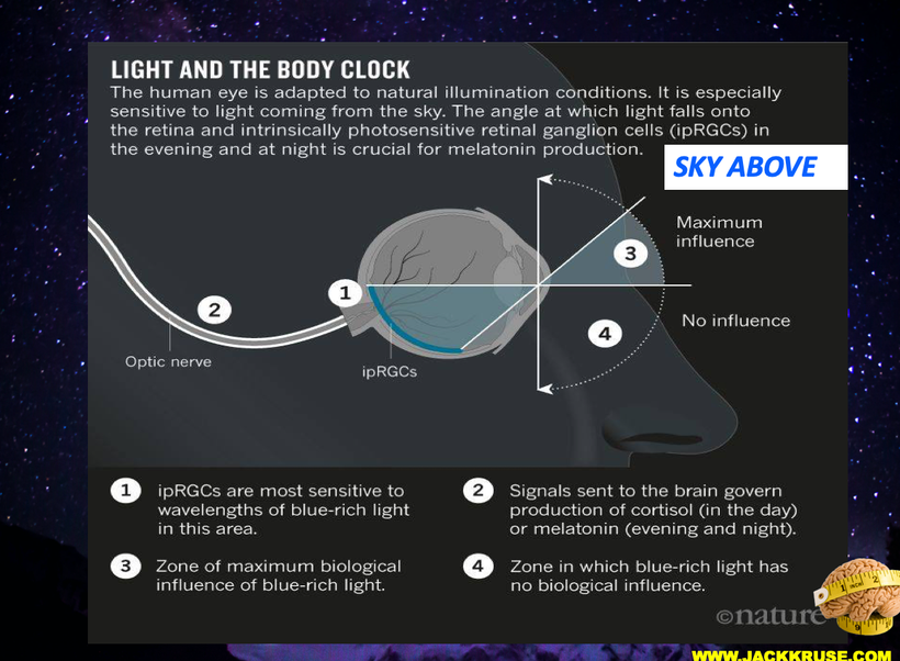
Solar light input into mammal TTFLs begins with dedicated non visual photoreceptors that elicit signaling that acts to induce (in fungi and mammals) or reduce (insects) the amounts of negative arm proteins mentioned above. In broad outline, Output occurs when the positive Arm heterodimer binds to DNA and activates expression of genes whose products do not impact the TTFL. The key take away is the clock gene actions are PROXIMAL to DNA translation and gene activation. This tells you that light inputs controls gene expression in mammals and it is not the other way around. Altering your genome will not improve your illness or disease if the light and dark environment is repair first.
HOW DOES TIME STAMPING WORK BY LIGHT AND DARK WORK?
Once enough modified protein products accumulate in the cytoplasm of a cell, they are transported into the nucleus where they inhibit the positive element from the promoter to stop transcription of clock genes. The clock gene is thus transcribed at low levels until its protein products are degraded, allowing for positive regulatory elements to bind to the promoter and restart DNA transcription. The negative feedback loop of the TTFL has multiple properties important for the cellular circadian clock. First, it results in daily rhythms in both gene transcription and protein abundance and size, caused by the delay between translation and negative regulation of the gene. The cycle’s period, or time required to complete one cycle, remains consistent in each individual and, barring mutation, is typically near 24 hours. This enables stable entrainment to the 24 hour light-dark cycle that Earth experiences from the sun & moon.
Additionally, the protein products of clock genes control downstream genes that are not part of the feedback loop, allowing clock genes to create daily rhythms in other processes, such as metabolism, within the organism. Light and dark cycles are the decentralized controllers of the TTFL.
THE TTFL USES MELANIN TO ELECTROCHEMICALLY TIME STAMP YOUR CELLS. This occurs in the retinohypothalamic pathways anterior to the SCN and it modifies what the SCN signals to all the other molecular clock genes it links to in tissues.
WHY IS MORNING LIGHT SO CRITICAL TO GET RIGHT?
CSP-1 (conidial separation 1) is a morning induced transcriptional repressor with a phosphorylation gated half-life is a key cog in driving EVENING gene expression in mammals. If you do not get AM sun your evening genomic expression will be AWRY. People have forgotten that leptin is released by fat cells and can only enter the hypothalamus under darkness after 4 hours. This should happen at night time. It cannot happen when CSP-1 is not created by AM light. These are the new recent insights into how circadian clocks in your eye and skin achieve phase-specific gene expression. This is how and why leptin resistance exists.
The negative element of the core circadian feedback loop is the frq or frequency gene. The frequency (frq) gene controls the morning-specific rhythmic transcription of a sense RNA encoding FRQ segment. As a result of this action, a long noncoding antisense RNA, qrf, is rhythmically transcribed in an evening-specific manner. It has been reported in the literature that the qrf rhythm relies on transcriptional interference with frq transcription and that complete suppression of qrf transcription impairs the circadian clock. The biological function of qrf transcription and its impact on the circadian clock are not understood in centralized science because centralized science has no light controls at night in labs.
CSP-1 expression is induced by light and glucose, and this finding suggests a rhythmic coordination of qrf transcription with metabolism. Because it is light and glucose we know POMC, ACTH, and melanin are the key to understanding CSP-1 biology. It also means that artificial light during the day or night is especially toxic when you know this is how the mechanism operates.
There are three type of melanins in humans and only one ACTH in humans. All three are used to time stamp the atomic lattice of cells to create an internal map of space time domains to be accurate measuring sticks for the flow of entropy inside of cells. This links melanin biology to Noether’s theorem directly. You have blogs on all these ideas now and it is time for you to link them all to comprend what I have been teaching your for 20 years. Light causes modern diseases.
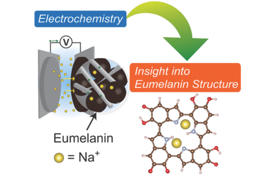
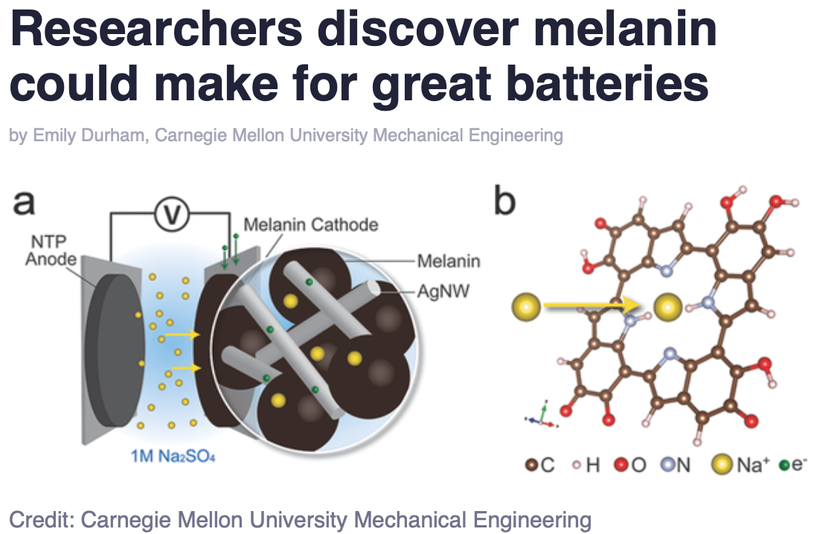
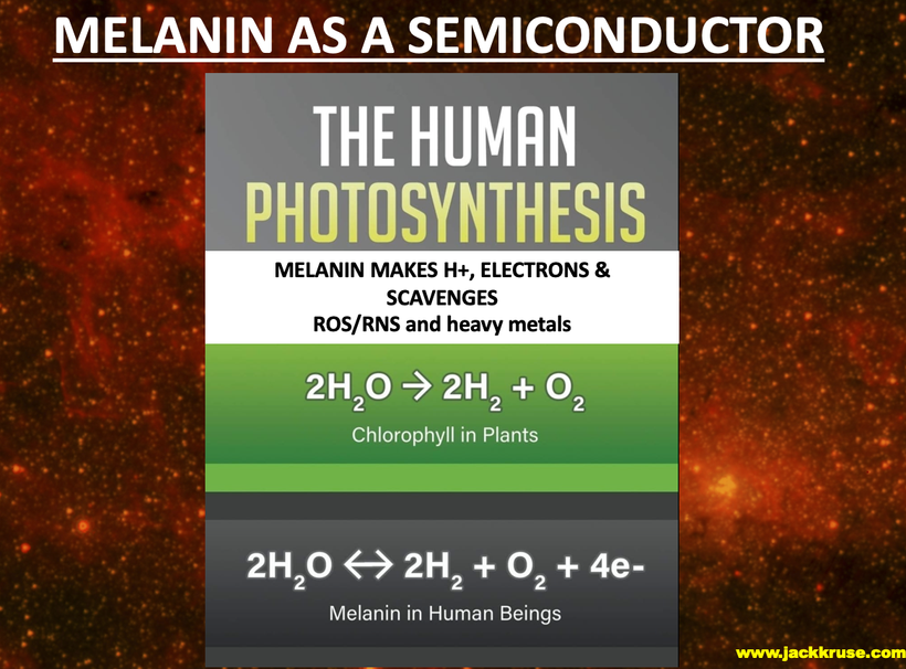
These 3 melanins are ALL extended heterogeneous biopolymers composed of molecular subunits with ambiguous macromolecular topology to modern centralized science. In the literature, an electrochemical fingerprinting technique has been described for melanin, which suggests that natural melanin pigments which contain indole-based tetramers seem to be always arranged into porphyrin-like domains to capture light and measure it in some way useful to the system.
Spectroscopy and density functional theory calculations suggest that sodium ions undergo occupancy-dependent stepwise insertion into the core of porphyrin-like tetramers in natural melanins at discrete potentials that time stamp the internal atomic lattice that allow it to act like a clock to measure the flow of entropy in the cellular system accurately just using light and dark as the feedback loops. It is fully decentralized because light and dark control this process. One is not more important than the other. A loss of melanin implies a loss of accurate time keeping inside the cell or tissue.
Lastly, in humans, the TTFL is a limit cycle, meaning that it is a closed loop that will return to its fixed trajectory even if it is disturbed by its environment, maintaining the oscillatory path on its fixed 24-hour period. It appears this is only true if the melanin structures it uses on surfaces and endogenously remain intact and are chronically replaced and renovated. If the endogenous electrochemical time stamping mechanism is damaged, the TTFL loses its periodicity and chronic modern disease results without any alteration to the DNA or RNA of a cell. These are diseases do not mimic genetic diseases like Tay Sach’s. These diseases are far more common than mutational diseases of DNA which are relatively rare. It means our circadian mechanism is open to the environment, and as such is not subject to calories measurement for metabolism because calories only is useful in closed thermodynamic loops.
CITES
1. Aronson BD, Johnson KA, Loros JJ, Dunlap JC. (1994) Negative feedback defining a circadian clock: autoregulation of the clock gene frequency. Science 263:1578-1584.
2. Belden WJ, Larrondo LF, Froehlich AC, Shi M, Chen CH, Loros JJ, Dunlap JC. (2007) The band mutation in Neurospora crassa is a dominant allele of ras-1 implicating RAS signaling in circadian output. Genes Dev 21:1494-1505.
3. Bell-Pedersen D, Shinohara ML, Loros JJ, Dunlap JC. (1996) Circadian clock-controlled genes isolated from Neurospora crassa are late night- to early morning-specific. Proc Natl Acad Sci U S A 93:13096-13101.
4. Cheng P, Yang Y, Heintzen C, Liu Y. (2001) Coiled-coil domain-mediated FRQ-FRQ interaction is essential for its circadian clock function in Neurospora. EMBO J 20:101-108.
6. Froehlich AC, Liu Y, Loros JJ, Dunlap JC. (2002) White Collar-1, a circadian blue light photoreceptor, binding to the frequency promoter. Science 297:815-819.
7. Gallego M, Virshup DM. (2007) Post-translational modifications regulate the ticking of the circadian clock. Nat Rev Mol Cell Biol 8:139-148.
8. Larrondo LF, Olivares-Yanez C, Baker CL, Loros JJ, Dunlap JC. (2015) Circadian rhythms. Decoupling circadian clock protein turnover from circadian period determination. Science347:1257277.
Xue Z, Ye Q, Anson SR, Yang J, Xiao G, Kowbel D, Glass NL, Crosthwaite SK, Liu Y.Nature. 2014 Oct 30;514(7524):650-3. doi: 10.1038/nature13671. Epub 2014 Aug 17.
Zhu Q, Belden WJ.J Mol Biol. 2020 May 29;432(12):3466-3482. doi: 10.1016/j.jmb.2020.01.009. Epub 2020 Jan 16.
12. Koike N, et al. Transcriptional architecture and chromatin landscape of the core circadian clock in mammals. Science. 2012;338(6105):349–354.
13. Li N, et al. The frequency natural antisense transcript first promotes, then represses, frequency gene expression via facultative heterochromatin. Proc Natl Acad Sci U S A. 2015;112(14):4357–4362.
14. Xue Z, et al. Transcriptional interference by antisense RNA is required for circadian clock function. Nature. 2014;514(7524):650–653.
POMC arose over 500 million years ago by an insertion of the melanocortin sequences into a prepro-endorphin gene. Evidence for this comes from structural identities with other opioid precursors in both the NH2- and COOH-terminal regions of POMC.
The phenomena of pro-opiomelanocortin (POMC) as a hormone precursor emerged gradually over time as observations slowly filled in pieces of the puzzle. Long before the concept of hormone precursors was realized, the bronzed skin color described by Addison in his patient with adrenal insufficiency (“melasma suprarenale”) gave perhaps the first hints of a connection between the hypothalamic, pituitary, adrenal (HPA) axis and skin color. A similar link between the pituitary and pigmentation came from the studies of Allen in 1916 and Smith in 1916 who both noted that immersing tadpoles in pituitary extract made their skins darker. In humans too, large doses of porcine pituitary extract also appeared to cause pigmentation in 1954, with this active extract of the pars intermedia of the pituitary henceforth termed “melanocyte stimulating hormone” or MSH.
In 1932, Cushing extended his clinical reports of a polyglandular syndrome caused by basophilic adenomas of the pituitary by linking this finding with adrenal hyperactivity. In the 1930s, work by Ingle and Kendall in the 1930s showed that administration of large amounts of “cortin,” a purified adrenal extract, produced atrophy of the adrenal cortex in rats. Importantly, they found that administration of the “adrenotropic principle” of the anterior pituitary was effective in preventing adrenal cortical regression following treatment with cortin. The first hints of a behavioral angle to POMC biology came from studies by Ferrari in the 1950s, when “stretching-yawning syndrome,” a bizarre crisis of muscular tone, occurred following central administration of MSH. Many other studies assessing the effects of central α-MSH on motivational processes followed, but it was not until 1976 that Panskepp observed for the first time that this peptide decreased food intake using light alone. What else might POMC do in a modern world run by artificial light now? Today, you get to see another perspective centralized science missed.
Viewed from the comfort and assured knowledge of the modern centralized molecular world, these observations and interventions could be considered overtly simplistic. However, I believe that these classic decentralized observations should be regarded as essential building core evolutionary building blocks, not only for our understanding of POMC peptide processing, but also for the work which subsequently tied together these seemingly diverse peptides.
Is this why did I had a ubiquitnation & mitochondrial series before the Quantum Engineering series on Patreon?
Yep.
In mammals, a master clock localized in the suprachiasmatic nucleus of the hypothalamus synchronizes cellular clocks in other central nervous and peripheral tissues with each other and with external time. At the molecular level, these clocks are based on an interregulatory network of clock genes, including the 3 Period genes (Per1–3), that control circadian rhythms (CRs) by rhythmic orchestration of 5–10% of the cellular transcriptome in a post translational tissue-specific manner. CRs time stamp genes after they are translated and cause diseases. I relayed this last month to the government of El Salvador in a speech I gave there.
Transcription-translation feedback loop (TTFL) is a cellular model for explaining circadian rhythms in behavior and physiology. Widely conserved across species, the TTFL is auto-regulatory, in which transcription of clock genes is regulated by their own protein products. Rev-erba is one and so is NF-kappa beta.
THIS IMPLIES TIME STAMPING CAUSES CHRONIC NEOLITHIC DISEASES.
What can we use as an example to see if light stress on out largest organ causes diseases below?
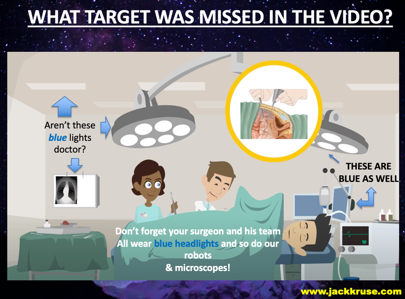
I posted the video above to show you how centralized edicational materials still miss the obvious elephant in the room. The lights used in surgery can cause the problem. the lights on laparascopic cameras are all high intensity blue lights, and it is already well know that other parts of the electromagnetic spectrum used in medicine can cause this problem without any previous surgery. Look at the picture below that I lifted from the video to show you how their own video on this topic misses the elephant in the room on adhesions. I even mentioned this to Huberman during my podcast when I told him everytime I open a skull I now worry about what I am doing because of my understanding.
Have you ever heard of adhesions of the gut?
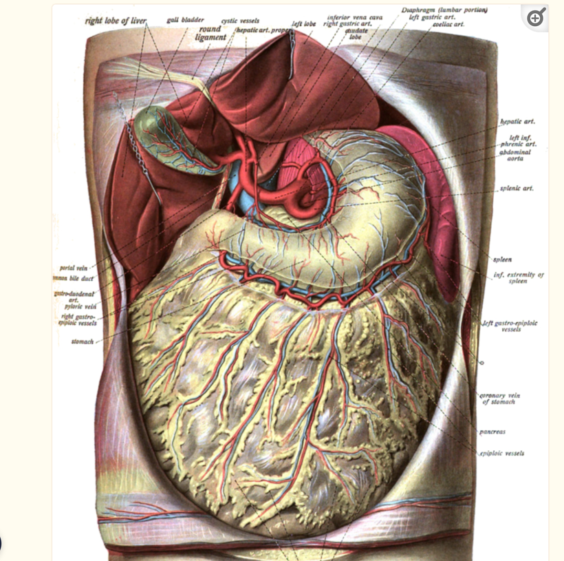
Abdominal adhesions commonly form after intra-abdominal surgery, radiation, and inflammatory processes in humans. In a subset of patients, adhesions lead to problematic symptoms such as abdominal pain, bloating, and bowel obstruction. The mechanisms of adhesiogenesis are not well understood by centralized science but are believed to involve mesothelial surface disruption with subsequent fibrinocoagulative and inflammatory signaling processes. I believe the reason centralized science has missed the boat on adhesions is because aberent light causes it. It is a biophysical disease at its core. Melanin in your omentum is a big deal to the gut clocks below. The melanin in the omentum links to the POMC genes in the skin of the rectus sheets on your abdomen.
WHAT ARE ADHESIONS?
Abdominal adhesions are fibrous bands that span two or more intra-abdominal organs and/or the inner abdominal wall (i.e. peritoneal membrane) which typically form after abdominal surgery. Adhesions may also form secondary to inflammatory conditions of the abdomen in the absence of prior abdominal surgery or as a sequela of abdomino-pelvic radiation. Although the majority of patients with intra-abdominal adhesions remain asymptomatic, a clinically significant subset of patients will develop “adhesive disease”, a symptomatic state ranging from mild and/or vague to highly distressing and even life-threatening symptoms.
Considering the fact that adhesions have no characteristic laboratory features and are not readily visible by currently available imaging methods, many cases of adhesive disease will go undiagnosed for prolonged periods of time, causing medical providers to find themselves in a diagnostic and therapeutic quandary. Patients, consequently, after extensive non-diagnostic testing and empiric treatments, may not only experience protracted symptoms and adverse medical outcomes, but can also suffer from significant emotional distress or demoralization, which in turn may be misdiagnosed as depression, anxiety, or a functional bowel disorder. This topic was going to be the headliner in 2019 in Vermont. That never happened.
WHAT IS THE LIKELY DECENTRALIZED CAUSE OF ADHESIONS?
Irradiating human skin with blue light in frequency bands that causes both type of blue light toxicity is likely the cause of abdominal adhesions. People with autoimmune conditions are at increased risk of adhesions and keloid formation on their skin. The link between all these conditions is the chronic irradiation of human skin with artificial blue light devoid of IR- A and UV frequencies with blue light. One must remember that isolated blue frequencies activates the blue light hazard in mitochondria powered by melanopsin signaling that cause pseudohypoxia in mitochondria. When will centralized medicine learn how light stress not from sunlight ruins mitochondrial biology? Melanin is a key battery in the skin and deep inside of our systems.
This type of isolated light irradiation can set up adults for epithelial cancer, myopia, low dopamine diseases, diabetes, and skin cancer as they age and their heteroplasmy rates grow larger faster as they age into adults formats. If you destroy the battery you have no energy left for health and disease is the result. Could it also be the cause of adhesions? Might this mean adhesions in the gut are a warning signal that surgeons should be paying attention to in patients?
It is for me. When i see arachnoid webs in the brain I know what it means to my patients.
Women with keloids on their cesarean scar have been found in prospective studies to have increased adhesions between the uterus and the bladder and between the uterus and the abdominal wall. This certainly supports my theory and my ideas on this topic. This is cited below.
In mammals, a master clock localized in the suprachiasmatic nucleus of the hypothalamus synchronizes cellular clocks in other central nervous and peripheral tissues with each other and with external time. At the molecular level, these clocks are based on an interregulatory network of clock genes, including the 3 Period genes (Per1–3), that control circadian rhythms (CR) by rhythmic orchestration of 5–10% of the cellular transcriptome in a post translational tissue-specific manner. This tells us that most human disease is not DNA/RNA based, it is mitochondrial based because circadian disease phenotypes mimic those of chronic diseases in centralized healthcare. Circadian biology and oscillations time stamp genes after they are translated and cause modern chronic diseases. How does it happen in humans?
Transcription-translation feedback loop (TTFL) is a cellular model for explaining circadian rhythms in behavior and physiology. It is widely conserved across ALL species, & the TTFL is auto-regulatory, in which transcription of clock genes is regulated by their own protein products. Rev-erba is one and so is NF-kappa beta. I’ve written about both of these things in the past on my blogs.
Rev-erbα, a nuclear receptor and circadian clock component, is a mediator of microglial activation and neuroinflammation in the connection between the CNS and human gut.
I believe people who get adhesions (Crohn’s, UC, SIBO patients) all have destroyed Transcription-translation feedback loop destuction due to an altered circadian mechanism. I propose that such a mechanism can be found within the molecular control of circadian rhythms and “Clock” gene biology and a lack of melanin in some key places in the spinal cord that connect the gut to the CNS.
All melanin’s can create a toxin when they breakdown in human tissue. For example, neuromelanin can reversibly bind and interact with amine containing neurotoxins, (e.g., MPTP(1-methyl-4-phenyl-1,2,3,6-tetrahydropyridine is an organic compound. It is classified as a tetrahydropyridine.) MPTP augments their actions in the terminal, eventually leading to the instability and degeneration of melanin-containing neurons due to oxidative stress and mitochondrial dysfunction.
In particular, a lack of neuromelanin and POMC translation by a lack of UV light appears to confer susceptibility to chemical toxicity by providing a large sink of iron-bound, heme-like structures in a pi-conjugated system. This system seemingly allows for stabilizing interactions including pi-stacking, sodium inclusion, as well as ligand binding to iron in cells. Given the progressive accumulation of neuromelanin components with aging. it seems melanin degradation corresponds with an apparent decrease in dopamine synthetic pathways in man. This raises an immediate question of whether melanin can also create or is capable of binding dopamine, the primary functional monoamine utilized in gut enterocytes and its corresponding gut neurons, should be raised by centralized research. To date, it has not been.
A number of physiological processes demonstrating circadian variation have been shown to involve ‘Clock genes’ in the suprachiasmatic nucleus (SCN), which then entrains a similar set of Clock genes in peripheral tissues such as the heart, brain, spleen, lung, liver, skeletal muscle and kidney. The intrinsic time-keeping system influences activity, such as sleep, temperature regulation, rates of metabolism, immune responses, blood pressure and hormone secretion. The function and availability of mediators involved in the inflammatory response, fibrinolytic and anti-coagulation pathways are all under the tight control of the molecular Clock system. These include IL-6, PAI-1, fibrinogen, fibroblasts and TNF-α. I am making the educated guess that disruptions in the ‘Clock system’ are central to the causal pathway of adhesion formation here. I am also implying this symptom is a gateway disease to many other diseases linked to altered cellular time stamping in humans.
The transcriptomic analysis of hippocampus from Rev-erbα-/- mammals has revealed a predominant inflammatory phenotype results in mammals and it suggests it comes from a dysregulated NF-κB signaling due to circadian dysfunction at its core. When one considers that melanin derivatives are always found in pathologic specimens it is not hard to come up with this linkage.
DEEP DECENTRALIZED SCIENCE THEY ARE MISSING
The brain gut connection requires pristine circadian rhythm control to stay healthy. Loss of Rev-erbα in primary astrocytes of the spinal cord of mammals had no effect on basal activation of the neurons that innervate the gut but do potentiate the inflammatory response to lipopolysaccharide (LPS). In vivo, Rev-erbα-/- in a mammalian model has exhibited enhanced hippocampal neuroinflammatory responses to peripheral LPS injection, while pharmacologic activation of Rev-erbs with the small molecule agonist SR9009 suppressed LPS-induced hippocampal neuroinflammation. This explains to us how a loss of circadian control allows the LPS of the microbiome to lead to a disease phenotype just with a loss of circadian control post translationally of DNA. This means DNA alteration is not the cause of a majority of modern human disease. This is 180 degrees from the current centralized perspective.
Rev-erbα deletion due to a loss of clock gene control clearly influences gut enterocyte function and neuronal health in a coordinated fashion. When this link breaks it makes the formation of adhesions post surgically much more likely. Lab experiments done have shown when gut neurons are conditionally cultured on media from Rev-erbα-deficient primary gut-glial cultures, it exacerbates oxidative damage in cultured neurons that innervate the gut. Rev-erbα-/- mammals have not only shown gut dysfunction in these models but they also exhibited significantly altered cortical resting-state functional connectivity. This finding tells me that many central neurological diseases maybe masquarding in centralized medicine clinics as circadian diseases of theTranscription-translation feedback loop(TTFL).
One neurologic disease in particular intrigues me with its skin & gut connection is ALS. ALS patients tend not to show up in low latitudes and they tend to be quite pale because they lack UV exposure POMC requires for translation of melanin. In human ALS, abnormalities have been found in mitochondrial structure, mitochondrial respiratory chain enzymes, and mitochondrial cell death proteins indicative of some non-classical form of programmed cell death. In ALS patients this happens in the spinal cord, skin, and their gut in pulsatile fashion over timescales. This is a hint to me that it might be a disease of time stamping of the TTFL masquarading as a neurological disease.
The creation of MPTP, which is lipid-soluble, readily penetrates the blood—brain barrier and enters the brain cells. Because it is amphiphilic, it is captured into acidic organelles, mostly lysosomes, of astrocytes. MPTP itself does not appear to be toxic, but its oxidized product, 1-methyl-4-phenylpyridinium (MPP+), is toxic. This has been linked in Parkinson’s Disease but I think it might link to ALS because the toxic MPP+ could affect defective mitochondria in neurons in the anterior motor horn from circadian rhythm damage in the gut. These malfunctioning mitochondria might contribute to neuronal death in ALS through the biophysical entity called the mitochondrial permeability pore (mPTP) activation.
The major protein components of the mPTP are enriched in mammalian motor neurons which also have abundant POMC genes. Early in the course of disease in ALS humans seem to present with mutant superoxide dismutase-1 enzymes in their mitochondria. The mitochondria in motor neurons undergo “trafficking abnormalities” which results in dramatic remodeling of mitochondria in ALS patients that results in the formation of mega-mitochondria (larger size) and coinciding with increased protein carbonyl formation and nitration of mPTP components. Anything that enlarges is a thermodynamic sign to me. In fact, lab experiments in nocturnal mammals have shown that genetic deletion of a major mPTP component called cyclophilin D which has robust effects in ALS by delaying disease onset & phenotype and extending survival of the anterior motor neurons.
When this process is disrupted the MPP+ action it appears to destroy the mPTP in a cascading manner in the spinal cord mitochondria and spreads like an infection would in the anterior motor neurons. I think the circadian disruption in the anterior motor neurons causes a paralysis like effect that we normally see in REM sleep. In ALS, the circadian disruption shows up when we are awake and it has gone unrecognized as a completely disrupted rhthym disorder because of how we perceive the disease and we have not looked at the data carefully enough. I happen to have one person with this disease who is doing something no one else has done with this diagnosis. He is improving his solar redox and improving mtDNA function by living in the tropics adjacent to a flat beach. So far he is doing a lot better than other ALS patients who aren’t paying attention to their light stamping problem. In fact, I believe some of these patients diseases are made worse when they travel outside of time zones. That seems to make the diseases more aggressive.
I think this model in mammals explains why anterior motor neurons can be destroyed in isolated fashion in the spinal cord when the circadian rhythms are disrupted in the nerves that innervate the gut but link back to their embryologic origins. The inflammatory cascade could start out in the splanchnic nerves below but as the spinal cord anterior motor neurons loses REV-erb alpha it could create an inflammatory cascade of LPS induced damage to “infect” other adjacent motor neurons by destroying their clock gene TTFL’s. This is how ALS might be progressing in these patients.
The Rev-erbα, mPTP in mitochondria, and TTFL should be thought of as as a key biophysical link between the circadian clock control and neuroinflammation in the CNS. People forget that the spinal cord is part of the CNS and so is their skin. Nerves link the gut to both.
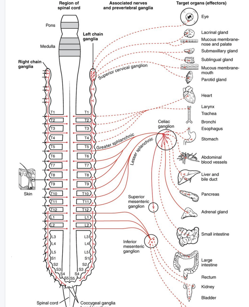
Look more carefully at the picture above then you did in QE #47. Note the skin is on the left of the picture and it is our largest organ and it has a somatotopic organization to the ENTIRE spinal cord and to our internal organs by way of nerves.
The Quantum Enginnering #47 blog has the key metric of why the eye, skin, PVN, and DLF of the brain stem sleep and pain areas give the input into celiac plexus to cause gut problems via the microbiome. The implications of this connection are massive in many other diseases besides bipolar disorder. I told you that there and I am doubling down on it here.
WHAT DID I SAY IN QE #47 AGAIN ABOUT THIS PROCESS?
I said, “The dorsal longitudinal fasciculus (fasciculus of Schutz) is a periaqueductal (area around ventricular system Aq above) ascending and descending fiber system arising from the hypothalamus and terminating to the autonomic nuclei of the pons and the medulla, conveying autonomic fibers from the brain to the gut in humans. It also conveys pain messages and is important in the sleep pathways of humans. These are usually altered in bipolar patients as well because of a lack of melanin in these areas.
The dorsal longitudinal fasciculus is found within the dorsal brainstem tegmentum. It passes through the periaqueductal gray matter and contains both ascending and descending fibers. The ascending fibers pass from the reticular formation (sleep region/insomnia) passing to the hypothalamus thus transmitting information related to the viscera in the thorax and abdomen. THIS IS HOW THEY PASS THE ANTERIOR MOTOR HORN CELLS AND THE GUT VISCERA!
People who travel a lot across time zones or people who use a lot of blue light or nnEMF at night or day in their cities are going to have massive sleep disturbances because they lack charge density in this spot. They mimic people with mental disorders like bipolar patients because they have broken the same rules of Nature. I view insomnia as a mental disorder in my decentralized medicine framework.
This topologic neuronal surface is a critical barrier to the brain health of humans. This is the pathway where metabolic syndrome, poor sleep, and fatty liver all come from fundamentally. This pathway could be in many blogs but I left it out this detail. Why? You already thought this stuff was too hard. Why add another level of complexity with neuroanatomy?
Many of these same findings are found in diabetics, bipolar disorder, and those with sleep disorders like insomnia. Patients with bipolar disorder tend to have all these symptoms at times that vary based on how defective their FM antennae are in their eyes & brains.”
DOES ANYONE SEE THOSE LINKS NOW IN THIS BLOG IN THE SERIES?
Now, go back and look at the lipofuscin blog in this series. That chemical comes from melanin degradation and is always associated with aging and light stress. Dopamine is made in the eye several ways by sunlight and it can be made in the gut by your microbiome due to the link to melanin. People forget that bacteria release 5000 times more light than eukaryotic cells and this light is capable of making dopamine from the aromatic amino acids in the gut. This is why serotonin and phenylalanine and tyrosine are stored in the enterochromaffin system in our gut.
The more and more you look into its biology, I feel more confident in saying that light is the most powerful drug for living systems. The light release is more important than the fuel in the diet for our health outcomes because food has light information built into photosynthesis. This stimulus leads to light release to start the optical signal cascade in the gut using aromatic amino acids as the mover in the GI tract. When you are blue light/nnEMF toxic you cannot repair the gut or sleep issues at all. This is why many people get gut adhesions without having had any surgery. The same thing will be true for pain thresholds and opiate use. I can tell you this. Your gastrointestinal system will never function optimally in a circadian mismatch (melanopsin/encephalopsin dysfunction) and that will bypass however good a diet is.
The study linked below in cite 6 is a longitudinal study on TBI following college football athletes across a sports season.
•Nanopore 16S rRNA sequencing of gut microbiome reveals changes after head injury.
•Serum biomarker GFAP increased during the competitive period of the season.
•S100β and SAA blood levels were positively correlated with Eubacterium rectale.
•Gut microbiota is suggested as a future biomarker for diagnosis following head & neck injury.
The blue light hazard/nnEMF links the gut to the brain, heart, and vascular damage leading to neurodegeneration that causes protein folding issues in blue light-damaged tissues as cites 4 and 5 show. Few are making these links in diseases like adhesions and ALS.
But I am.
CITES
Van Goor H. Consequences and complications of peritoneal adhesions. Colorectal Dis. 2007 Oct;2(9 Suppl):25–34.
Menzies D., Ellis H. Intestinal obstruction from adhesions; how big is the problem? Ann. R. Coll. Surg. Engl. 1990;72:60–63.
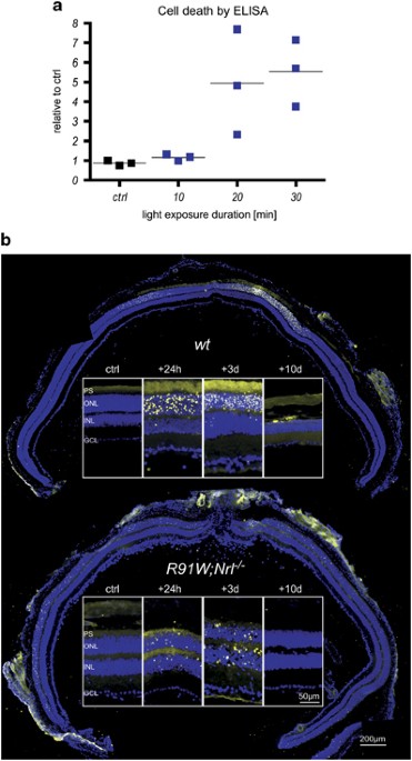
Little is known about the mechanisms underlying macular degenerations, mainly for the scarcity of adequate experimental models to investigate cone cell death. Recently, we generated R91W;Nrl−/− double-mutant mice, which display a well-ordered all-cone retina with normal retinal vasculature and a…
Concussions, both single and repetitive, cause brain and body alterations in athletes during contact sports. The role of the brain-gut connection and …
Allen BM. Extirpation experiments in Rana pipiens larvae. Science 44: 755–757, 1916. doi: 10.1126/science.44.1143.755.
Smith PE. Experimental Ablation of the Hypophysis in the Frog Embryo. Science 44: 280–282, 1916. doi: 10.1126/science.44.1130.280.
Lerner AB, Shizume K, Bunding I. The mechanism of endocrine control of melanin pigmentation. J Clin Endocrinol Metab 14: 1463–1490, 1954. doi: 10.1210/jcem-14-12-1463
.Ingle DJ, Kendall EC. Atrophy of the Adrenal Cortex of the Rat Produced by the Administration of Large Amounts of Cortin. Science 86: 245, 1937. doi: 10.1126/science.86.2228.245.
Panskepp J, Reilly P, Bishop P, Meeker RB, Vilberg TR, Kastin AJ. Effects of alpha-MSH on motivation, vigilance and brain respiration. Pharmacol Biochem Behav 5, Suppl 1: 59–64, 1976. doi: 10.1016/0091-3057(76)90329-4.
The auditory nerve transmits auditory information up a series of nuclei to the cortex where perception occurs. These nuclei include 1) cochlear nucleus, 2) superior olivary nuclei, 3) lateral lemniscus, 4) inferior colliculus, and 5) medial geniculate nuclei.
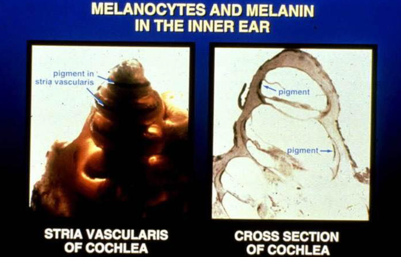
Melanin pigment is normally present in the outermost layer of the retina of the eye, the inner ear adjacent to capillaries in stria vascularis near hair cells, and vestibular organs. In the skin, it is in the basal layers. In the gut, it is found in the enterochromaffin cells. Significant reduction in melanin pigment in mammals is associated with embryonic miswiring and disruption of visual and auditory functions. This has implications for human diseases like autism which usually affects all sensory inputs to the thalamus. In this way, it mimics what I wrote about Autism in this series in QE#45. The consequences for the visual system include abnormal development of the retina and misrouting of optic pathways into the brain impairing visual acuity, eye movement, and stereovision. Lack of melanin pigment in the inner ear is associated with greater susceptibility to noise damage and poorer localization of sound in space.
Auditory sense in mammals is unique in another way and may be critical to why melanin exists where it does. Unlike other cells within the brain, hair cells within the Organ of Corti of the cochlea do not have axons. Neurons within the spinal ganglion have peripheral axons that synapse at the base of the hair cell soma. These axons make up the auditory nerve. Most (90%) of auditory nerve fibers receive their input from the inner hair cells. Thus, the inner hair cells facilitate a majority of auditory processing. My bet is this arrangment is how sound waves are converted to light waves that the auditory nerve can process.
The melanin sheets in the ear are proximal to the auditory nerves. When melanin is hypoxic it can break down into noradrenaline and dopamine. The mammalian auditory brain stem, and in particular the medial nucleus of the trapezoid body (MNTB) (Wynne and Robertson 1996), receives extensive adrenergic input. This tells me that the sheet in the cochlea is creating neurotransmitters by melanin degradation in the auditory pathways as the slide below shows.
The mammalian auditory brain stem receives profuse adrenergic innervation, whose function is currently poorly understood by centralized science. Noradrenaline increases high-frequency firing at the Calyx of Held synapse during development by inhibiting glutamate release.
The calyx of Held synapse plays an important role in the auditory system, relaying information about sound localization via fast and precise synaptic transmission, which is achieved by its specialized structure and giant size. During development, the calyx of Held undergoes anatomical, morphological, and physiological changes necessary for performing its functions. The large dimensions of the calyx of Held nerve terminal are well suited for direct electrophysiological recording of many presynaptic events that are difficult, if not impossible to record at small conventional synapses. This unique accessibility has been used to investigate presynaptic ion channels, transmitter release, and short-term plasticity, providing invaluable information about basic presynaptic mechanisms of transmission at a central synapse.
DECENTRALIZATION ANSWER to why all senses have melanin sheets between them and the environment?
Why does the spiral cochlea have this huge melanin sheet? Melanin shows up in mammalian tissue because of the melanin concentration hormone. Melanin-concentrating hormone (MCH) was originally isolated from salmon pituitaries (think of Huberman fish gaff on melanopsin now) where it induces the aggregation of melanin granules in melanophores, which results in pale skin color. MCH sequence is conserved in ALL MAMMALS analyzed to date, including mice, rat, rabbits, and humans. In mammals, the neurons that synthesize and release MCH are present mainly in the hypothalamus and nearby areas. Is the cochlea near the hypothalamus? No. But the melanin stimulatory hormone is very prominent in the brainstem and the cochlear is very close to that. Lack of melanin pigment in the inner ear is associated with greater susceptibility to noise damage and poorer localization of sound in space.
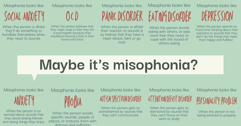
I think a relative lack of melanin in different areas of the cochlea is the cause of misophonia. Misophonia is a disorder in which certain sounds trigger emotional or physiological responses that some might perceive as unreasonable given the circumstances. Those who have misophonia might describe it as when a sound “drives you crazy.” Their reactions can range from anger and annoyance to panic and the need to flee. If your centralized health professional doesn’t know about misophonia, they may take all that information and try to fit it with something they do know about. Here are some diagnoses you might hear before a health professional finally acknowledges your misophonia:
Melanocytes are distributed in the stria vascularis and spiral ligament regions and recent evidence illustrates the distribution of melanin in the human cochlea with the lower relative amount of melanin observed in the basilar turns compared to apical turns. Sensorineural hearing loss caused by presbycusis (old age heteroplasmy), maternal-fetal rubella infection, aminoglycosides, and noise exposure disproportionally impacts the basal turn with variable losses of spiral ganglion and hair cells reflected in a down-sloping audiogram. Melanin is sparse in those regions. The location of melanin and its known function as a free radical scavenger may explain some patterns of sensorineural hearing loss and highlight that melanin has special abilities in the sensory pathways of mammals.
Might it have to do with the binary code of biology and the fidelity of sound that I raised in the Kruse for Dummies lecture? I believe it does. Look at how many areas sound inputs are sent. If we are built to semiconductor sound waves this implies we need a big equalizer in our ear no?
Since this synapse is massive in all mammals it has been studied. What do we know? Researchers have studied multiple mammal species and to the extent that a comparison is possible, the time course for development embryologically matches earlier experiments performed in rats, cats, mice, or gerbils, suggesting that the development of this synapse is highly conserved across all mammalian species. There is another surprise. The essential steps to build hearing occur largely before the onset of hearing, supporting the view that sensory activity does not play a major role in the formation of this synapse. So the sound stimulus is unnecessary to morphology. This is a clue that morphology likely links back to the binary code in morphogenesis.
Do you know what the Calyx of the Head is in humans? It is the big equalizer mentioned above.
Function. The calyx of Held is a part of the auditory system, connecting the globular bushy cells (GBCs) of the anteroventral cochlear nucleus to the principal neurons of the medial nucleus of the trapezoid body (MNTB).
Most of the MCH-positive fibers in mammals have been detected throughout the brainstem and the cochlea is very close to it in mammals. Interestingly, there was a profuse MCH innervation of brainstem areas that are involved in the control of REM sleep.
The calyx is a giant glutamatergic terminal formed by the main axon of globular bushy cells (Fig. 2, GBC). These cells have their cell body in the cochlear nucleus contralateral to the MNTB and receive large axosomatic terminals from the auditory nerve (endbulbs of Held).
The calyx of Held is probably the largest synaptic terminal in the brain, forming a unique one-to-one connection in the auditory ventral brainstem. During early development, calyces have many collaterals, whose function is unknown.
During embryonic and postnatal development, the calyx of Held undergoes a significant transformation in order to possess morphological and functional properties necessary for performing its major role in relaying acoustic information from the environment to our brain.
SUMMARY
The implication of this blog are that hearing, tinnitus, and acoustic changes can all be changed by solar exposure on the skin. If this is true is there evidence that heavily melanated human skin leads to less acoustic disease?
There is. There is a ton of data I provided you below.
Many studies have shown that blacks have a 40%–70% lower prevalence of hearing loss than do whites.
In a previous cross-sectional study, Lin et al. (below in cites) demonstrated an association between certain Fitzpatrick skin phototypes and lower odds of hearing loss among Hispanics, which they suggested might be due to differences in melanocytes between darker-skinned and lighter-skinned individuals. Melanocytes are known to be present in the inner ear, and results from evolutionary studies have suggested that darker-colored individuals tend to have more internal (nonskin) melanin. Furthermore, studies in humans have demonstrated a positive association between number of melanocytes in the skin and in the inner ear.
Melanocytes have antioxidant functions that might serve to protect against reactive oxygen species that are associated with the death of inner ear hair cells in noise-induced hearing loss . Furthermore, cochlear melanocytes serve as intermediate cells in the stria vascularis and are important in the generation of the endocochlear potential. All this tells me that getting a tan is a great way of maintaining your acoustic system and lowering your risk for both acoustic diseases like tinnitus and misophonia, ocular diseases like retinopathy and thalamic diseases like autism. Get your skin in the game. Decentralize your biology to get back what you’ve lost.
J Kil, GH Kageyama, MN Semple, LM Kitzes, Development of ventral cochlear nucleus projections to the superior olivary complex in gerbil. J Comp Neurol353, 317–340 (1995).
K Kandler, E Friauf, Pre- and postnatal development of efferent connections of the cochlear nucleus in the rat. J Comp Neurol328, 161–184 (1993).
BK Hoffpauir, JL Grimes, PH Mathers, GA Spirou, Synaptogenesis of the calyx of Held: Rapid onset of function and one-to-one morphological innervation. J Neurosci26, 5511–5523 (2006).
VC Wimmer, H Horstmann, A Groh, T Kuner, Donut-like topology of synaptic vesicles with a central cluster of mitochondria wrapped into membrane protrusions: A novel structure-function module of the adult calyx of Held. J Neurosci26, 109–116 (2006).
DK Morest, The growth of synaptic endings in the mammalian brain: A study of the calyces of the trapezoid body. Zeitschrift Anat Entwicklungs127, 201–220 (1968).
Lin FR, Maas P, Chien W, et al.. Association of skin color, race/ethnicity, and hearing loss among adults in the USA. J Assoc Res Otolaryngol. 2012;13(1):109–117.
Lin CS, Zak FG. Studies on melanocytes. VI. Melanocytes in the middle ear. Arch Otolaryngol. 1982;108(8):489–490.
Dubey S, Roulin A. Evolutionary and biomedical consequences of internal melanins. Pigment Cell Melanoma Res. 2014;27(3):327–338.
Wolff D. Melanin in the inner ear. Arch Otolaryngol Head Neck Surg. 1931;14(2):195–211.
LaFerriere KA, Arenberg IK, Hawkins JE Jr, et al.. Melanocytes of the vestibular labyrinth and their relationship to the microvasculature. Ann Otol Rhinol Laryngol. 1974;83(5):685–694.
Nofsinger JB, Liu Y, Simon JD. Aggregation of eumelanin mitigates photogeneration of reactive oxygen species. Free Radic Biol Med. 2002;32(8):720–730.
Meyskens FL Jr, Farmer P, Fruehauf JP. Redox regulation in human melanocytes and melanoma. Pigment Cell Res. 2001;14(3):148–154.
Henderson D, Bielefeld EC, Harris KC, et al.. The role of oxidative stress in noise-induced hearing loss. Ear Hear2006;27(1):1–19.
Takeuchi S, Ando M. Inwardly rectifying K+ currents in intermediate cells in the cochlea of gerbils: a possible contribution to the endocochlear potential. Neurosci Lett. 1998;247(2-3):175–178.