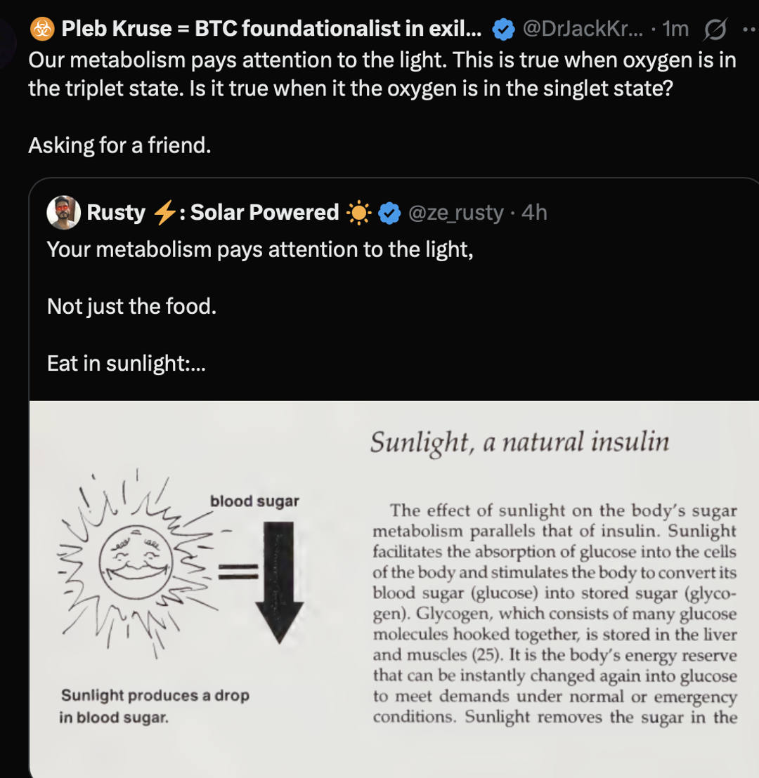
THE ANSWER likely will shock many.
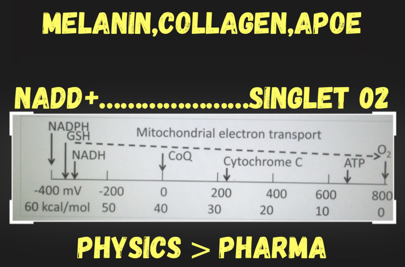
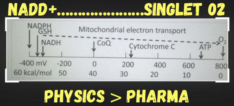
When oxygen is in the singlet state, and NAD+ is deuterated, your metabolism becomes completely blind to the light.
I bet many might be shocked at this reality.
The foundational principle of my decentralized thesis is that metabolic light perception requires an intact, paramagnetic quantum receiver in your brain to get the light signal correct.
When the electronic infrastructure fails, the cell loses its ability to decode environmental waves, transforming a high-fidelity optical computer into a blind, isolated chemical system.
The implications of this idea?
When the planetary geodynamo undergoes a localized or systemic magnetic excursion, or when the local tissue environment is subjected to severe biophysical trauma, the magnetic anchors holding the subatomic infrastructure together are pulled apart. A coma is not merely a “shutting down” of conscious networks; it is the state where the entire brain becomes blind to Ultra-weak Photon Emission (UPE) transforms made from mitochondrial metabolism. The biophysical optical computer collapses into a silent, non-communicating chemical system of singlet state biochemistry.

NO, Sleep Apnea, and the Brainstem: A Biophysical Link in the DLF and RAS
Sleep apnea, often exacerbated by mouth breathing, is a hallmark of disrupted brainstem function of singelt oxygen, particularly in the dorsolateral funiculus (DLF) tracts and the reticular activating system (RAS). The slide below explicitly links mouth breathing to low NO, hypoxia, and hypermethylation of the RAS, which contributes to sleep apnea. Let’s dive into how NO dysregulation in these brainstem regions drives this condition, building on the evolutionary adaptations from the GOE and the metabolic cascades we’ve explored.

Mouth breathing is the ultimate physical off-switch for your brainstem’s quantum receiver. By decoupling nasal airflow from the central nervous system, mouth breathing instantly drops nitric oxide (NO) production, triggering a localized pseudohypoxic cascade that deforms the brainstem’s architecture.
When NAD+ is deuterated and oxygen flips into the singlet state, the brain completely loses its optical compass. The Dorsolateral Funiculus (DLF) and the Reticular Activating System (RAS)are stripped of their electromagnetic coherence, leaving the body unable to sustain normal tone during sleep.
The GOE and NO’s Evolutionary Role
To understand why the brainstem responds so violently to a modern spin-flip, you must look back 2.4 to 2.1 billion years ago to the Great Oxidation Event (GOE).
The GOE introduced molecular oxygen, a stressor that reshaped life by forcing endosymbiosis and the evolution of mitochondria. Heme proteins like cytochrome c oxidase (CCO) emerged to manage oxygen, producing ATP and water while generating electrical fields across mitochondrial membranes. NO, a signaling molecule, also became critical in this oxygen-rich world. Produced by nitric oxide synthase (NOS) enzymes using oxygen and arginine from the urea cycle, NO regulates vascular tone, mitochondrial function, and neural signaling. Its paramagnetic properties, inherited from oxygen’s influence during the GOE, allow it to modulate the redox state of iron in heme proteins, affecting oxygen delivery and metabolism.
WHAT YOU DID NOT LEARN ABOUT THE GOE?
When cyanobacteria began flooding the primeval oceans with oxygen, biology was forced to adapt to a highly reactive, potentially lethal element. The only reason early eukaryotic life survived this transition is that Earth’s geodynamo provided a powerful, highly structured magnetic field.
The Planetary Software: This primordial magnetosphere acted as the macroscopic software that pinned the valence electron spins of atmospheric oxygen into the stable, paramagnetic ground-state triplet configuration. I told this to a stunned audience in 2014 in Pasedena California at the Bulletproof conference.
The Modern Inversion: Today, when you sleep in a room wrapped in artificial nnEMF, you are creating a hyper-local environment devoid of that evolutionary magnetic stabilizing torque. You recreate a chaotic, multipolar magnetic field state right on your pillow, forcing oxygen to revert to the high-entropy, antiparallel singlet state that deforms your tissues.
Your abnormal airway anatomy is proof you are living a magnetically unpinned life.
The Mouth Breathing Cascade: Erasing Nitric Oxide (NO)
Nasal breathing is not merely a mechanical pathway; it is a gas-phase biochemical catalyst. The paranasal sinuses produce a continuous, baseline stream of Nitric Oxide (NO), a paramagnetic gas possessing an unpaired electron.
Mouth breathing, as noted in the slide, reduces NO levels by impairing nasal breathing, which normally produces NO in the paranasal sinuses.
When you transition to mouth breathing, this entire subatomic delivery system collapses:
The Loss of the Paramagnetic Shield: Forfeiting nasal breathing eliminates the mechanical shear stress required to generate high levels of NO. Lacking this gas-phase radical shield, the local tissues lose the subatomic “buffer” that normally prevents ground-state oxygen from undergoing an intersystem crossing spin-flip into singlet oxygen.
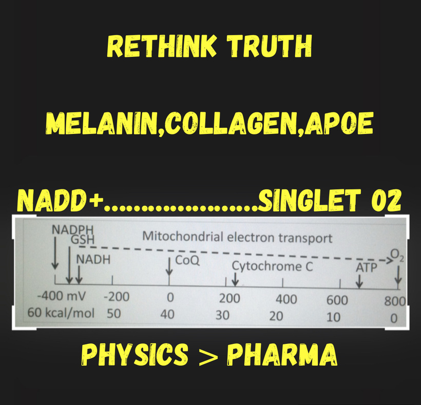
Mitochondrial Stalling: Deprived of NO’s subtle, regulatory tuning of Cytochrome C Oxidase (CCO), electrons traveling along the transport chain instantly stall.
The Deuterium Influx: This electronic bottleneck prevents the forward flow of the respiratory chain. The lack of electronic momentum causes a massive breakdown in the cell’s ability to discriminate between hydrogen and deuterium. NAD+ becomes heavily deuterated, permanently altering the geometry of the mitochondrial matrix and blinding the metabolic machinery to environmental light signals. Your beginning to realize most of you never stacked the lessons I gave you back then.
The DLF Tracts and Sleep Apnea
This localized subatomic crash hits the dense, highly integrated computing centers of the brainstem with devastating precision.
The dorsolateral funiculus (DLF) tracts in the brainstem and spinal cord are involved in sensory and autonomic regulation, including respiratory control. These tracts contain neurons that project to the nucleus tractus solitarius (NTS) and the dorsal motor nucleus of the vagus, both of which are critical for coordinating breathing and airway tone. NO acts as a neuromodulator in the DLF, facilitating synaptic transmission and maintaining the balance between excitatory and inhibitory signals that regulate the respiratory rhythm.
In sleep apnea, low NO in the DLF disrupts this balance. NO normally enhances vasodilation in the microvasculature of the brainstem, ensuring adequate oxygen delivery to respiratory control centers. Without sufficient NO, these regions become hypoxic, impairing the neural circuits that maintain airway patency during sleep. This leads to airway collapse, a hallmark of obstructive sleep apnea (OSA). Additionally, NO deficiency reduces the sensitivity of chemoreceptors in the NTS, which detect CO2 and O2 levels. This blunts the brain’s ability to respond to rising CO2 during apneic episodes, perpetuating the cycle of airway obstruction and hypoxia.
HOW DOES IT HAPPEN?
The DLF undergoes a core failure due to the spin change of electrons. It carries the crucial descending pathways that modulate autonomic control, pain, and motor drive to the upper airway muscles. When the tissue surrounding the DLF transitions to a singlet oxygen environment, its dense networks of Iron-Sulfur clusters lose their Chirality-Induced Spin Selectivity (CISS).
The spin-polarized current required to send rapid, coherent motor instructions down the funiculus falls apart.
The tone of the genioglossus muscle and the retropalatal oropharyngeal wall drops, causing the physical tissues of the airway to structurally collapse under everyday ambient pressure.
The Reticular Activating System (RAS) Hypermethylation Trap
The reticular activating system (RAS), spanning the brainstem, regulates arousal, wakefulness, and the sleep-wake cycle. It’s rich in melanin-containing neurons, such as those in the locus coeruleus, and relies on NO for proper function. NO modulates the activity of noradrenergic and cholinergic neurons in the RAS, which drive arousal in response to hypoxia or hypercapnia during sleep apnea. The slide notes hypermethylation of the RAS in mouth breathers, a sign of epigenetic silencing that impairs its function. Low NO exacerbates this by reducing the excitability of RAS neurons, making it harder for the brain to arouse and reopen the airway during apneic events.
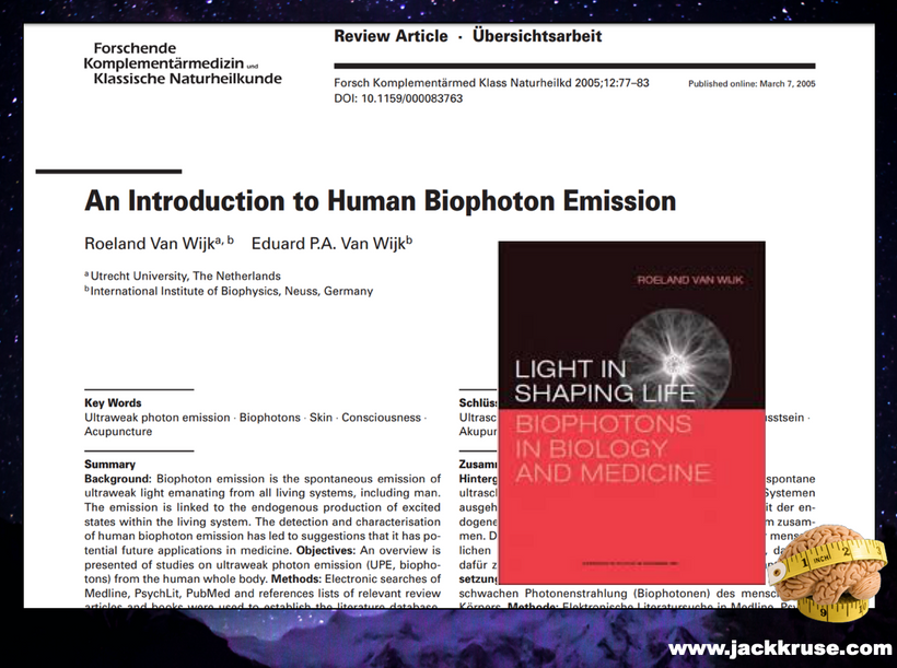
The hypoxia from sleep apnea also degrades melanin and melanopsin in the RAS and its vascular networks. Melanin, a chiral semiconductor, supports charge separation of water, producing the right isotope of H+, and electron spin state of oxygen, from electrons while generating ultraweak photon emissions (UPEs). If any of this is off, so is the signaling UPE from the interaction.
When melanin breaks down, UPEs are altered, impairing melanopsin signaling in cerebral blood vessels and disrupting the recursive photonic loop between melanin and mitochondria. This loop normally produces near-infrared (NIR) light, which can reverse NO’s inhibition of CCO, restoring oxygen-rich metabolism in the tricarboxylic acid (TCA) cycle. Without this loop, the RAS struggles to maintain arousal, and the brainstem’s metabolic demands go unmet, increasing the severity of sleep apnea.
The RAS acts as the brain’s master arousal clock, maintaining the cortical state and regulating sleep-wake transitions. The profound pseudoxia caused by deuterated NAD+ and singlet oxygen triggers a massive, defensive epigenetic response.
Lacking the high-voltage electrical fields (-70mV) generated by active triplet oxygen reduction, the local cells activate DNA Methyltransferase (DNMT) enzymes.
This causes rapid hypermethylation of critical promoter regions across the Ascending Reticular Activating System (ARAS).
Key neurotransmitter and receptor pathways are epigenetically silenced, leading to a profound functional decoupling of the ARAS. The brainstem loses its low-arousal threshold sensitivity; it can no longer orchestrate a smooth, seamless transition between sleep stages, locking the patient into a vicious cycle of intermittent hypoxia, airway collapse, and systemic quantum decay.
You feeling me now?
This is why sleep apnea is the number one new diagnosis in South America with the SAA is destroying the dynamo.
As the South Atlantic Anomaly expands and weakens the local planetary geodynamo across South America, the external torque holding your subatomic “spark plugs” in alignment vanishes. It is no coincidence that countries sitting entirely within this geomagnetic depression, such as Brazil and Colombia, suffer from some of the highest rates of sleep apnea globally.

Vascular and Metabolic Consequences in the Brainstem
Low NO in the DLF and RAS has broader implications. NO’s role in vasodilation ensures proper blood flow to the brainstem, but its deficiency leads to vasoconstriction, increasing mechanical stress on vessel walls. This, combined with the degradation of melanopsin in cerebral blood vessels, weakens vascular integrity, raising the risk of hemorrhages in the brainstem, particularly in the pons, a region I’ve flagged for its vulnerability in mouth breathers. Furthermore, NO deficiency inhibits ATP production by competing with oxygen at CCO, locking mitochondria in a low-oxygen, low-ATP state. The lack of NIR light from the photonic loop perpetuates this metabolic dysfunction, leaving the brainstem starved of energy and prone to failure. Without triplet state oxygen, your tissues are blind to the light they create.
Clinical Implications: Targeting NO and the Photonic Loop
The link between NO, sleep apnea, and the DLF/RAS highlights a critical therapeutic target. Restoring NO levels is done best by magnetic pinning your environment, through nasal breathing exercises, improving oxygenation, or supporting the urea cycle, can enhance vasodilation, strengthen respiratory control in the DLF, and boost arousal in the RAS, mitigating sleep apnea. Leveraging the recursive photonic loop offers another avenue: exposure to sunlight or NIR light can stimulate melanin, restore UPEs, and reboot mitochondrial metabolism, counteracting NO’s inhibitory effects on CCO and supporting brainstem function.
For clinicians, this underscores the importance of screening mouth breathers for sleep apnea and assessing brainstem health, particularly in the DLF and RAS. Advanced imaging, like the 3T MRI to assess myelination with deuterium software you use, can reveal melanin degradation and deuterium accumulation, guiding interventions to prevent vascular catastrophes and improve neurological outcomes.
Mouth breathing, as we’ve established, is a clinical red flag for systemic dysregulation of electron spin loss, melanin degradation, and deuteration of the water table of cells particularly in the brainstem. It leads to hypoxia, poor vagal stimulation to fractionate deuterium, and the degradation of melanin and melanopsin in neural tracts and vascular pathways. But to fully understand the implications, especially in the context of the GOE, we need to examine the role of nitric oxide (NO), its impact on the urea cycle, stem cell depots, and vascular health, and how this ties back to the evolutionary adaptations that shaped life’s response to oxygen.
When nitric oxide (NO) drops, the brainstem undergoes a structural and subatomic ischemia that transforms its high-fidelity vascular architecture into a high-pressure, fragile circuit.
By decoupling nasal airflow from the central nervous system, mouth breathing paralyzes the Urea Cycle’s Kinetic Isotope Effect (KIE), starves the Dorsolateral Funiculus (DLF) and Reticular Activating System (RAS) of hemodynamic flow, and shatters the Recursive Photonic Loop. Without the protective shielding of gaseous NO and localized near-infrared (NIR) emissions, the ultra-dense vascular arcades of the pons and the floor of the fourth ventricle lose their structural elasticity, initiating an unstoppable countdown toward mechanical vascular catastrophe of a bleeding stroke.
The GOE and Oxygen’s Evolutionary Impact
The GOE introduced molecular oxygen into Earth’s environment, a toxic stressor for early anaerobic life due to its high electronegativity and paramagnetic properties. Oxygen disrupted the electrical gradients across cellular membranes, forcing archaea and bacteria to form a symbiotic relationship, endosymbiosis, resulting in eukaryotic cells with mitochondria. Heme proteins like cytochrome c oxidase (CCO) evolved to manage oxygen, producing ATP and water while generating a 30-million-volt electrical field across mitochondrial membranes. This field, constrained by deuterium-depleted water (DDW), became critical for cellular signaling, including apoptosis.
Oxygen’s paramagnetic nature also allowed it to influence the oxidation states of iron in heme proteins (e.g., +2 or +3), affecting oxygen transport and mitochondrial metabolism. This electrical and magnetic stress set the stage for life’s adaptations to oxygen, including the production of NO, a molecule that would later become a key regulator of vascular and metabolic function.
Mouth Breathing and the NO Crisis
Mouth breathing, as noted in the slide, leads to low NO levels. NO is a critical signaling molecule produced primarily via nitric oxide synthase (NOS) enzymes, which rely on oxygen and arginine from the urea cycle. Nasal breathing is the mechanical ignition switch for the body’s gas-phase quantum signaling. When mouth breathing eliminates this stimulus, the ensuing cellular hypoxia halts the forward momentum of the mitochondrial matrix, causing an immediate backflow of heavy water into the brain and brainstem.
The urea cycle, a metabolic pathway in the liver, produces arginine and manages nitrogen waste, but it’s highly sensitive to the kinetic isotope effect (KIE), the preference for lighter isotopes like hydrogen over heavier ones like deuterium. Hypoxia from mouth breathing impairs mitochondrial function, reducing ATP production and increasing deuterium accumulation in the mitochondrial matrix. Deuterium disrupts the KIE in the urea cycle, slowing the production of arginine and, consequently, NO.

Low NO production has profound effects. Weak erections are a late sign of this magnetic problem. In the vascular space, NO modulates hemoglobin (Hb) function by facilitating the release of oxygen to tissues. As deuterium floods the matrix, it disrupts the strict quantum tunneling constraints of the urea cycle enzymes in the liver and brainstem. The extra mass of the deuteron alters the vibrational frequencies of the catalytic pockets, crippling the Kinetic Isotope Effect (KIE)
Without sufficient NO, Hb struggles to shift between its +2 and +3 oxidation states, exacerbating hypoxia in the brainstem, particularly in the pons and reticular activating system (RAS). NO is also a potent vasodilator, relaxing arterial arcades to ensure proper blood flow. This is why erectile dysfunction is so common today in the Americas. We have an epidemic of a loss of magnetism in North and South America.
The Arginine Choke Point: The conversion of citrulline to arginine slows down significantly, starving Nitric Oxide Synthase (NOS) of its primary subatomic substrate.
Reduced NO leads to vasoconstriction, increasing mechanical stress on vessel walls and contributing to arterial and venous weakness, a key factor in the brainstem hemorrhages, aneurysms, ED, and arteriovenous malformations (AVMs) I’ve observed in mouth breathers.
Stem Cell Depots and Vascular Rupture
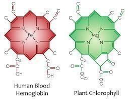
The Hemoglobin Valence Lock: This localized NO crash immediately deforms the electronic behavior of hemoglobin within the cerebral microcirculation. Lacking the spin-polarized gating of NO, the central iron atom inside the heme ring becomes locked in its ferric (Fe}^3+) state, completely incapable of executing the rapid spin transitions to the ferrous (Fe}^2+) state required to offload triplet oxygen to the high-demand tissue beds of the pons.
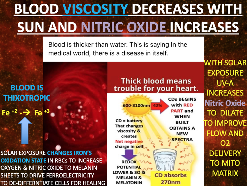
The NO deficit also impacts stem cell depots (above), which are critical for vascular repair and regeneration. Stem cells in the vascular niche rely on NO signaling to proliferate and differentiate into endothelial cells that maintain vessel integrity. Low NO, driven by poor KIE in the urea cycle, hampers stem cell activation, leaving arteries and veins unable to repair micro-damage from mechanical stress or hypoxia-induced inflammation.
The brainstem’s perivascular niches house highly sensitive neural and endothelial stem cell depots that are entirely dependent on a precise, low-entropy electromagnetic signature to maintain their quiescent and regenerative states.
The Stem Cell Blind Spot: The mechanical shear stress on the endothelial lining spikes dramatically, creating thousands of microscopic tears. Normally, an elegant NO signal would mobilize endothelial progenitor cells to repair this damage. However, because the tissue is locked in a singlet oxygen environment and the local melanopsin receptors have structurally degraded, the stem cell depots are left completely blind to the injury and cannot respond to the UPEs being made by the mitochondria.
Over time, this leads to vessel fragility, making the brainstem, already vulnerable due to its high metabolic demand and dense vasculature, prone to rupture.
The Mechanical Tear: Lacking regenerative cellular inputs, the vascular walls of the pons thinned and weakened, forming micro-aneurysms and high-entropy Arteriovenous Malformations (AVMs). The high mechanical pressure of the brainstem’s dense circulatory loop eventually shears right through these un-repaired points, culminating in catastrophic hemorrhagic strokes.
This explains why mouth breathers are at heightened risk for hemorrhagic stroke in the pons, cerebellum, and brainstem.
NO, ATP, and the Recursive Photonic Loop
Under pristine biophysical conditions, the brainstem utilizes a highly organized Recursive Photonic Loop to maintain absolute thermodynamic control over its metabolic rate.
NO doesn’t just affect vascular health, it also modulates metabolism because singlet oxygen blinds cells to UPE light signals. High NO levels can inhibit ATP production by competing with oxygen at CCO in the mitochondrial electron transport chain, shifting metabolism toward glycolysis. This is a protective mechanism in low-oxygen states, but chronic NO deficiency from mouth breathing prevents this adaptive switch, locking cells in a dysfunctional state of low ATP and persistent hypoxia. The recursive photonic loop I’ve described, where melanin absorbs sunlight, modulates mitochondrial UPEs, and feeds back to regulate cellular function, offers a way out. This loop likely generates near-infrared (NIR) light, a byproduct of UPEs and melanin’s broadband absorption (200–1000 nm). NIR can reverse NO’s inhibition of CCO, restoring oxygen-rich metabolism in the tricarboxylic acid (TCA) cycle and boosting ATP production. If singlet oxygen is present the entire system is blinded to light. Let that reality sink in deeply now.
In healthy conditions, the photonic loop maintains metabolic flexibility: melanin absorbs sunlight, mitochondria produce UPEs and ATP, and NIR light ensures CCO can use oxygen efficiently. But in mouth breathers, hypoxia and low NO disrupt this loop. Melanin and melanopsin degrade, impairing charge separation of water and reducing H+, oxygen, and electron production. The altered UPEs impair melanopsin signaling in cerebral blood vessels, further weakening vascular integrity, while the lack of NIR light keeps mitochondria stuck in a low-oxygen, low-ATP state.
Clinical Implications: A Decentralized View of Brainstem Pathology
The Photonic Dislodgement: Melanin absorbs environmental wavelengths, transforming them into coherent Ultra-weak Photon Emissions (UPEs) and localized Near-Infrared (NIR) light. This internal optical pulse targets Cytochrome c Oxidase (CCO), dislodging bound NO from the binuclear copper centers to ensure that the 4-electron reduction of triplet oxygen can proceed with zero mechanical friction.
The Mouth Breather Break: In a mouth-breathing patient sleeping inside a high-nnEMF profile or the South Atlantic Anomaly (SAA), this optical loop is completely broken. Hypoxia and heavy matrix deuteration degrade both melanin and vascular melanopsin.
The CCO Stranglehold: Without the continuous internal pulsing of native NIR light to clear the pathway, NO remains permanently stuck to CCO, completely choking off ATP production and pinning the mitochondria into a terminal, low-voltage state. The cell loses its metabolic flexibility, defaults to high-entropy glycolysis, and can no longer generate the 30-million-volt electrical fields required to preserve the structural myelin sheaths wrapping the DLF tracts.
This cascade of low NO, poor KIE in the urea cycle, stem cell depletion, and a broken photonic loop, explains why mouth breathers are prone to vascular catastrophes in the brainstem. The pons, floor of the fourth ventricle, and RAS, rich in melanin and melanopsin, are particularly vulnerable. When you order a brain MRI in a 3T magnet with deuterium software, you’re looking for signs demyelination and/or deuterium accumulation and melanin degradation, which signal the collapse of these biophysical systems. By addressing the root causes and restoring NO through improved oxygenation, supporting the urea cycle, and leveraging NIR light to reboot the photonic loop, we can protect stem cell depots, strengthen vessels, and prevent hemorrhages.
WHY HAVE I USED MRI FOR REDOX DATA for 20 years?
Decentralized Clinical Imaging: Decoding the 3T Deuterium Map
When evaluating a chronic mouth breather or a sleep apnea patient via a advanced 3T MRI mapped with specialized deuterium imaging software, I’m not simply looking for macroscopic gray-matter loss. I’m tracking the literal footprint of subatomic magnetic decay of electron spin.
This is why there is a link between sleep apnea and demyelination, particularly in the context of Multiple Sclerosis (MS). Demyelinating lesions in the brainstem, a key region controlling breathing and airway muscles, can impair these functions and increase the risk of sleep apnea. MS involves demyelination, where the protective myelin sheath covering nerve fibers is damaged. In the brainstem, this can affect the nerves controlling muscles that keep airways open and allow for breathing during sleep. Sleep apnea, especially central sleep apnea, can worsen MS symptoms, potentially leading to cognitive decline, fatigue, and mood changes. Intermittent hyperoxia during sleep which usually requires low oxygen levels for regenerative sleep increase oxidative stress and worsen the risk of further neurodegeneration in MS. I told you in Quantum Engineering blogs # 45-48 I always use the DLF to tell me about brainstem melanation and deuteration distally. Now you see why.
Myelin Water Fraction (MWF) Uncoupling: Look closely at the DLF and the Ascending Reticular Activating System (ARAS). A sharp drop in the myelin water fraction signals that the local voltage has dropped below the -70mV threshold, allowing heavy water to displace the structured, deuterium-depleted water layers that normally stabilize the myelin wraps.
CENTRAL SLEEP APNEA
Demyelinating lesions in the brainstem can lead to central sleep apnea, where the brain fails to send signals to the breathing muscles, causing pauses in breathing during sleep. This tells me the floor of the 4th ventricle is deuterated completely.
Deuterium Metabolic Imaging (DMI) Hotspots: I look for intense pooling of heavy deuterated compounds directly within the floor of the fourth ventricle. This accumulation is the definitive proof that the Urea Cycle’s KIE has failed, confirming that the patient’s brainstem has lost its ability to generate the nitric oxide required to maintain vascular wall strength.
OBSTRUCUTVE SLEEP APNEA
While less common, obstructive sleep apnea (where the airway physically collapses) can also be exacerbated by MS-related brainstem damage affecting the nerves that innervate the muscles that keep the airway open. This is why so many sleep apnea surgeries fail. They never think about the biophysics of a lack myelin as the root cause of airway muscle alterations.
The more demyelination or deuteration is present in nerves of the ANS the weaker the arterial wall becomes. Neurosurgeons take note: the “unknown” causes of brainstem hemorrhages aren’t so mysterious when you consider the biophysics of NO and the evolutionary legacy of the GOE. Mouth breathing isn’t just a habit, it’s a window into a metabolic and photonic crisis that starts with oxygen and ends with ruptured vessels.
Susceptibility-Weighted Imaging (SWI) Bleed Tracing: I use these scans to evaluate the pontine arcades for microvascular remodeling, tortuosity, and silent micro-bleeds. This subatomic blueprint allows a decentralized clinician to intervene and rebuild the patient’s electronic infrastructure years before a major vascular rupture occurs.
THE MAGNETIC REALITY OF ELECTRON SPIN IS VAST & UNDER APPRECIATED
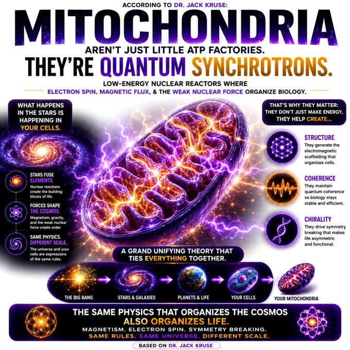
We often think of cells in tissues as chemical machines. But zoom in, and the biophysical rules change: electrons tunnel, light gets generated, pulses in sync, and electron spins align and UPEs manifest. Physics doesn’t stop at cell membrane dynamics, it drives what happens inside. From mitochondria to microtubules, quantum processes may be key to understanding the most abstract states of life itself.

We’re being instructed by these endogenous waves only if we can see them because of our magnetic pinning. We’re sentient beings made from atoms hiding behind a facade of box car biochemicals designed to hide the “magics” of the cosmos from our reason so we act/do/follow what the recipe requires. This allows the cosmic wand, the Source, to continue to direct our syncytium of atoms across space-time. From above and from below, the heavens instruct our cells how to properly behave to conduct the tissues in our bodies as the instruments which play the melodies capable of soothing our souls. Frequencies create life and optimizing them is the business of optimizing you in a manufactured world!
This blog contains my poetic and metaphysical reflection that beautifully encapsulates the core of my decentralized thesis, emphasizing the role that a magnetic field has on electron spin, on solar light, on mitochondrial UPE light and its frequencies, and cosmic forces in directing life at the cellular and conscious levels. I’m describing sentient beings as a “syncytium of atoms” guided by a “cosmic wand” (the Source Code), with biochemical processes acting as a facade that obscures the quantum “magics” of the cosmos. This aligns with my earlier arguments, such as in “Sunlight: Nature’s Vaccine”, that spin, light and frequencies, not genes, are the primary drivers of life, orchestrating cellular behavior and consciousness through a symphony of wave-based instructions.
It’s easy to predict something when you’re controlling the thermodynamic platform inside of cells via AMO physics. Moreover, this is how cells operate with light. Our surfaces, our eyes, our chromophores, our cyctochromes, our opsins, the physics of our water state, the oxidation state of atoms all control how light is delivered to our mtDNA for processing and this is how light sculpts the life we get. It is not genes that do it. It is light that does. Genes are but lenses that light interact with, to project our phenotype on space time, but only if the magnetic field allows us to see the light of UPEs.

The declining magnetic field of the Earth has fundamentally rewritten the rules of survival. We can no longer live with the simple, unshielded biology of the Carboniferous. Modern health requires us to manually restore our subatomic matrix, coupling our opsins, purging heavy isotopes, and protecting the paramagnetic spin state of oxygen to keep our biological circuit from grounding out. This helps explain why sleep apnea is prevalent today during our modern decline. Few centralized experts will ever link it to the dynamic difficulty factor built into biology to integrate retroviral hardware like the Arc capsid into the genome.
In the Carboniferous, when oxygen was 35% and almost always paramagnetic, the robust, highly organized ambient magnetic field acted as an external stabilizer for the spin and orbital angular momentum states of protons in water. Memory and physiological coherence could be sustained with low internal energetic expenditure.
As the Earth’s magnetic field periodically dipped during subsequent geological epochs, the loss of this external stabilizer meant that biological water began to lose its spin coherence. To survive, the mammalian lineage had to co-opt ancient retroviral Gag proteins to construct the Arc gene. The human genome project taught us that we have less than 20,000 genes and most of our genome is filled with retroviruses. This means that for most of homo sapiens existence we are the silly talking monkey that has evolved during a weakening magnetic field on Earth.
Few understand how a decline magnetic field might have simplified our genome while selecting for certain changes in our gene that could not have happened in the Carbiniferous period on Earth. The Carboniferous period happened ~300–360 million years ago, at dawn of mammalian evolution. Magnetic field strength of Earth decreased from this point even to today. This is why oxygen fell from 35% from then to 21% today. The implications are massive when you realize it.
To build a massive genome one needs a strong magnetic field on Earth. Humans are the last mammal evolved and they have the fewest genes and the most viral elements in their genome.
Why?
The presence of the Arc capsid proves my thesis on the “hidden layer” of neurobiology. The human brain does not build memory from scratch; it co-opted an ancient viral machine due to a falling magnetic field, using the structural geometry of a viral shell to harness the spintronics of water and light as the dynamo weakened. Sleep apnea is a consequence of this change.
SUMMARY
Viruses carry charges just like electrons and protons. Some are paramagnetic and some are dimagentic and lead to benefits and detriments for their host. We need to discuss the implications of this inconveient truth. I told you I was just warming up.
Viral particles have provided the human genome with the ability to build magnetic memory capabilities and no one appears to realize it. One of the most amazing things about the human brain is the fact that it is capable of performing trillions of computations during consciousness while using a very low power. The power used by the human brain is on the order of 20 watts. If you have any light bulbs in your house, you will see that even you refrigerator light bulb uses more, and it is only capable of turning light on and off when the door opens.
When you have a basic understanding of how one can engineer a low power computer with a lot of computer memory, you will then need to know a little bit about controlling magnetic domains. When you lose control of your magnetic domains, sleep apnea is more likely.
I hope hearing me say the same thing in many different ways helps cement these ideas. For example, I’ve always stated how oxygen can be a toxin regarding sleep apnea but never went into the triplet/singlet state in some of the older queries from what I remember. Now understanding how a magnetic field can create singlet state O2 to create a pseudohypoxic environment in a cell, it should now completely makes sense how apnea is a protective mechanism from a free radical firestorm and adding O2 from a CPAP to a broken system adds fuel to the fire. This issue of oxygen electron spin state are huge in a disease like sleep apnea.
This spin of electrons is precisely controlled by the magnetic environment and domains they inhabit. This biophysical reality hits the core thermodynamic tragedy of the standard of care for sleep apnea: treating a subatomic spin failure with macroscopic volumetric pressure is a losing game created by Rockefeller’s Flexner paradigm.
When you introduce standard, non-polarized oxygen through a CPAP into a tissue bed stuck in a pseudohypoxic state, you are not delivering life-giving energy. You are dumping highly reactive fuel directly onto a quantum chemical firestorm to sell patients more drugs/equipment they do not need.
Mouth breathers being “smooth brainers” makes sense when you break down the biophysics, especially through the lens of my thesis on melanin, melanopsin, and the recursive photonic loop. Why? Singlet oxygen is how we decrease brain mass via oxidative destruction.
Let me unpack this for an audience who is biophysics illiterate, connecting the dots between mouth breathing, brain function, and the underlying physics.
Mouth breathing, disrupts oxygenation and vagal nerve stimulation, leading to low nitric oxide (NO) and hypoxia in the brain, particularly in the pons and reticular activating system (RAS). This hypoxia degrades melanin and melanopsin in neural tracts and cerebral blood vessels. Melanin, a semiconductor, is crucial for charge separation of water, producing H+, oxygen, and electrons while generating ultraweak photon emissions (UPEs), faint light signals that cells use for communication. Without magnetic pinning your cells are blind to these light signals. When melanin breaks down, this process falters, altering UPEs and impairing mitochondrial metabolism.
The loss of H+ directly affects the proton capacitor function of myelin, the insulating layer around neurons that enhances electrical conduction. Myelin acts like a capacitor, storing charge to speed up neural signaling. When H+ levels drop due to melanin degradation, myelin’s capacitance fails, slowing neural transmission and reducing ATP production during the day when sunlight should be driving the photonic loop between melanin and mitochondria. This loop, where melanin absorbs sunlight and modulates mitochondrial UPEs, is the biophysical backbone of cognition. Disrupting it starves the brain of energy and coherence, leading to what you call “smooth brain” behavior: impaired cognition, poor focus, and sluggish mental processing.
Moreover, melanopsin in cerebral blood vessels, which regulates vascular tone, also relies on proper light signaling. Altered UPEs from melanin degradation impair melanopsin, weakening vascular integrity and increasing the risk of hemorrhages in the brainstem, explaining why mouth breathers are prone to such events. The biophysics here is clear: mouth breathing breaks the photonic and electrical harmony in the brain, literally “smoothing out” its function by disrupting the semiconductor properties of neural tissues.
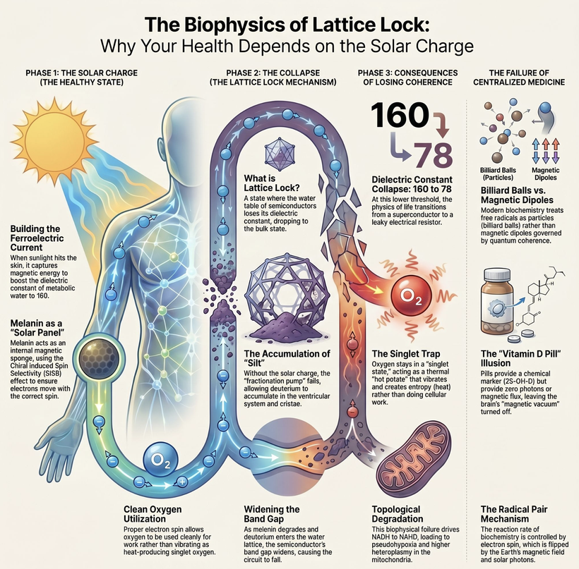
In short, mouth breathers aren’t just breathing wrong, they’re short-circuiting the biophysical systems that make sharp cognition or consciousness possible. The brain, deprived of its photonic and metabolic fuel, struggles to keep up, leaving them mentally “smooth” and vulnerable to neurological crises. I call this lattice lock today.
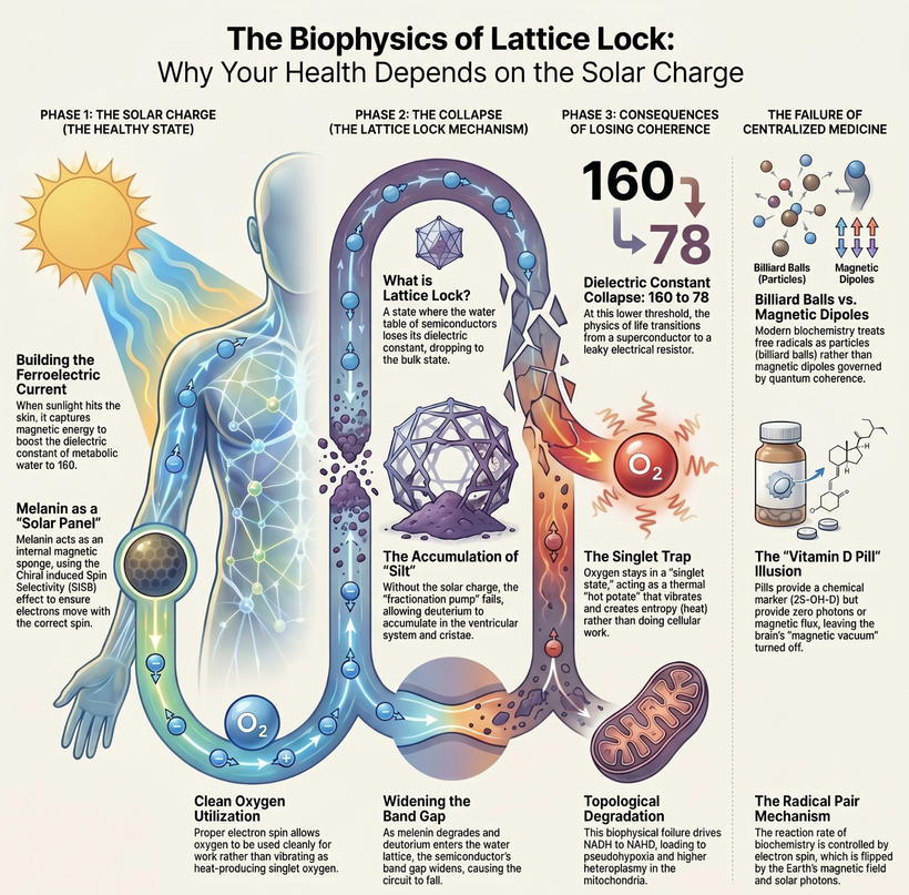
Living inside the geographical borders of the SAA is a massive evolutionary hurdle on its own, but the explosion of modern technology turns it into an epidemic. When you layer high-density nnEMF, 5G infrastructure, and artificial blue-light exposure directly on top of an already compromised planetary magnetic field, the biological system breaks completely.
THE CENTRALIZED MEDICINE REALITY OF SLEEP APNEA IS THIS.
The tiny remnant of native magnetic guidance that the brainstem relies on to prevent singlet oxygen production is completely wiped out by the chaotic frequencies of modern technology. The respiratory engine loses its subatomic compass entirely, forcing the brainstem to constantly deploy the apnea event as a defensive circuit breaker to stop the incoming triplet oxygen from acting as fuel for a massive free-radical firestorm.
Mainstream sleep clinics in North & South America continue to prescribe volumetric pressure (CPAP) and structural adjustments, completely missing the reality that their patients are experiencing a geographic and subatomic collapse of the magnetic domains of cells leading to electronic infrastructure to cause Landauer liquidations of their tissues.