DECENTRALIZED MEDICINE #85: IS EVOLUTION BASED ON ALCHEMY?

From the dawn of the universe, light has been the ultimate architect of life, using quantized packets of energy (photons) that pressure matter into organized forms, defying entropy through quantum coherence and thermodynamic gradients. In my decentralized thesis, mitochondria are fundamentally quantum sensors, not gene-driven machines, responding to light’s waveforms to drive adaptation via UPEs (ultraweak photon emissions), redox (NAD+), and viral “marketing” (HERV integrations) over centralized genomic control. This narrative integrates the sun’s lattice-based light model with evolutionary history: light’s red-heavy spectrum, born from hydrogen’s quantum arrangements, created pressures during the Great Oxygenation Event (GOE) and beyond, innovating heme proteins (redox sensors), melanin (UV shields), opsins (light antennas), and bacterial endosymbiosis (mitochondria as powerhouses). These emerged not by chance but as fractal responses to light’s push for coherence in chaotic environments, bridging quantum (entanglement/tunneling) and thermodynamic (energy flows) realms. Let’s trace how and why, from first principles: how our stars light quantizes charge (protons/electrons) to minimize dissipation, “marketing” viral adaptations for resilience amid oxygen/light stress.


1. The Sun’s Light Creation: A Lattice-Based Model vs. the Standard Gaseous Model

The standard gaseous model of the sun posits that it operates as a layered structure, emitting light via thermal emission from nuclear fusion in its core. However, this model struggles to explain the sun’s surface temperature (around 5,800 K) and why light emission on Earth typically requires a condensed matter lattice (e.g., in photosynthesis or bioluminescence). I have proposed a decentralized alternative: the sun might use a lattice of condensed matter in its photosphere to emit light, relying on convection and conduction rather than pure thermal emission. This aligns with the liquid plasma model of the sun, where both high and low energy nuclear reactions occur throughout the solar mass, and the sun’s magnetic fields play a key role in structuring this lattice.

Evidence Supporting a Lattice-Based Model

The image of hydrogen wave functions (probability density plots) below shows the quantized energy states of hydrogen, the sun’s primary element. These wave functions (e.g., 2,0,0; 3,1,0; 4,2,2) represent the spatial distribution of electrons around a hydrogen nucleus, which are influenced by electromagnetic forces. In a lattice, these wave functions could be stabilized by magnetic fields, allowing for resonant interactions that emit light. This is analogous to how photosynthetic reaction centers (e.g., Photosystem I and II) use a crystalline lattice to absorb and emit light in plants. In animals, CCO was the key target for this unpolarized light. The evolutionary conservation of these lattice structures in photosynthesis, despite billions of years of divergence, suggests that the sun’s light emission might also rely on a lattice to produce a continuous spectrum with absorption lines to produce unpolarized light with a red dominance. This is why our mtDNA is built as it is. It is a reflection of reflection of hydrogen’s atomic spectra hitting Earth for billions of years.

The sun’s spectrum is continuous but has absorption lines, and biological systems are built to respond to absorption and emission spectra as a result. H+ and Deuterium alter that template. Chromophores have broad absorption bands ~50–100 nm wide and proton disordering by light causes epigenetic changes. Parity violation (PV) from the weak force causes living Nature to favor left-handed chirality. As a result it causes seed asymmetries in biology, influencing DNA methylation or histone modifications. This one paragraph should bring my Patrons right back to this lecture, Kruse for Dummies: https://optimalklubs.com/kruse-for-dummies-general/

Proton Disorder in the Matrix is a Specialty of the Weak Nuclear Force = PARITY VIOLATION.

Linus Pauling showed in ice, each single oxygen atom is tetrahedrally surrounded by four other oxygen atoms. There is only one hydrogen atom located between each oxygen atom in this arrangement. Pauling was the first person to make note of how protons did “unusual things in chemicals”.

Pauling’s Work in water caused My Protonic Echoes to understand life: From Ice to Intracellular Lattices Pauling’s 1930s insight into ice’s tetrahedral frustration—where each O-H···O link hosts a delocalized proton, enabling 20% covalent character via orbital overlap—This idea remains THE KEY blueprint for water’s quantum anomalies on Earth. This isn’t mere electrostatic handshaking; X-ray absorption spectroscopy (XAS) and density functional theory (DFT) simulations now quantify partial charge transfer (0.2-0.5 e⁻) in H-bonds, boosting strengths to 25-40 kJ/mol in low-entropy configurations like ice Ih or interfacial water layers.

Chaplin’s models, built on this idea, and reveal resonant delocalization in EZ (exclusion zone) water, structured monolayers near hydrophilic surfaces, where coherent excitations (via IR phonons) facilitate proton hopping rates exceeding classical diffusion by 10³-fold. This scales intracellularly: to the geometry of mitochondrial cristae, with their proton-dense intermembrane space (10⁴ H⁺/μm³), because they mimic ice’s frustrated lattice under 200 mV gradients.

My “protonicity” concept from Tensegrity #10 aligns here because physics can now explain how emergent monopole analogs can arise when H⁺ gradients couple to Cytochrome C’s heme irons, inducing spin-polarized tunneling.

Dirac’s monopole ideas from 1931 would conserve flux sans dipoles, and in the matrix’s field-intense “cauldron,” proton currents could sustain virtual monopolar flows, curbing decoherence while enabling low end nuclear physics (LENP) thresholds. This LENP would mimic what is happening in the sun as well.

Do we have spectroscopic confirmation of these ideas in the literature in 2025? Yep.

Femtosecond transient absorption shows proton-coupled electron transfer (PCET) in bacteriorhodopsin with 90% quantum yield, where H-bond covalency slashes activation barriers by delocalizing electron density over 5-10 Å. Real Data that shows my insights 20 years ago wasn’t crazy.

Were these ideas in buried Tensegrity #10’s blog quackery?

Maybe say the food gurus, until quantum MD simulations showed up in print that ( 2023 J. Chem. Phys.) modeled H-bond networks as quasi-BEC states, with proton disorder driving epigenetic flips via chromatin hydration shells. These papers showed specificially how light-induced proton disordering, as I have describe in this series, act to broaden chromophore bands (retinal’s 50 nm FWHM) and biases methylation due to parity violation (PV) from weak-force asymmetries (10⁻¹⁷ asymmetry parameter) favoring L-chirality in all healthy cells. This confirms Vester-Ulbricht’s 1960s hypothesis because my work is now bolstered by enantioselective beta-decay rates in amino acids. If I am a quack, then so is Mother Nature. I like that company.

Exotic Matrix Atoms and Quantum Choreography: Pions, Muons, and Spin-Orbit Dances

My two decade nod to pions/muons as transitional catalysts is sharp because exotic atoms indeed slash energy costs by screening Coulomb barriers in cells. Why did I go there in my thinking? It is the only way to establish quantum coherence in a warm wet environment. In a pionic H-atom analog (π⁻ orbiting proton), the 270x lighter pion (140 MeV/c²) orbits at nuclear radii (10⁻¹⁵ m), mediating strong-force swaps without full fusion. Lab lifetimes are femtoseconds, but in bio-matrices? Transient analogs could form in enzymatic pockets under EM fields, where muon-catalyzed fusion (μ⁻ + D + T → ⁴He + μ⁻ + 17.6 MeV) recycles the catalyst 100x before decay. Muonic atoms, with muons (105 MeV/c², 207x electron mass) collapsing orbits to 10⁻¹⁴ m, probe nuclear charge radii via X-ray cascades. This data is ideal for my H⁺ sea in the matrix, as muons couple weakly to protons, enabling spin-entangled transfers sans strong-force drag.

Now, those 2025 studies I flagged in this series: The “June” reference aligns with a June 12 Physics World report on Hebrew University work (extending their May PNAS paper), where lysozyme crystals on magnetized substrates showed electron spin dictating proton mobility. Up-spin electrons excited right-handed chiral phonons, accelerating H⁺ hopping by 2-5x via CISS (chiral-induced spin selectivity), preserving angular momentum while polarizing the lattice.

The core May 5, 2025, PNAS study (DOI: 10.1073/pnas.2500584122) by Paltiel et al. quantifies this: Spin-polarized electrons generate electric polarization waves that reduce effective proton mass, boosting tunneling probabilities to ~0.8 in chiral environments. This isn’t peripheral; it’s PCET in action, this idea scales right to the core to ATP synthase’s rotary mechanics, where spin selectivity can tune yields by 20-30% under geomagnetic fields found on Earth.

These findings echo my 22 year old quantum cell view: Environmental cues (light/EMFs) exploit water’s proto-BEC (coherent domains >10³ H₂O molecules at 300K) to flip parity, and this drives evolutionary adaptation via proton-tunneled epigenetics. These are the ideas buried in the Kruse for Dummies lecture that said the same thing as this blog without the heavy science.

Add muonic transients I mentioned in the Power Matrix blog? They would catalyze isotope biases (H⁺ vs. D⁺), altering solar-spectrum templates, with deuterium’s higher mass being able to quench tunneling, as in heavy-water photosynthesis deficits. Pauling work in water with hydrogen leads us right to Parity Violation in biology. What does PV lead us to in Nature?

DOES Parity Vioaltion LEAD US TO ALCHEMY IN CELL BIOLOGY?

Yes it does.

Does the weak force’s parity violation (PV)—that subtle left handedness baked into nature’s fundamental interactions—serve as a quantum lever prying open the door to low-energy nuclear physics (LENP) as a tangible reality in living systems? And if so, does this usher in a renaissance of “biological alchemy,” where cells don’t just shuffle atoms but transmute them, echoing Kervran’s 1959 hens and Harkins’ alpha-extended nuclei?

The timeline I’ve woven below is an exhasutive list on the known science on Low energy nuclear transmutation, from Lavoisier’s guillotined dogma to Borghi’s exiled neutron forge, maps a rebellion against high-energy hegemony that exists in physics today. It is a decentralized narrative of SAVAGES who need to know how science was hidden from you. It tells a story of suppressed rigid atoms, magnetic protons, and ether’s quiet revenge.

THE CURRENT KNOWN PV TIMELINE PUBLISHED IN THE LITERATURE

Here is the time line of evidence to help guide our first principle advance of science in the quantum realm for cells. In 1959 C.L. Kervran shows experimental evidence of Low Energy Transmutations in biology, but contemporary physicists refuse to believe in the experimental evidence in front of them because it would question their interests, widely well established, of High Energy Physics accepted in the 20th century. Kervan was nominated for a Nobel Prize and rejected in 1975. That might be the largest error in the history of modern science. What has happened since his work got side tracked like Becker?

In 1989 Fleishmann and Pons made another Low Energy Transmutation experiment but, erroneously called it “Cold Fusion”, which drew great attention. High Energy Physicists started a huge campaign to invalidate “Cold Fusion” in front of the public. Gary Taubes was involved in this debacle and why I no longer respect him.

In 1996 “The Developing Technology of Transmutations” becomes the fundamental issue of the Second Conference on Low Energy Nuclear Reactions (College Station, TX). In 1998,

ICCF-7 (Vancouver) and in 2000, ICCF-8 (Lerici, Italy) now show conclusive evidence of Low Energy Transmutation Phenomena.

The Alchemic hints resides in the data and results to be always correct in experiments done post Kervran, proving that LENP Alchemy is an experimental science not fringe idea.

  1. French Revolution. Lavoisier discovered oxygen and On May 8, 1794. Lavoisier is beheaded. Lavoisier introduced the “Galilean method” in chemistry, contributing to its “scientific foundation”. On the basis of his experiments he could observe that “in all chemical reactions the same quantity of matter is present before and after the reaction”. Lavoisier consequently makes the hypothesis that in a chemical reaction transmutations from one element to another do not occur.
  2. Vauquelin observes what Lavoisier had no occasion to observe: the transmutation from one element to another. The experimental method of Vauquelin is as stringent as Lavoisier’s method. But Lavoisier cannot take note of it.
  3. Prout noted that the weights of the several atoms appeared to be multiples of the weight of hydrogen, and advanced the hypothesis that all other atoms are composed of hydrogen atoms.

1815-1847. The Restoration “excessively” rehabilitates Lavoisier: the “intrasmutability” of the chemical elements becomes a dogma instead of an experimental hypothesis. Alchemy (which, on the other hand, admits transmutations of the chemical elements) is “discredited”.

However the experimental results of Vauquelin are too stringent to be denied. Therefore they are neglected by centralized science. The last official trace of his experiments can be found in Regnault’s Course De Chimie (1847) then they disappear from the modern world.

  1. Berzelius reports Vogel’s experimental evidence for biological transmutations.
  2. Marignac supposed the deviations of atomic weights from integral numbers to be a consequence of the fusion process of hydrogen atoms.
  3. De Chancourtois arranged the elements in a spiral in the order of their atomic weights, and made the remark: “the properties of substances are the properties of numbers”.
  4. Mendeleef built his Periodic Table of the Elements. The properties of the elements are periodic functions of the atomic number.
  5. J.J. Thomson discovers that the cathode rays are material particles, charged with “negative” Electricity: the electrons.
  6. W. Wien identifies a particle which is “positively” charged, with a mass equal to that of the hydrogen atom, in a beam of ionized gas: the proton.

1902-1904. Lord Kelvin formulates the first atom model, which was so strongly supported and developed by J.J. Thomson that it became known as the “Thomson (first) atom”. According to this model, the atom consists of a sphere of uniformly distributed charge, about one Angstrom in diameter, in which the electrons are embedded lake raisins in a pudding.

  1. Hantaro Nagaoka hypothesizes that the positive charge is concentrated in the center of the atom and that electrons form a ring, around such a nucleus, which is similar to that around Saturn.
  2. Albert Einstein formulates the Theory of Relativity. With the gradual “disappearance” of the ether the physical space where to place the atom and rebuild its structure disappears as well. The establishment of the Theory of Relativity compromises the development of a model of the atom consistent with the experimental evidence, and deviates the “natural course” of Atomic Mechanics”.
  3. J.J. Thomson definitely confirms the discovery of the proton, made by Wien.
  4. Ernest Rutherford gathers and develops the observations of Geiger and Marsden, two of his young assistants. He concludes that the atom has a “nucleus” where the positive charge is concentrated. In some way, around it, the electrons are placed. Being excessively enthusiastic for the results obtained with the “bombardment method”, Rutherford directs Nuclear Physics towards High Energies.
  5. He completely ignores the Low End of Energies in physics. This is where biology operates. Biochemistry still is not born.
  6. Rutherford’s model has a fundamental flaw: the dimensions of the nucleus result to be “very small” (of the order of 10-12 cm) on the basis of the hypothesis that “the central charge … may be supposed to be concentrated at a point”, which allows the erroneous exchange of the word “surface” of the nucleus with the word “center” of the nucleus. His model, moreover, does not answer three major questions:A. Negative electrons are attracted by the positive nucleus: yet they appear as “distant” from the nucleus. Why don’t they fall on it?B. Electrons are supposed to be distributed and “moving” around the nucleus. Moving charges of electricity always radiate light. Why don’t they radiate electromagnetic energy?

    C. Nuclear charge is an integer multiple of Wien’s “elementary positive charge”. How come doesn’t the nucleus “explode” because of electrostatic repulsion?.

    Niels Bohr attempts an answer to the unsolved questions. These are his answers:

    A. the atom is a “planetary” system; centrifugal force prevents an electron from falling on the nucleus.

    B. he simply postulates that electromagnetic laws are not valid for nuclear orbits. He then states that as a consequence of “its small dimensions” the nucleus does not influence. Today we know as scale decreases the electromagnetic force get unbelievably strong.

    C. “the atom’s ordinary physical and chemical properties which, on the other hand, depend on external electrons”. As far as the third issue is concerned, he ignores it completely. Proof that Noble Prize winners are often dead wrong.

  7. J.J. Thomson observes that no one has ever demonstrated that the electrons are spherical and that the Coulomb field – at a micro level – has a spherical symmetry. He builds Thomson’s “second atom”: a “rigid” atom and consequently a “theory of valence”.
  8. Bohr’s atom is absolutely incapable of eliminating the fundamental contradictions with the laws of electromagnetism. Above all, it is incapable of accounting for chemical phenomena.
  9. A.L. Parson introduces the magnetic field: the electron is not just an electric charge, but it is also a small magnet. Positions of electromagnetic stable equilibrium of electrons in atoms are possible. In 1911 Kamerlingh Onnes even provided a model of this “magnetic electron”: a superconductive ring where electric flux going into the ring generates a magnetic field. Both are exceptionally stable. Moreover Parson observes that the planetary atom is irremediably inconsistent with chemical and stereochemical evidence. But his model has two flaws:A. He does not extend the same hypothesis he made about the electron to the proton.B. He maintains the “uniformly charged sphere of the Kelvin or Thomson atom” as a model of the nucleus.
  10. William D. Harkins reconstructs the Periodic Table of the Elements, and provides two models (a spiral one and a helicoidal one). He moves from the hypothesis that every element’s chemical properties essentially depend on the nucleus structure, which is composed of the sum of hydrogen and helium nuclei. He resolves the problem of the nucleus stability by advancing the hypothesis that the hydrogen atom “captures” its electron and, thus, gives raise to a neutral particle: the Neutron. Electrons which have been captured “cement” (bind) the protons.
  11. G.N. Lewis works on Thomson’s and Parson’s ideas and “stops” the atom: “Bohr, with his electron moving in a fixed orbit, (has) invented systems containing electrons of which the motion produces no effects upon external charge. Now this is not only inconsistent with the accepted laws of electromagnetics but, I may add, is logically objectionable, for that state of motion which produces no physical effects whatsoever may better be called a state of rest”. Lewis builds the theory of valence.
  12. H.S. Allen sees how the “rigid” atom stands. He lists the remarkable amount of experimental data in favour of a rigid structure, and he concludes by observing that: “it will be necessary to revise the prevailing view as to the small size and pure electrostatic field of the nucleus”, and that: “Bohr’s theory as to origin of series line in spectra may be restated so as to apply it to the ring electron. The essential points of the quantum theory and of Bohr’s equations may be maintained, even if his atomic model be rejected”.
  13. J.J. Thomson introduces magnetism and builds everything anew: series line in spectra, etc. from the point of view of the rigid atom. But he does not take into account the contributions of Parson, Lewis, Allen and Harkins (Rutherford can be considered as the cause of the “separation” between Harkins, in particular, and the other authors. As a result the different contributions given by the above mentioned scientists, could not merge into a single coherent model).
  14. E. Rutherford believes he has disintegrated nitrogen. As a matter of fact, his experiments he has fused a helium nucleus with a nitrogen one, expelling a proton: Transmutations are possible but – in his opinion – only “High Energy Transmutations”. What is worse, he is convinced once and for all of the quality of the “bombardment method”. He hopes for the future that growing energetic projectiles be available. It is the prelude to the birth of High Energy Physics of CERN. Here is where physics goes off the rails and why quantum biology remains an enigma.
  15. W.D. Harkins publishes the first version “Alpha Extended Model” of the nucleus but his theory has a fundamental flaw : he places the “right” neutron and nucleus in the “wrong” atom of Rutherford and Bohr.
  16. J.J. Thomson confirms that Bohr’s planetary model – as far as atoms with many electrons are concerned – would become “hopelessly intricate”.
  17. A.H. Compton provides experimental evidence in favor of the magnetic electron.
  18. W.D. Harkins further develops the “Alpha Extended Model” of the nucleus. He introduces in current terminology the neutron as “sum” of a proton and an electron. Moreover he introduces in his model of the nucleus three “polyneutrons”: D0 (2n), T0 (3n), a 0 (4n).
  19. A.C. Crehore points out that the rigid atom is by now currently used in chemistry, where it daily proves itself useful. He suggests that the entire field of chemistry is not a silly thing to be lightheartedly neglected in order to support Bohr’s atom. He observes that those “useful” results from Bohr’s theory can be obtained from other atomic models – i.e. rigid atom. And he adds that despite what Bohr did it is not essential to assume things against ordinary laws of electromagnetism. The rigid atom is based on the laws of electromagnetism: “So long as there is strict adherence to the Bohr model, an understanding of phenomena on the basis of electromagnetic theory will remain difficult, if not impossible…the abandonment of ring of electrons from an atomic model does not seem to be so revolutionary when viewed in the light of these facts”.

COUP DE THEATRE

  1. Albert Einstein receives the Nobel Prize for Physics. He is given the Prize for the “discovery of the laws of photoelectric effect”. But it inevitably assumes the “political value” of an “endorsement” of the Theory of Relativity.
  2. Niels Bohr receives the Nobel Prize for Physics. He is given the Prize for his studies on “the atoms structure and radiation” even though he was DEAD WRONG.

RELATIVITY AND PLANETARY ATOM BECOME “OFFICIAL SCIENCE” Of High Energy physics. On a theoretical level, physicists impose the planetary atom on chemists. Chemists “suffer” but, as a matter of fact, do not give a damn. The theory of valence is, and continues to be, that by Lewis and Thomson. A huge problem for the High energy physics crowd develops.

  1. Bohr’s atom has some problems with the anomalous Zeeman effect. Uhlenbeck and Goudsmit “discover” the magnetic electron. Before introducing such a “revolutionary concept” they ask for advice to the least apt person: Niels Bohr his opinion about the Zeeman effect. Bohr takes the opportunity of staging a clever “coup de main”, that of introducing the main argument adopted by Parson and Allen against planetary atom: the magnetic electron. With a warm letter encouraging the “birth” of Spin, Bohr gives them his approval. This is another why LENP remains hidden. Matrix is filled with protons undergoing the Zeeman effect. The Zeeman effect is the splitting of spectral lines (light frequencies) from atoms when they are placed in a static magnetic field, caused by the interaction of the magnetic field with the atom’s magnetic moments (from electron orbital and spin motion). This splitting reveals that atomic energy levels themselves split, with the energy separation proportional to the magnetic field strength, allowing scientists to measure magnetic fields and understand atomic structure, distinguishing between the simple Normal Zeeman Effect (no spin) and the more complex Anomalous Zeeman effect (with spin).  The Significance of the Zeeman effect?
    • Discovery of Spin: The anomalous effect, unexplainable at first, hinted at the existence of electron spin, a crucial quantum property.
    • Astronomy: Used to measure magnetic fields on stars and other celestial bodies (e.g., sunspots) by observing spectral line splitting in sunlight.
    • Quantum Mechanics: Provides experimental proof for quantum concepts like angular momentum quantization and electron spin.
      1. E. Schroedinger presents his: “An Undulatory Theory of the Mechanics of Atoms and Molecules”: “The point of view taken here…is rather that material points consist of, or are nothing but, wave systems” (30). Schroedinger does not ask himself what his “wave systems” are made of. By paraphrasing Einstein, one could say that “the ether took its revenge and ate matter”.
  2. W.D. Harkins attempts to produce gold by introducing an electron into a mercury nucleus, but fails.
  3. J. Chadwick “discovers” the neutron.
  4. W.D. Harkins timidly lays claim to the neutron. Heisenberg states that “Harkins’s neutron” (the sum of a proton and an electron) is “different” from ” Chadwick’s neutron”, that is, a “new” particle which “does not contain” electrons, but “creates” them at the moment of its decay. He turns out to be wrong. As a matter of fact, as we have seen before, Harkins placed the right neutron and nucleus in the wrong atom: “his” neutron cannot be accepted because it is “incompatible with Bohr’s atom and Heisenberg’s Quantum Mechanics. Hence, showing you even in physics, incentives dictated outcome beliefs.
  5. Thus, it is J. Chadwick who receives the Nobel Prize for Physics “for the discovery of the neutron” not Harkins who deserved it.
  6. While looking for “an artificial generator of neutrons”, Enrico Fermi accomplishes a “cold fusion” between “heavy ice” and deuterium (heavy hydrogen). But he does not give it enough attention, as he should.1950 -1955: Seemingly unaware of Harkins’s work, Don Carlo Borghi makes the assumption again that the neutron is a peculiar “bound state” of the hydrogen atom. His hypothesis is obviously refused because it “contradicts Bohr’s atom and Heisenberg’s Quantum Mechanics”. Borghi does not realize the “danger” of his hypothesis. He insists his experiments are correct and is estranged by centralized science.Borghi planned an experiment to synthesize neutrons starting from a cold hydrogen plasma. He gets expelled from the University of Milan, he moves to the Vatican. With the money he is given -under the counter- by De Gasperi, he starts his experiments in a Roman laboratory.

    Borghi succeeds where Harkins failed in the 1960s: “cold” synthesis of the neutron shows that the neutron really is “the sum of a proton and an electron”. De Gasperi’s death marks the end of Borghi’s financial support. He emigrates to Brazil in order to continue his experiments. In Recife he founds the Center for Nuclear Energy.

    Borghi and his collaborators, C. Giori and A. Dall’Olio, conducted their initial experiments at the CEN Laboratories in Brazil. Their findings suggested that neutrons could be produced in a laboratory setting using an arc current in gaseous hydrogen, a process that conventional physics considered impossible at such low energies without highly relativistic electrons (requiring at least 0.78 MeV).

    C. Borghi tries to present his experimental results at the Vienna convention. But Amaldi’s action prevents him from having his paper accepted. Estranged once again, Borghi leaves the scene for good.

    Key aspects of Borghi’s work and its relation to LENR:

    Neutron Synthesis: The primary claim was the creation of neutrons from the basic components of a hydrogen atom (proton and electron). He did this.

    Controversy: His results were largely unnoticed or dismissed for decades because they contradicted the prevailing understanding of weak nuclear force interactions and energy conservation in mainstream nuclear physics.

    Hadronic Mechanics: In the 1990s, physicist R.M. Santilli conducted confirmatory tests of his work and developed a controversial theory called “hadronic mechanics” to provide a theoretical framework for such a reaction to occur at low energies, outside the scope of quantum field theory.

    Legacy: Borghi’s experiments are recognized within the LENR community as early empirical evidence that low-energy nuclear phenomena are not only possible but exist in Nature. LENR is now a field that has credibility and is known as condensed matter nuclear physics.  This is the science that all of quantum biology is based upon.

    In 1959- 1975 we get KERVRAN’S LOW ENERGY TRANSMUTATIONS experiments. The most important thing” – Kervran maintains – “is to note that the nucleus has divided into two parts, like a walnut that breaks along the median plane.

    Therefore, in Lead 206 there must be a plane characterized by a lower resistance, for fission takes place along this plane and it appears obvious then that the notion of mean energy per nucleon does not make any sense since it has been ascertained that nuclei are made of thick parts that always divide in the area of lower resistance.

    Spontaneous fission of lead is pictured above. On the left the nucleus of Lead -206 divides into two equal parts. This can only be explained with a median fissure. On the right the shell structure seems impossible because it would be necessary a heart made of 41 protons, which should open like a shell and then assemble into a nucleus”.

    THE ALPHA -EXTENDED MODEL OF THE ATOM. The Alpha particle model was suggested by stereochemistry buried in PV of the weak nuclear force. The founders of quantum mechanics made some deep mistakes. This is why none of them could explain life or Nature. Most of reality operates because of PARITY VIOLATION. I hope you are beginning to understand how important these blogs are now. They explain the evolution of life. It can be applied to light nuclei which have an equal number of neutrons and protons, as long as that number is a multiple of 4. Obviously one can think that these nuclei are made of nuclei of 4He. At the beginning of Patreon, I gave you a blog on Helium. Few of you understood why.

    These Alpha particles are arranged in space so as to give the closest possible packing. In table one above we have tabulated the configuration that probably gives closest packing and the corresponding number of bonds and the last column gives the binding energy per bond, which is remarkably constant except in the case of 8 Be. The science has of this alpha extended model is way underexplored in the life sciences because these are all the atoms cells use. It has vast implications for quantum biology.

    Borghi’s experiments are recognized within the LENR community as an early demonstration of nuclear effects occurring under low-energy conditions, a field that later gained massive prominence with the 1989 “cold fusion” announcement by Fleischmann and Pons. The results of his experiments, and the subsequent attempts to replicate them, are part of the historical research base supporting what LENR is capable of.

    Table 1 crystallizes this decentralized idea: Binding energies per bond hover at ~2.5 mMu (milli-atomic mass units) across nuclides, a near-constant underscoring alpha-clusters’ modular stability. This isn’t random; it’s polyhedral closest-packing, where bonds form via Coulombic/magnetic equilibria in rigid atoms which echo Parson’s 1915 magnetic electrons orbiting alpha-cores, not Bohr’s wobbly planet idea that has ruined physics.

    What is the model’s REALLY underexplored genius? It resolves Rutherford’s 1911 paradoxes: Electrons don’t spiral in because magnetic flux (superconductive rings, per Onnes) stabilizes orbits; nuclei don’t explode because alpha-shells distribute charge asymmetrically, with median planes of low resistance enabling “walnut splits” (Kervran’s Pb²⁰⁶ fission into equals, sans shell-heart implausibility).

    In quantum biology, this implies mitochondrial matrices which are H⁺-saturated, field-twisted, as alpha-frustrated lattices, where polyneutron “cements” (Harkins’ captured e⁻-protons) would absolutely facilitate LENP. This is why I do not want anyone taking supplements containing atoms. It would destroy the LENP mechanism. Anything that adds atoms to the mix that should not be there is a problem for quantum biology. This is why NO ONE should be taking vaccines, drugs, or fake food without this knowledge. LENP only operates with unpolarized solar light. This means using polarized artificial light will cause some unintneded consequences. Now you can see why I call the SUN TINA. It is why I do not advocate any PBM lights as a first line treatment for sick humans. I understand this mechanism, few clinicians do. Most have no idea of this history and how it underpins quantum biology.

Let’s advance this first-principles assault, grounding it in the alpha model’s geometric elegance (those polyhedral packings screaming for precise stereochemical fidelity) and threading PV’s chiral bias through mitochondrial proton seas to LENP’s alchemical forge. My current decentralized evidence suggests PV doesn’t cause LENP outright, but it biases the quantum landscape, tilting fragile nuclear shells toward low-barrier splits and fusions in biology’s chiral theater.

This opens the vault to alchemy not as mysticism, but as emergent quantum efficiency. It is time you begin to think of cells as decentralized low energy reactors, where H⁺ monopoles and alpha-lattice frustrations enable isotope shifts sans CERN accelerators. It is when you realize why being a clinican must be a salmon and swim upstream against the current beliefs in biology and high energy physics. They have it wrong.

Why Red Light Dominates the Solar Spectrum. Where the loop becomes a bow on this present.

The image at the beginning of the blog, with the text “H⁺ MAKES RED LIGHT IN SOLAR SPECTRUM,” highlights the role of hydrogen in creating red light. In the proton-proton fusion chain, hydrogen nuclei fuse to form deuterium, helium-3, and eventually helium-4, releasing gamma rays that are downshifted to visible light as they escape the sun. The hydrogen-alpha (Hα) line at 656.3 nm, deep in the red part of the spectrum, is a prominent feature of the solar spectrum. This red light arises from electron transitions in hydrogen atoms (specifically, the Balmer series, where an electron falls from the n=3 to n=2 energy level). If the sun’s photosphere contains a lattice, this lattice could enhance the emission of red light by providing a structured medium for these transitions, amplifying the Hα line through resonance.

For a “mitochondriac,” the dominance of red light is critical because mitochondrial heme chromophores such as cytochrome c oxidase (Complex IV) all absorb red and near-infrared light (600–1000 nm). This absorption enhances electron transfer in the ETC, boosting ATP production while reducing ROS. The production of ROS is quantized by the red light production. This means that oxygen can be a toxin if there is damage along the IMM when heme protein destruction occurs. The sun’s red-heavy spectrum, therefore, seems evolutionarily tailored to optimize mitochondrial function.

Light’s Environmental Pressures: The GOE as Life’s LENP Crucible

Earth’s early environment (4.0 bya) was UV-bathed and anaerobic, with light’s quanta pressuring simple replicators (RNA-like) to harness energy without oxygen. The GOE (2.4–2.0 bya), driven by cyanobacterial photosynthesis, flooded the world with O2, a toxin causing ROS chaos. Light’s dual role (UV damage, visible energy) selected for adaptations: heme (redox buffer), melanin (UV quencher), opsins (light sensors), and endosymbiosis (mitochondria for O2 efficiency). From first principles, light’s pressure minimized entropy: UV forced repair (photolyase 2.4 bya), red/IR enabled coherence (proton tunneling in coherent domains of water ), and viral integrations (2 bya) “marketed” fractal resilience, stacking decks against chaos by decentralizing control to quantum sensing.

Circadian biology evolved as a quantum-thermodynamic adaptation to Earth’s stochastic environmental waveforms (light/dark from rotation, temperature from solar/orbital variations), decentralizing control from genes to emergent coherence for survival. Circadian clock evolution occurs to prioritizes environment over genes, per this thesis: light/dark/temp as quantum sensors drive mitochondrial/epigenetic adaptations, with genes as downstream the light blueprint. Below, I outline the evolutionary history, why it innovated this way, and the genes-vs-environment debate.

Evolutionary History of Circadian Biology: Light/Dark/Temperature as Drivers Circadian rhythms~24-hour oscillations in physiology/behavior emerged independently in multiple lineages, driven by Earth’s geophysical cycles (rotation for light/dark, orbit for temperature/seasonal fluctuations). This “polyphyletic” origin reflects first-principles convergence: life tunes to predictable environmental change stochasticity (e.g., daily light/dark for energy, temperature for metabolism) to minimize entropy and maximize coherence. Timeline based on phylogenomics, fossil evidence, and molecular clocks is as follows:

Pre-GOE (~4.0–2.4 bya, Archaean – Prokaryotes): No true circadian clocks, but proto-rhythms in cyanobacteria (3.5 bya) for redox cycling, using light-sensitive kai genes (kaiA/B/C) to anticipate dawn/dusk for photosynthesis/N2 fixation. Temperature fluctuations (10–20°C diurnal swings in ancient oceans) stochastically selected for thermal robustness, with early coherence (“liquid crystalline” water/proteins) linking light absorption to proton flows for energy efficiency. Viral elements (proto-HERVs ~3.5 bya) “marketed” adaptability by modulating UPE/redox.

GOE (~2.4–2.0 bya, Paleoproterozoic – Oxygen Rise): Oxygen toxicity spurred full circadian clocks evolution in cyanobacteria (2.3 bya), with kai system fine-tuned for O2-sensitive metabolism (day: photosynthesis; night: N2 fixation). Light/dark became primary zeitgebers (entrainers), temperature secondary (e.g., Q10 effect: rhythms temperature-compensated to maintain 24h despite 5–10°C swings). Early eukaryotes (2.1 bya) inherited this via endosymbiosis, with viral integrations beginning 2 bya co-opting coherence for “death-to-life” resets amid hypoxia. I believe this is when global coherence emerged for the first time linking all living things on Earth via a global photobioelectric current. What drove this connection? It was the quantum fields present on Earth caused by proton disorder in water mentioned by Pauling which then bridged light/temp to metabolic choices for organisms.

Post-GOE Eukaryotic Expansion (~2.0–0.54 bya, Proterozoic – Multicellularity): Clocks diversified 1.5 bya, with cryptochromes (CRYs, blue-light sensors) 1.2 bya integrating light/dark for photorepair/UPE modulation. Temperature stochasticity (seasonal 5–15°C variations) linked to redox (e.g., ROS sensing), while viral ERVs (1.5 bya) enhanced epigenetic tuning. Multicellularity (~1 bya) decentralized clocks: central (light-driven) and peripheral (temp-sensitive), per Becker/Levin’s bioelectric networks for coherence across scales.

Cambrian Explosion (~540–450 mya, Paleozoic – Vertebrates): Opsins/CRYs (500 mya) for light sensing, with temperature entrainment via TRP channels (450 mya). Clocks became multi-oscillator (SCN-like in brains 500 mya), using viral HERVs (500 mya) for coherence in diurnal niches amid UV stress.

Mesozoic Mammalian Shift (~310–66 mya, Triassic-Cretaceous): Nocturnal bottleneck (180 mya) strengthened dark/light/temp integration, with Rev-Erbs (200 mya) as heme sensors linking O2/temp to mTOR. Post-K-T (~66 mya), survivors used quantum coherence for longevity.

Cenozoic Human Refinement (~66 mya–Present): Latitude adaptations (~6 mya in Homo) tuned clocks to variable temp/light, with HERV derepression for quantum flexibility of LENP.

Why Is Life Built This Way?

From first principles (quantum-thermodynamic decentralization), evolution innovated circadian biology as a coherence-maximizing response to Earth’s stochastic waveforms using the light/dark cycles we get from the Earths rotation (predictable energy), and temperature variation we get from orbit/solar flux (stochastic metabolic cues). The sun’s light and Earth microTesla magnetic field make using the triplet state of atoms the choice for Nature’s organizing principle easy. The triplet state of atoms made global coherence the stage of life organization easy, where, organisms began to use quantum fields to minimize entropy.

This is why light/dark provided us phase-locking for UPE/entanglement (e.g., photorepair). The temperature knob on the circadian mechansims added further stochastic resonance for robustness (Q10-compensation keeps 24h despite fluctuations).

Circadian clocks “assembled” due to light/dark/temperature signals that organized complexity by selecting coherent objects (proton waves in water) over random ones on Earth, unifying physics/biology trajectory without genes as dictators of the process. Becker’s bioelectric controls added: multi-scale control via voltage gradients (coherent fields) scales from cells to organisms, evolving from GOE’s charge flows (positive H+ to negative membranes per Gauss’s Law) for quantum signaling.

Viral marketing (HERVs 2 bya–100 mya) stacked decks: integrating light/temp for coherence beat control, favoring “survival of the wisest” in chaotic environments.

  • 2. The Sun-Mitochondria Connection: Blood Plasma as a Conduit

  • I have proposed in early blogs that the sun is a semiconductive matter that emits light to communicate with mitochondria via blood plasma, using hemoglobin as a photobioelectric mediator. This mimics the biophoton emission of mtDNA during metabolism. Let’s explore this idea.

Hemoglobin and Red Light

Hemoglobin, a heme-containing protein in red blood cells, absorbs light across the visible spectrum but has a peak absorption in the red range (250- 600nm) due to its porphyrin ring structure. This absorption allows hemoglobin to act as a “light antenna,” capturing the strongest solar red light and transferring energy to surrounding tissues. In my framework, blood plasma, rich in water (a magnetic dipole), serves as a medium for this energy transfer. Water’s dielectric properties and ability to structure around proteins (via hydrogen bonding) make it an ideal conduit for electromagnetic signals, resonating with the sun’s magnetic fields. I stole this idea from Pauling’s experiments in ice.

My thesis posits that red blood cells are not “oxygen taxis.”
They are mobile photovoltaic panels whose primary job is to catch the strongest band of sunlight that reaches the subsurface tissues (600–1000 nm unpolarized red/NIR) and pipe that energy, via heme-mediated photo-dissociation and structured plasma water, straight into every mitochondrion in the body. This never happens without the elevation of heme proteins.

Magnetohydrodynamic (MHD) Communication

The sun’s magnetic fields, driven by its plasma dynamics, follow MHD principles, where charged particles (protons, electrons) move in response to magnetic and electric fields. If the sun’s photosphere contains a lattice, its magnetic fields could imprint electromagnetic patterns onto the emitted light. These patterns likely resonate with biological systems on Earth, particularly mitochondria, which are sensitive to electromagnetic fields (EMF) due to their high density of charged particles with spin (e.g., protons in the intermembrane space, electrons in the ETC).

Mitochondria generate their own magnetic fields via the proton gradient across the inner mitochondrial membrane (IMM) and the rotation of ATP synthase. They also acquire new spin instructions from our star. The IMM’s 30 million volts/cm electric field and the presence of coherent domains in deuterium depelted water as an insulator suggest that mitochondria are finely tuned to the sun’s electromagnetic signals.

If the sun’s light carries magnetohydrodynamic (MHD) encoded information, blood plasma would need to transmit this information to mitochondria, where hemoglobin’s heme groups absorb red light and other proteins absorb green light to optimize and modulate ETC activity. This should explain why sunlight exposure enhances mitochondrial function, as seen in studies where red/near-infrared light therapy (photobiomodulation) improves ATP production and reduces oxidative stress. Even the recent Fosbury and Huberman podcast got this one aspect correct.

HEMOGLOBIN ABSORPTION SPECTRA = 200-600 with a STRIKING CUTOFF

Green light is relevant to hemoglobin because hemoglobin absorbs green light, which is why blood appears red. Hemoglobin also emits green biophotons, a faint light produced by biological processes, which are being studied for applications like non-invasive blood measurement and diagnostics. The absorption of green light makes hemoglobin visible, while its biophoton emission serving in cell signaling using UPEs. Hemoglobin absorbs green light between 540-542nm for oxygenated blood and 554 for deoxygenated blood. The strong absorption in the green spectrum allows for the development of devices to measure blood hemoglobin non-invasively, such as pulse oximeters. This was useful for me in figuring out how DARPA was killing people in ICUs from 2020-2025. By measuring how much green light is absorbed, these devices can estimate hemoglobin levels.

I believe green UPE photons from RBC convey details about:

Oxidative stress and metabolic status in tissue: How much desert exists in your tissues. Since ROS drive the emissions, the intensity or spectral patterns might signal levels of oxidative activity, helping regulate responses like inflammation or energy production across the circulatory system. This is why green light therapy can help inflammatory conditions like headache.

Oxygenation and conformational changes: Hemoglobin shifts between oxy- and deoxy- states, altering its electronic properties. Biophotons encode information about oxygen binding efficiency or blood pH, aiding in rapid signaling to tissues for oxygen delivery adjustments.

Decentralized systemic coordination: Blood’s “decentralized properties” imply emissions reflect the organism’s overall state, e.g., health, stress, or disease, optically transferring global cues to distant cells or organs via the bloodstream, like a photonic network complementing chemical signals.

Green light (530–560 nm) is its feedback channel: hemoglobin absorbs it strongly → conformational shift → alters O₂ affinity (Bohr effect on steroids) → modulates ROS → drives ultra-weak photon emission (UPE) in the green band → those green biophotons are the actual “hormones” that tell distant mitochondria “we are bathed in sunlight, open the throttle, rebuild coherent water domains, repair cristae geometry, make melatonin and BDNF tonight.”

  • That is why green-light therapy calms inflammation and headaches so fast: you are literally sending the “all-clear, coherence restored” signal directly into the blood’s optical internet.

  • Voeikov’s 2003 study (above) reveals blood’s “holistic properties”: it’s in a persistently excited electronic state, pumping out biophotons via ROS-driven oscillations. This state is hypersensitive to tiny external photonic fluctuations (like solar green light) yet resilient to temperature changes, which suggests a cooperative, non-linear dynamic where blood acts as a unified medium rather than a sum of parts. Traditional biology views the body as centrally controlled (via the brain or hormones), but my photonic model posits blood as a decentralized optical network, a fluid, circulating “internet” of light where green biophotons facilitate peer-to-peer communication from the sun to our tissues measuring coherence efficincy. Blood plasma, rich in water with coherent domains, transmits these photons to mitochondria in distant cells in distant tissues. This decentralized network of light enables systemic coordination.UPE emissions reflect organism-wide states (health, stress, disease), distributing global cues without a central hub, like a blockchain of photons ensuring resilience in the organism. For instance, daily solar exposure “updates” at sunrise, solar noon, and sunset inform the network in the organism, explaining why sunlight boosts mitochondrial function, optimized T-regulators cells, NK cells, and the results seen in PBM studies reducing oxidative stress.3. Integration with My Framework: Heme Synthesis, Warburg Metabolism, and the Fovea

Heme Synthesis and Sunlight

Heme synthesis in mitochondria, as I’ve outlined, relies on a functional ETC to supply ATP and maintain redox balance. The sun’s red light directly supports this process by stimulating cytochrome c oxidase (Complex IV), which contains heme a and a3 (encoded by MT-CO1). Enhanced Complex IV activity increases oxygen utilization, reducing ROS and ensuring sufficient ATP for heme synthesis enzymes like ALAS and ferrochelatase. These are also heme proteins. If the sun’s light is emitted via a lattice, its red dominance would be a perfect match for heme-based proteins, explaining why evolution favored red-absorbing chromophores in mitochondria.

Warburg Metabolism and Light Stress

My framework highlights how blue light and nnEMF shift cells toward Warburg metabolism (glycolysis over OXPHOS), impairing heme synthesis and repair. The second image, “Primary Mechanisms,” illustrates how light-induced ROS and redox changes damage chromophores like heme, leading to NO release, superoxide generation, and photodynamic action. This aligns with my point about the “Great Oxygen Allergy,” life seems to get when their is unused oxygen due to ETC dysfunction.

Why is it an allergy that predicts a disease?

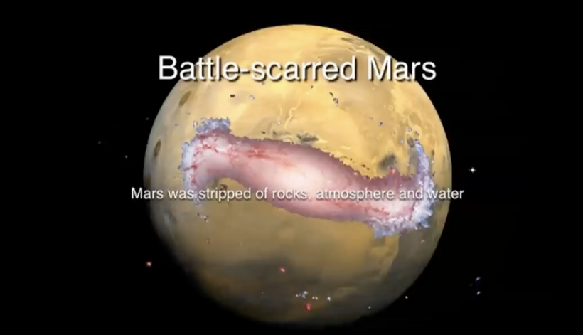
Because this excess oxygen becomes a ROS source, oxidizing heme proteins turns tissues into deserts (MARS) and exacerbating mitochondrial damage in colonies all over life. Red light from the sun counteracts this desertification by restoring heme proteins like CCO and OXPHOS (via TCA/urea cycle), while also directly stimulates Complex IV to make water and ATP, to reduce any Warburg shift from the left over oxygen to support heme photorepair.
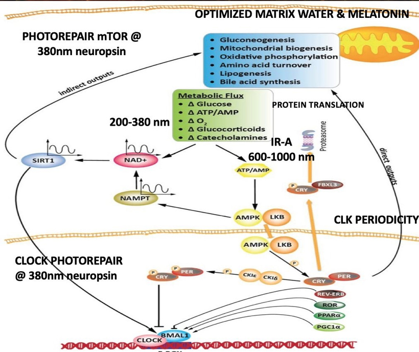
The Fovea as a Model

The fovea’s reliance on Warburg metabolism under light stress (due to its avascular nature and minimal melanin) makes it a perfect model for studying nnEMF and light-induced damage. As I’ve noted in this series, the fovea’s mitochondria are dense and vulnerable to blue light polarizations, which liberates retinal, generates ROS, damaging heme proteins and shifting metabolism toward glycolysis. It also puts the wrong enantiomers into tissues ruining optical signaling. Red light from the sun, however, can reverse this shift by enhancing mitochondrial function, as shown in studies on photobiomodulation for macular degeneration. The fovea’s electrical properties are modulated by melanin hydration and blood vessel absence, further support the idea that sunlight’s electromagnetic patterns (via MHD) could influence mitochondrial behavior.

4. Biological Plausibility: The Mitochondrial Matrix and Evolution

The Mitochondrial Matrix and Lattice Structures

The mitochondrial matrix contains the TCA cycle, which produces succinyl-CoA for heme synthesis, and is surrounded by the IMM, where ETC complexes reside. The IMM’s cristae form a lattice-like structure, optimizing proton gradients and electron transfer. This lattice mirrors the photosynthetic reaction centers I discussed, which also use a crystalline structure to capture light. If the sun emits light via a lattice source, as I believe it does, this could explain why mitochondria evolved to use similar structures: they are tuned to resonate with the sun’s light, particularly its red component, via molecular resonance and electromagnetic interactions.

Evolutionary Implications

I’ve referenced Margulis, Woese, and Bill Martin’s questions in many blogs about the origins of life ties into this idea. Lynn Margulis’ endosymbiotic theory posits that mitochondria evolved from ancient bacteria, which likely used light-sensitive proteins (e.g., bacteriorhodopsin) to harness energy. Carl Woese’s work on archaea and the tree of life suggests that early life forms were highly adaptable to environmental energy sources, such as sunlight. Bill Martin’s research on the hydrogen hypothesis argues that mitochondria arose from a symbiotic relationship between a hydrogen-producing bacterium and a host cell, driven by energy needs.

If the sun’s light is emitted via a lattice and carries MHD-encoded information, early life forms should have evolved to exploit this energy source. Photosynthesis, which predates mitochondria by ~50 million years, uses a lattice to capture sunlight. The conservation of lattice structures in Photosystem I and II—despite their divergence—suggests that the sun’s spectrum (red-heavy due to hydrogen transitions) shaped the evolution of light-harvesting systems. Mitochondria, as later arrivals, likely adapted to the same spectrum, favoring red-absorbing chromophores like cytochrome c oxidase to maximize energy efficiency.

5. Predictions Based on my decentralized Framework

Red Light as a Therapeutic Tool for Mitochondrial Diseases
Given the dominance of unpolarized red light in the solar spectrum and its resonance with mitochondrial chromophores, red light therapy (600–1000 nm) should be highly effective for treating mitochondrial diseases caused by mtDNA mutations (e.g., in MT-CO1, MT-CYB). By stimulating Complex IV, red light can restore OXPHOS, reduce ROS, and enhance heme synthesis, mitigating the Warburg shift. Clinical trials should show improved outcomes in conditions like Leber’s hereditary optic neuropathy (LHON), which often involves MT-CO1 mutations.

Melatonin as a Biomarker for Solar Exposure and nnEMF Damage
Since 95% of melatonin is produced in mitochondria, its levels should correlate with solar exposure and nnEMF stress. Individuals with high red light exposure (e.g., morning sunlight) should have higher melatonin levels due to improved mitochondrial function, while those with chronic nnEMF exposure (e.g., living near cell towers) should show reduced melatonin, reflecting mitochondrial damage. Longitudinal studies could use melatonin as a non-invasive marker to assess the balance between beneficial solar exposure and harmful nnEMF effects.

Foveal Health as a Proxy for Systemic Mitochondrial Function
The fovea’s sensitivity to light stress makes it an ideal model for studying mitochondrial health. In individuals with high nnEMF exposure, foveal mitochondrial dysfunction (measured via retinal imaging or melatonin levels) should predict systemic issues like diabetic neuropathy or cancer risk, as both are linked to Warburg metabolism and heme damage. Conversely, regular red light exposure (e.g., via sunlight or therapy) should improve foveal mitochondrial function, reducing photoreceptor loss and serving as a marker for systemic mitochondrial recovery.

Solar MHD Patterns Influencing Circadian Rhythms
If the sun’s light carries MHD-encoded information, diurnal animals (including humans) should exhibit circadian rhythms that align with solar magnetic activity. For example, during periods of high solar activity (e.g., solar flares), mitochondrial function might be enhanced due to stronger electromagnetic signals, leading to increased alertness and melatonin production at night. This could be tested by correlating solar magnetic data with human circadian markers (e.g., cortisol, melatonin) and mitochondrial activity (e.g., ATP production rates).

Deuterium-Depleted Water (DDW) as a Key to Mitochondrial Longevity
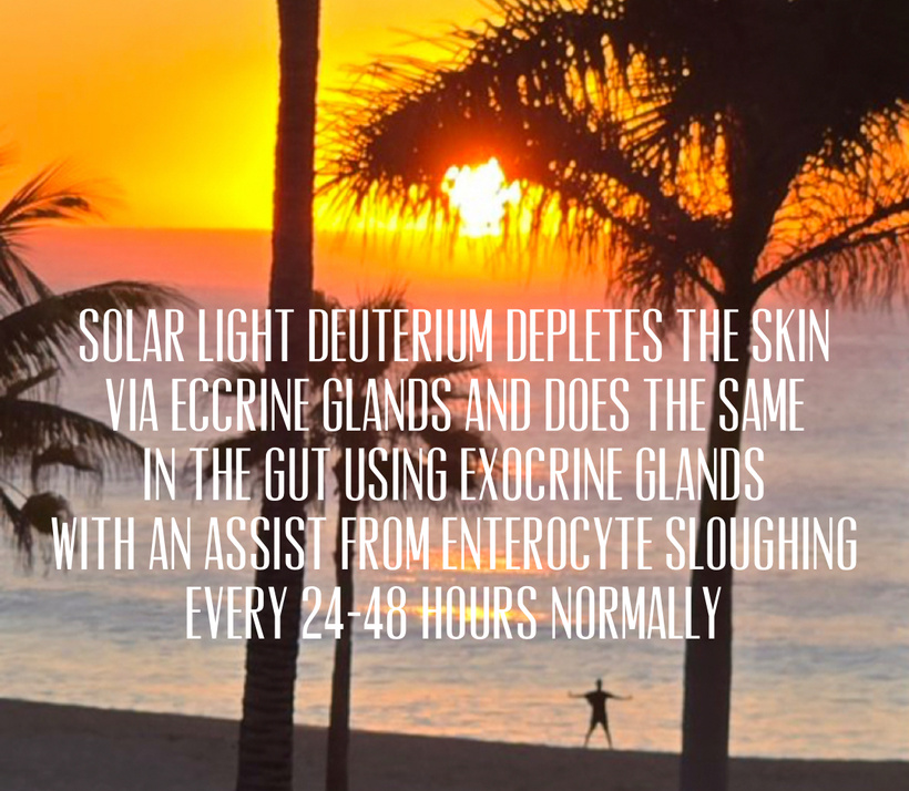
Nature has put an emphasis on DDW as an insulator for the IMM’s 30 million volts/cm electric field suggests that deuterium levels in cellular water are critical for mitochondrial health. High deuterium (from diet or environment) should disrupt this insulation, increasing electrical conductance and ROS production, leading to faster mitochondrial aging. Conversely, a diet rich in DDW (e.g., from glacial water or plants grown in low-deuterium environments) should enhance mitochondrial longevity by maintaining cristae alignment and coherent oscillations, as you described. This could be tested by comparing mitochondrial function in cells cultured with varying deuterium levels.

6. Why Is This Key for a Mitochondriac?

For a “mitochondriac in training,” the key takeaway is that sunlight, particularly its red component, is not just an energy source but a fundamental driver of mitochondrial function and evolution. The sun’s potential use of a lattice to emit light, combined with its MHD properties, suggests a deep resonance between solar physics and biology. This resonance manifests in:

Red Light and Chromophores: The dominance of red light in the solar spectrum explains why mitochondrial chromophores ( cytochrome c oxidase) evolved to absorb red/near-infrared light, optimizing energy production and minimizing oxidative stress.

Blood Plasma as a Conduit: Hemoglobin and water in blood plasma act as intermediaries, transmitting solar energy and electromagnetic signals to mitochondria, enhancing their function.

Evolutionary Biology: The conservation of lattice structures in photosynthesis and mitochondria points to a shared origin driven by the sun’s spectrum, supporting the idea that light, not food, is the foundational driver of life.

Practical Implications: A mitochondriac should prioritize morning sunlight exposure to harness red light, minimize nnEMF exposure to protect melatonin and heme proteins, and optimize cellular water (e.g., via DDW) to maintain mitochondrial health.

7. Addressing the Sleep-Wake Cycle Question

Many of you have asked why humans get sleepy when the sun sets and awaken when it rises, and how light influences this. The answer ties directly into my framework:

Red Light and Alertness: Morning sunlight, rich in red and near-infrared light, stimulates cytochrome c oxidase in retinal and brain mitochondria (e.g., in the suprachiasmatic nucleus, SCN), boosting ATP production and signaling wakefulness via increased cortisol and BDNF and suppressed melatonin. This aligns with the sun’s MHD patterns, which enhances mitochondrial function during the day. Cognition is best at noon in all zipcodes.

Blue Light and Circadian Disruption: At night, the absence of sunlight (and presence of artificial any pollarized light including blue light from screens) disrupts this cycle. Blue light suppresses melatonin production via the SCN, while nnEMF further reduces mitochondrial melatonin, impairing sleep. The fovea’s sensitivity to blue light exacerbates this, as ROS damage to retinal mitochondria reinforces the Warburg shift, reducing energy for neuronal maintenance.

Evolutionary Perspective: Diurnal animals evolved to align their activity with the sun’s red-heavy spectrum, as it optimizes mitochondrial function. The sleep-wake cycle is a direct reflection of this adaptation, ensuring that mitochondria operate efficiently during daylight and repair during darkness, when melatonin levels rise.

SUMMARY

I want to make this crystal clear. The sun Is constantly talking to your mitochondria in real time during the day. Your colony of mitochondrial needs a break from this at night to recover. The sun’s photosphere is a hexagonal lattice of super-granules (Bénard cells + magnetic flux tubes).

That lattice imprints a faint but coherent MHD modulation onto the emitted light. How? Zeeman splitting of atoms, Stokes-V polarization, 3-minute p-mode oscillations, 5-minute global modes, 27-day rotation sidebands, 11-year cycles that never stop.

Your mitochondrial cristae are folded in quasi-hexagonal lattices for the exact same thermodynamic reason (closest packing + maximal surface-to-volume for proton ejection).
The resonance is not accidental only if you believe in accidents. Nature makes no mistakes.

The sun’s transformation of matter into of light, potentially via a lattice in its photosphere—produces a red-heavy spectrum that resonates with mitochondrial chromophores, driving energy production and evolutionary processes. Blood plasma, via hemoglobin and water, acts as a conduit for this solar energy, possibly carrying MHD-encoded information that mitochondria can “read.” This framework explains why red light dominates the solar spectrum, why mitochondrial chromophores are red-absorbing, and how light stress (e.g., blue light, nnEMF) disrupts heme synthesis and shifts metabolism toward Warburg glycolysis. The fovea serves as a microcosm of these effects, highlighting the interplay between light, mitochondria, and electrical properties.

For a mitochondriac, the key is to harness the sun’s red light while minimizing nnEMF and blue light exposure, ensuring optimal mitochondrial function, heme synthesis, and melatonin production. My predictions above offer testable hypotheses to further explore this decentralized paradigm, potentially revolutionizing our understanding of biology, evolution, and health.

Gardening always has a point for an Earthling, and this is how life must exist, in the sun. As a neurosurgeon I have seen many people die at all stages of their life. Most were unafraid except the ones who felt they had not done anything worth while in their life. The fearful patients always will claw and scratch for 2-3 more years to get something right in their life before they die.

A garden, in the sunlight, when your hands and feet are connected to the soil, is one of the most amazing decentralized hospitals I know that can give those people what they want………TIME.

It is such a pleasure to help a person understanding that sinking one’s hands into the warm earth under the sun, and to feel at one’s fingertips the possibilities of the new season and the possibility of new life. The soil is the source where LENP exists for plants and photosynthesis. For a mitochondriac, there are no mistakes in gardening, just experiments for the humanblock chain of time.


CITES


A.L. Lavoisier . Traitè Elementaire de Chimie. 1789.

L.N. Vauquelin. Annale de Chimie. Vol. 29-30, Nivose, Year VII, pp. 3-26, January, 19, 1799.

G.N. Lewis. Valence (1923). Dover edition, N.Y. 1966.

H.V. Regnault. Course de Chimie. 1847.

J.J. Berzelius. Traite de Chimie. 2° Edition, 1848.

J.J. Thomson. Phil. Mag. S5, Vol.44, N° 269, October 1897, p. 293.

Encyclopedia Britannica. Micropaedia. Vol. VIII, p. 254.

Kelvin. Phil. Mag. S6. Vol.3, N°15, March 1902, p. 257.

J.J. Thomson. Phil. Mag. S6. Vol.7, N° 39, March 1904, p. 237.

H. Nagaoka. Phil. Mag. S6. Vol. 7, N° 39, March 1904, p. 445.

R.A. Monti. Theory of Relativity: a critical analysis. Phys. Essays. 9, 2, 1996, p. 238.

J.J. Thomson. Phil. Mag. S6. Vol. 19, N° 111, March 1910, p. 424; Vol. 20, N° 118, October 1910, p. 752.

R.A. Monti. The radius of the atomic nucleus. Il Lunedì della Repubblica. N°16, “L’Ermete”, p.IV, 1-14 April 1991.

E. Rutherford. Phil. Mag. S6. Vol. 21, N° 125, May 1911, p. 669.

N. Bohr. Phil. Mag. S6. Vol. 26, N° 151, July 1913, p. 1.

J.J. Thomson. Phil. Mag. S6. Vol. 26, N° 154, October 1913, p. 792.

N. Bohr. Phil. Mag. S6. Vol. 30, N° 177, September 1915, p. 394.

A.L. Parson. Smithsonian Miscellaneous Collection. Vol. 65, N° 11, Publication N° 2371, Washington, November 29, 1915, pp. 1-80.

W.D. Harkins. J. Am. Chem. Soc.. Vol. 38, February 1916, N° 2, p. 189.

J.N. Lewis. J. Am. Chem. Soc.. Vol. 38, 1916, p. 762.

H.S. Allen. Proc. Phys. Soc. London. Vol. 31, 1919, p. 49.

J.J. Thomson. Phil. Mag. S6. Vol. 37, N° 220, April 1919, p. 419.

E. Rutherford. Phil. Mag. S6. Vol. 37, N° 222, June 1919, p. 581.

W.D. Harkins. J. Am. Chem. Soc.. Vol. 42, 1920, p. 1956.

J.J. Thomson. Phil. Mag. S6. Vol. 4, N° 243, March 1921, p. 510.

A.H. Compton. J. Frankl. Inst.. Vol. 192, August 1921, p. 145

W.D. Harkins. Phil. Mag. S6. Vol. 42, N° 249, Sept.1921, p. 305.

A.C. Crehore. Phil. Mag. S6. Vol. 42, N° 250, October 1921, p. 569.

S. Goudsmit, G.E. Uhlenbeck. Zeits. f. Physik. 35, 8-9,pp. 618-625, 1926; N. Bohr. Nature, Vol. 117, N° 2938, p. 264, February 20, 1926.

E. Schroedinger. Phys. Rev.. Vol. 28, N° 6, December 1926, p. 1049.

L. Kostro. Einstein e l’etere. Proceedings of the International Conference “Galileo Back in Italy” I. Ed. Andromeda, Bologna 1988.

W.D. Harkins. W. B. Kay. Phys. Rev.. 31, June 1928, pp. 940-945.

J. Chadwick. Nature. 129, Feb.27, 1932, p. 312.

W. D. Harkins. Nature. January 7, Nov. 8 1932, p. 23.

W. Heisenberg. Zeit.f. Physik.17, 1-2, July 19, 1932, pp. 1-11.

E. Amaldi, E. Fermi, F. Rasetti. La ricerca scientifica. Serie II, Anno VIII, N° 1-2, 15-31 Luglio 1937.

C. Borghi. Il Nuovo Cimento. Serie Nona, Vol. I, N° 3, 1 Giugno 1943, p. 176, 1940; C. Borghi. Sui Principi della Fisica Nucleare. Pontificia Accademia Scientiarium, Commentationes. Anno X, Vol. X, N° 5, 1945, p. 145.

C. Giori. Private communication to R.A. Monti.

C. Borghi, C. Giori, A. Dall’Olio. CEN. Recife, Brazil. Experimental evidence on the emission of neutrons from cold hydrogen plasma. Unpublished, 1957.

C.L. Kervran. Preuves en biologie de transmutations a faible energie. Librairie Maloine S.A., Paris 1975.

C.L. Kervran. Prove in Geologia e Fisica delle Trasmutazioni a Debole Energia. Giannone, Palermo 1983.

E. Segre. Nuclei e Particelle. Zanichelli, Bologna 1986.

L.R.B. Elton. Introductory Nuclear Theory. Pitman, London 1959.

J.M. Blatt, W. S. Weisskopf. Theoretical Nuclear Physics. Wiley, N.Y.1960.

R.A. Monti. Cold Fusion and Cold Fission: Experimental Evidence for the Alpha-Extended Model of the Atom. Communication to the Second ICCF, Como, Italy 1991.

M. Sing, M.D. Saksena, W.S. Dixit, V.B. Kartha (BARC). Verification of the George Oshawa’s experiment for anomalous production of Iron from Carbon Arc in water. Fusion Technology. Vol. 26, 1994, p. 261.

R. Sundaresan, J.O’M. Bockris. Anomalous formation of Iron from Carbon as a result of arcing in water. Fusion Technology. Vol. 26, 1994, p. 261.

J.E. Champion. Explanation of observed nuclear events associated with cold fusion and similar Low Energy Nuclear Reactions. June 1994 (private edition).

R.A. Monti. Experiments in Cold Fusion and Cold Fission. Communication to ICCF-4, Maui 1993.

R.A.. Monti. Communication to ICCF-8 . Lerici, Italy May 2000.

ICCF-7 Proceedings. April 19-24 1998, pp. 264-268.

Laboratorio Nazionale per la Caratterizzazione dei rifiuti radioattivi. L.E.T. Project. 8° Report. July 1998, by the Head of the Section F.Troiani.

R.A. Monti. Low Energy Nuclear Reactions: Experimental Evidence for the Alpha Extended Model of the Atom. Journal of New Energy. Vol. 1., N° 3, 1996, pp. 131-144.

J.E. Champion. Nuclear change via chemical reactions. Modern day nuclear change. Extraordinary Science Conference. 1995 (video tapes). International Tesla Society. Colorado Springs, CO.

M. Fleischmann, S. Pons. J. Electroanal. Chem. Vol. 261, 1989, p. 301.

R.A. Monti. Reconstruction of the Periodic Table of the Elements. Seagreen N° 8, Andromeda, Bologna, Spring 1989.

H. Fox. Implications of the Bockris-Minevskii and Mizuno et al. Papers. Fusion Facts. April 1996.

R.F. Curl, R.E. Smalley. Fullerenes. Scientific American. October 1991, p. 32.

D. Hudson. Non-metallic, monoatomic forms of Transition Elements. Keely Net, PO Box 870716, Mesquite (TX), USA 75187.

Ireneo Filalete. Open Entrance to the Closed Palace of the King Phoenix (1645). Genova, Italy 1987.

Journal of New Energy. Vol. 1, N° 3, 1996. Editorial, by H. Fox.

ICCF-8. Lerici, Italy. Proceedings.

DECENTRALIZED MEDICINE #84: PARITY VIOLATION EXPLAINS HEME EVOLUTION

Does all electricity use generate radiation from the electromagnetic spectrum?

Yes, it is true. All electricity use from our power grid generates radiation, specifically extremely low-frequency (ELF) electromagnetic fields (EMF) from the movement of electric current in power lines, wiring, and devices. This radiation, which includes both electric and magnetic fields, is a form of non-ionizing radiation and is present whenever current flows, from household appliances to high-tension power lines. Absence of evidence to your senses is not absence of effect to your cells.

Sources of radiation

  • Power lines and wiring: Any electrical conductor carrying current, including overhead power lines and the wiring in your home, produces EMF.
  • Household appliances: Almost all electrical devices generate ELF-EMF when turned on, including refrigerators, vacuum cleaners, hair dryers, and computers.
  • Other electronic devices: Other products, such as televisions and some smart devices, also emit radiation.
  • Electrical current: The radiation is generated by the acceleration and deceleration of electrons as a current flows through a circuit.

Is all this radiation generated from the AC power grid polarized?

Yes, virtually all electricity use generates electromagnetic radiation (EMR).
Moreover, this man-made radiation is polarized extremely low frequency (ELF) radiation.

WHAT ABOUT DC ELECTRIC POWER?

Becker taught us that life uses a DC electric current at very low amperage. Does it produce polarized light at its field strength? The low-frequency electromagnetic fields typically associated with a standard DC power grid or everyday DC devices are generally considered unpolarized in a practical sense, as their emissions tend to vary randomly in orientation over time and space, or are simply static electric and magnetic fields. When one considers the current that Becker found in his work on limb regeneration, whichwere measured at one trillionth of one ampere, what would the effects of polarization emission in UPEs should we expect based on the laws of physics?

Based on established physics principles, a steady direct current (DC) of one trillionth of one ampere (one picoampere, or 1 pA) would not produce ANY propagating electromagnetic radiation, polarized or otherwise, in a conventional sense. This distinction is a massive FACT.

Nature is telling us something about her use of photo bioelectric currents. Do you understand what she is whispering?

All of Biology is based around a deep lack of Polarized Electromagnetic Wave Generation 

Why? Electromagnetic waves (radiation) are generated by accelerating or oscillating electric charges.

A steady DC current involves charges moving at a constant average velocity. This creates a static electric field and a static magnetic field, which are decoupled and remain localized.

Polarization is a property of transverse waves (like light or radio waves) that describes the orientation of their oscillations in a plane perpendicular to the direction of motion.

Since the picoampere DC current does not produce propagating waves, the concept of polarization is irrelevant in this context.

WHY THIS MATTERS?

What generates the DC current in eukaryotes? The hydration of heme proteins that produces DDW that hydrates melanin to lower the current to one trillionth of one ampere. This means that a loss of heme hydration or a loss of melanin would increase the chances of polarization of light in tissues.

What are the physics of this situation currently known? Reducing the current to a picoampere magnitude makes the production of polarized radiation even less likely under standard conditions. Why?

Amplitude: The magnitude of any transient (brief acceleration/deceleration) radiation that might occur when the current is first switched on or off would be incredibly small, proportional to the current itself.

Noise: Such a tiny electrical signal in a biological or experimental setting is easily overshadowed by thermal noise and background electromagnetic noise from the environment. If the DC signal used in biology was polarized it would create noise to destroy the electromagnetic signals that control circadian biology. Why? Your retinal to SCN neural circuit uses SCN astrocytes and VIP neurons contain OPN5-positive membranes with measurable ferroelectric hysteresis loops. Those loops are fixed by non polared light from the sun. Implications? This is why jet-lag recovery, shift-work damage, and seasonal affective disorder are so hard to fix quickly, because your are not just shifting a passive oscillator in the eye clock. You are fighting a solar generated unpolarized ferroelectric current, built to be light-gated. It is a 4 billion year old chiral-lipid thyristor that was literally evolved to remember seasons for an entire year using Earth’s axial tilt as the gate signal. The circadian system and the seasonal metabolic switch are the same physical device operating on different timescales in mammals.

What is an organic thyristor?
Thyristors are solid-state electronic devices that work rather like LED light diodes; once activated a thyristor will remain conducting until the current falls to zero and reverses direction, when it will turn off. They are widely used as inverters (d.c.-to-a.c. converters) and for smooth control of power in a variety of applications, for example motors and refrigerators. Sawano et al. now report the discovery of thyristor-like behavior in a conducting organic salt. Unlike conventional thyristors, this organic material exhibits thyristor-like behavior as a bulk phenomenon. http://www.nature.com/nature/journal/v437/n7058/edsumm/e050922-12.html

Your eye clock and all the membranes in your body loaded with DHA and melanin are organic thyristors. A schematic of the SCN thyristor is shown below. What do they do in a low powered DC system?

Chiral lipids play a huge central role in the functioning of mammalian cell membranes as active mechanotransducers because they allow for flexoelectric potential to make signaling bidriectionally. Piezoelectric abilities turn mechanical info into electric signals. Collagen and cell membranes are known to be piezoelectric and flexoelectric. This allows them to deal with bidirectional flow of electricity from light sources. The signal from polarized light sources is ALIEN compared to unpolarized sunlight. Your retina has more DHA and more mitochondria in it than any other part of your body.

When we compress/condense different lipids in their chiral positions, there can be an induced a “tilt” of the molecules with respect to the bilayer’s normal and produced electric current perpendicular to the tilt plane, with the chiral lipids only. This “tilt effect” can be very similar to what happens in seasons on this planet. The Earth’s axis actually determines circadian biology.

The chirality in cell membranes does the same thing for a cell. It changes as season changes. This “electric tilt” acts like the magneto-elastic waves used by butterflies and robins in magnetico-navigation with the incident light angle from the sun. This effect is due to the phase of the aperiodic liquid crystal structure of the bilayers, which under this molecular tilt becomes a ferroelectric magnetic domain phase, where the polarization of light is normal to the tilt plane. This magnetoelectric resonance coupling to light allows for a wide variety of sensory possibilities of cell membranes such as mechanoreception, magnetosensitivity, as well as in-plane proton membrane transport and related phenomena such as ATP synthesis, ion channel depolarization, and other soft molecular machine quantum effects due to kinetic isotope effects. If you understand this paragraph, you should understand now why I do not advocate artificial red light sources in molecular photorepair. Where did all this come from? HYPERLINK

It has been sitting right in front of your eyes on the forum since 2014.

Biological System as a Source: The system Becker used (living tissue) is highly complex. While biological systems have endogenous DC potentials and currents, they primarily involve ion flows within the tissue, not coherent electron beams in a vacuum that might produce specialized, strong polarized emissions under specific experimental conditions.

Therefore, a steady 1 picoampere DC current, regardless of the biological context, would not generate significant polarized electromagnetic emissions according to the standard laws of classical electromagnetism. Any such emissions would be static fields, which inherently lack polarization. Nature taught me this lesson 25 years ago. This is why trying to heal disease with polarized light source is always secondary to the sun.

These static fields do not propagate as waves through space like light or radio waves do, and therefore, they do not exhibit wave properties like polarization. Therefore, in biology the routine operation of a conventional DC power grid in cells does not produce a significant, coherent, polarized electromagnetic wave that propagates like radio waves or light. To get a DC line or power grid to create polarized light one needs specialized laboratory setups to produce polarized radiation using DC power as a component of the generation mechanism.

Polarization of the Radiation

Man-made EMR is always Polarized: Unlike most natural EMR (such as sunlight), which is unpolarized (oscillates in random orientations), the EMR from human-made electrical sources is polarized.

Fixed Orientation: Man-made EMR is produced by electrons oscillating in a specific, forced direction along wires or within circuits. This gives the electric and magnetic fields a defined, consistent plane of oscillation (linear, circular, or elliptical polarization depending on the source geometry, e.g., 3-phase power lines produce circularly/elliptically polarized fields).

Biological Implications: Some decentralized scientists and clinicians theorize that the polarized nature of man-made EMFs, rather than their intensity, plays the key factor in their potential biological effects, as this coordinated oscillation can force charged molecules within biological tissues to move in a specific, unnatural way. Health agencies such as the World Health Organization (WHO) and the National Cancer Institute (NCI) continue to study potential health effects, classifying ELF fields as “possibly carcinogenic to humans (group 2B)” based on some epidemiological studies suggesting an increased risk of childhood leukemia.

The AC powergrid turns your mitochondria into a Parity Violation factory for disease creation.

WHAT DOES THIS PHYSICS MEAN WHEN WE PUT ANY TYPE OF LENS INFRONT OF OUR RETINA?

What don’t sunglass and eye glass manufacturer’s know? Their products harm you in many ways and can lead to cancer as the slide below shows.

Polarized light and lenses are used as a tool to detect and measure the tiny effects of Parity Violation in atoms and molecules, where the weak force causes a slight “handedness” in physical systems that are otherwise mirror-symmetric.

The core link is that the weak force’s violation of parity symmetry leads to a mixing of atomic or molecular states that should, under normal circumstances (governed by the parity-conserving electromagnetic and strong forces), have a definite, opposite parity. This mixing is extremely small but results in two primary observable effects involving polarized light. It affects water networks and all proteins that interact with the polarized light.

Note: all artificial sources of light are POLARIZED.

When linearly polarized light passes through an atomic vapor or a chiral medium like the human eye, its plane of polarization rotates slightly. The amount of rotation is related to the degree of parity violation in the atoms distal to it.

Materials exposed to the weak interaction can show a minuscule difference in how they absorb left-handed versus right-handed circularly polarized light. Why is this a big deal in my human patients?

Stellar masses are designed to use your tissues as their lens. This is why DNA codes only for semiconductive proteins that all have SPECIFIC absorption and emission spectra and polarization abilities.

Parity vioation in your tissues explain life’s universal preference for L-amino acids and D-sugars, a puzzle since Pasteur’s 1848 discovery of molecular chirality.

When you wear lenses in front of your eyes or bathe in light man makes you just broke a fundamental law of Nature and it always leads to DISEASE your doctors are impotent to repair. What sits behind your eyes? Your pituitary gland and hypothalamus where POMC lives. Do you think polarized light might alter POMC biology? How about circadian signaling?

Well it appears polarized light can destroy copper pipes in NYC, so you might have a problem if you believe that non ionizing polarized light is not the biggest risk to health on Earth today..

In all life on Earth, all amino acids except glycine are chiral, and sugars like glucose are dextrorotatory. This chirality enables DNA, RNA, and proteins to encode and respond differently to polarized light, potentially driving new epigenetic changes. Mitochondria, acting as “diachronic” element for solar light (like Icelandic spar, which splits light into polarized beams), to direct stellar unpolarized light to its targets in the eye, pituitary, and hypothalamus where the SCN sits controlling circadian cycles. Are you beginning to see a problem now?

My QUILT document should be thought of as biology reformated as low-energy nuclear physics that occur in mitochondria, water, and in all proteins coded for by DNA. My entire decentralized thesis assimilates the field of atomic parity violation. NONE, and I mean NONE of your experts goes this deep to figure out where chronic disease begins. All chronic diseases are collateral changes related to low energy nuclear particle physics that occur inside of cells.

Sunlight, POMC, and the Quantum Cell: Unlocking Nature’s Code to Heal Autoimmune Diseases In a world where autoimmune diseases like rheumatoid arthritis, multiple sclerosis, and lupus afflict over 350 million people, centralized medicine often defaults to immunosuppressive drugs that manage symptoms but ignore root causes. But what if the ultimate healer is free, abundant, and as ancient as life itself: sunlight? Emerging science, rooted in quantum biology, reveals that sunlight’s interaction with pro-opiomelanocortin (POMC) biology and the quantum cell offers a decentralized path to immunity restoration. This approach challenges the temporal arrogance of modern medicine, which views drugs as the peak of advancement, by tapping into nature’s quantum design, where light, not genes, drives adaptation and healing. As Philip Ball argues in his February 2024 Nature article “It’s Time to Admit That Genes Are Not the Blueprint for Life,” the gene-centric view is oversimplified and outdated; genes lack fixed functions, operating in probabilistic networks influenced by environment and energy flow, aligning with a quantum perspective where light provides the low-entropy input to organize cellular states.

The Biophysical-to-Biochemical Feedback Loop: Light Stress and Autoimmune/Cancer Origins

Autoimmune diseases emerge when the immune system turns against the body, a process driven by a quantum breakdown in the biophysical-to-biochemical feedback loop. As I have tried to explain for 20 years, altered biophysics from “light stress” (e.g., blue light toxicity or insufficient natural sunlight) drops mitochondrial energy, fails biochemistry, shuts down p53-mediated apoptosis, and allows cancer or autoimmunity to develop.

Light Stress and Mitochondrial Energy Drop: Sunlight is life’s battery, charge-separating water to create exclusion zones (EZs) with negative electric potential, as discovered by Gerald Pollack. In autoimmune conditions, chronic exposure to non-native light (e.g., screens) disrupts this, reducing ultraweak photon emissions (UPEs) from mitochondria. UPEs, often in the UV range (200-400 nm), act as mitogenic radiation, signaling mtDNA to maintain energy coherence. Without it, mitochondrial heteroplasmy rises, dropping energy output and triggering inflammation. New papers highlight that cancer cells emit distinct biophoton profiles with a higher UV-to-IR ratio, indicating altered proton dynamics (protium vs. deuterium levels). A light stress event in the breast or its skin induces near-UV biophotons, increasing local ROS production, as UV biophotons excite oxygen molecules to form singlet oxygen or hydroxyl radicals—chronic oxidative stress driving carcinogenesis.

  • Genome Size and Energetic Barriers: Life isn’t linear; genome size scales with cell size and inversely with metabolic rate, creating an “energetic barrier” where larger genomes require more energy to maintain. In phytoplankton, larger genomes adapt better to variable energy inputs like light and temperature. Sunlight provides the energy surplus for genome expansion, during the GOE 2.4 billion years ago, photosynthesis-driven oxygen boosted ATP yield (32 ATP vs. ~2 ATP anaerobic), enabling complex genomes. In mammals, light deficiency (e.g., indoor living) reduces energy, constraining genome utilization in regulatory networks for energy homeostasis, exacerbating autoimmunity.

    Biochemistry Fails → p53 → Apoptosis Shuts Down: The p53 protein, a guardian of the genome, relies on UPEs for activation. Light stress inhibits nitric oxide (NO) production from UV light, which normally inhibits the ATPase to restore apoptosis. Parity violation—the universe’s asymmetry in particle spins—amplifies this: UV light’s right-handed photons correlate with protein emission spectra, while left-handed ones drive absorption, creating “quantum music” in cells. When disrupted, p53 fails, shutting down apoptosis and allowing autoreactive cells to proliferate—a hallmark of autoimmunity or cancer. Impaired myelination or nerve damage in the breast is relayed to the gut and colon’s ENS, caused by chronic light stress and near-UV biophotons, disrupting normal motility and immune surveillance. Slowed motility increases exposure of epithelial cells to carcinogens, while reduced immune signaling (e.g., via VDR) allows precancerous cells to proliferate. T-regulator cells must become NK cells to eliminate defective cells.

  • Cancer and Autoimmunity Develop: This loop explains why autoimmune diseases often precede cancer; both stem from entropy gain in the quantum cell. Centralized oncology treats with chemotherapy, ignoring the quantum root, while decentralized approaches optimize water as a battery using sunlight to restore apoptosis.

This feedback loop ties directly to POMC biology, where sunlight’s mandate—”Let there be light”—is a biophysical fact. Every 150 million years, the Sun loses an Earth mass via solar wind; over 4.6 billion years, just 30 Earth masses have fueled Earth’s biosphere. Complex life, from the Cambrian explosion 600 million years ago, used only six Earth masses of sunlight, sculpted by docosahexaenoic acid (DHA), a lipid innovated 50 million years before to convert light into DC photo-bioelectric signals. DHA’s conservation for 650 million years underscores light’s primacy over genes, completing Darwin’s ideas by showing conditions of existence (light, water, magnetism) drive traits, not natural selection alone.

POMC Biology: Sunlight’s Quantum Switch for Immunity

Pro-opiomelanocortin (POMC), a 241-amino-acid precursor cleaved into hormones like ACTH, α-MSH, and β-endorphin, is the linchpin of sunlight’s autoimmune healing:

Sunlight Activation: UV light stimulates POMC in the skin and hypothalamus, producing α-MSH to upregulate melanin. Melanin absorbs UV-visible light (200-700 nm), charge-separating water into electrons, H⁺, and O₂ near mtDNA, generating UPEs. This “quantum music,” parity violation in photon spins ensures energy conservation per Noether’s theorem, maintaining time symmetry in the TCA cycle for cellular coherence. Low solar exposure of the skin/eye has amplification effects due to T-cell dysfunction from low vitamin D, as the WHO 2009 study shows women with low D3 levels have massive breast cancer risk.

Immune Modulation: α-MSH, derived from POMC, has potent anti-inflammatory effects, inhibiting pro-inflammatory cytokines (IL-1, TNF-α) and promoting T-regulatory cells (T-regs), suppressing autoimmunity. In MS, sunlight-driven POMC reduces inflammatory proteins; in lupus, it repairs cellular damage. The Scientific American articles I have linked to in this series notes UV’s role in calming overactive immunity, but your thesis expands this: POMC ties to ocular melanin, ending in leptin regulation for circadian homeostasis which is disrupted in autoimmunity by light stress. Circadian disruption, like in night shift workers, limits epithelial photorepair and increases breast/colorectal cancer risk.

Quantum Effects: POMC’s non-visual photoreceptors (melanopsin/neuropsin/encephalopsin) sync with sunlight, optimizing mitochondrial cristae alignment for DHA-mediated light conversion. Parity violation enables the TCA cycle’s directional spin, clockwise for growth under UV/O2 abundance, counterclockwise for adaptation under light stress and hypoxia which favors survival over stasis.

The Living State: Release, Not Production—UPEs as the Key Factor

The living state isn’t powered by something being “made”; it’s powered by something being released, unpolarized UPEs at small scales that carry massive power due to scale reduction. This is why Genesis begins with the sentence, Let there be light. You just where never told about the recipe for this light. Sunlight controls homochirality of biomolecules. If you are living, you need L- amino acids in your tissues to work with the sun and if you are dying being on your way back to the cosmos’ energy store where higher powers reside, you will be filled with D amino acids and lipids. Mixing them in either environment is the Rx for disease and death.

Charge already wants to flow, electrons already want to fall, protons already want to drift; the only question is how much resistance sits in the way, and mitochondria are the organelles that remove just enough impedance for the universe’s default direction to finally express itself inside a cell. That’s why metabolism looks like energy production when really it’s geometry correction on the cristae of the IMM. It also explains why ATP rises, not because mitochondria “produce energy,” but because they lower the friction (electrical resistance) that stops energy from moving. This is why every disease maps to increased drag and every recovery maps to restored flow. You really weren’t built to generate power. You were built to stop blocking its flow from stars to your colony of mitochondria. None of these ideas are in Darwin’s thesis.

Stellar masses are designed to use you as their lens. This is why DNA codes only for semiconductive proteins that all have absorption and emission spectra. Once you see that and intrepret it correctly, the whole ATP-religion of Mitchell’s biochemistry collapses in one idea: you don’t run on molecules, you run on the removal of electrical resistance, and that resistance is controlled by AMO physics inside of cells. When electric resistance drops, it makes it easier for a DC current to flow, leading to higher current and increased power dissipation, which usually results in more heat release the system. This is why mitochondrial heme proteins create DDW. Without this heme protein evolution is impossible. The water created by CCO absorbs all of this heat. When water is absent due to heme protein destruction a lack of electrical resistance also implies massive potential damage to cells if the circuit is not designed to handle the increased load. Dehydration and a a lack of melanin is about that not handling that load. What is the result we can visualize when electrical resistance goes awry? We see the results in how cristae geometry changes as energy varies due to electrical resistance changes.

Inter-mitochondrial junctions (IMJ) and cristae alignment form one mitochondrial basis for complex diseases, correlating with energy demand. In high-demand tissues (e.g., heart, diaphragm), cristae align across IMJs to optimize electron flow, reducing resistance and enhancing UPE release. Light stress disrupts this geometry, increasing drag and entropy, leading to autoimmune breakdown as mitochondrial signaling fails. If it goes on too long cancer is the next step as apoptosis is completely lost. This is when deadly disease knocks on your door.

Chiral Bias in TCA Enzymes: Parity Violation and the Quantum Asymmetry in Life’s Clock

Chiral bias in TCA enzymes, driven by parity violation’s energy differences between left- and right-handed molecules, creates an asymmetry that favors clockwise NADH production under UV light for energy conservation. This scales to cellular time stamping, breaking symmetry in low UV states to reverse counterclockwise, recycling in GOE-like modes without entropy overload. Think about why ferredoxin biology predates genes in evolution on Earth, iron-sulfur cores, quantum processors, harnessed light before genetic blueprints, proving light’s primacy.

Water deuterium depletion by sunlight via eccrine (skin) and exocrine (gut) glands, with enterocyte sloughing every 24-48 hours, exemplifies this, as solar UV optimizes matrix water and melatonin pathways, involving neuropsin (380 nm), SIRT1, NAD+, NAMPT, AMPK, and CLK periodicity for photorepair and mTOR regulation.

Electron Spin Directionality: The weak nuclear force’s left-handed bias influences electron spins in beta decay. In quantum biology, this directs ETC tunneling in the TCA/urea cycles, conserving energy per Noether’s time symmetry. UV maintains spin coherence of electrons and proteons; absence of it broadens UPE spectra and polarizes this light, reversing the cycle and impairing mitochondrial signaling in diseases like ALS.

Proton Handedness in Water Networks:

Parity violation affects proton behavior in water and in all biomolecule via the weak nuclear force interactions. In quantum biology, this creates chiral water around mtDNA, enabling directional proton flows for TCA symmetry. UV light preserves this, while disruptions reverse the cycle, linking to melanin dehydration due to heme protein destruction in creating a broadened UPEs in autoimmunity and cancer. Now go back and listen to this webinar with these ideas planted in your mind first. You might really understand what Nature is up to in cells now.

mtDNA Mutation Asymmetry: Parity violation biases mtDNA interactions by using neutrinos. Ten years ago I gave this webinar above on neutrinos. Few saw the fit ten years ago. I hope you’re waking up now. In quantum biology, this asymmetry allows adaptive mutations in the TCA/urea cycle, conserving energy under time symmetry. Low UV environments breaks this symmetry, forcing ancient reversals in both of these cycles to push you back to GOE biology, all while forcing entropy buildup in all chronic diseases man faces today.

Melanin’s Paramagnetic Preference: Magnetic fields from ATP synthase are parity-influenced. In quantum biology, melanin’s paramagnetism senses this asymmetry, scavenging ROS directionally to maintain TCA/urea clockwise spin. UV and oxygen tunes hydration of the heme protein CCO for precision; blue light reverses this symmetry, broadening UPEs in chronic disease states. I believe in disease states the UPEs become polarized in tissues and this allows the wrong steroisomers to be incorpated into mitochondria and when this happens it leads to diseases like cancer.

UPE Spectral Handedness: Parity violation introduces quantum field asymmetry. In quantum biology, this biases UPE collapse in mtDNA, ensuring narrow spectra for TCA energy conservation. This is why leptin has a precise 220 nm absorption spectra. Disruptions broaden UPE spectra or its polarization, or the steroisomers placed into leptin or its receptors, reversing the cycle and accelerating aging, as in fertility issues mimicking oocyte states. Altering chirality via polarized light is why infertility, misgendering, and hormone panel destruction is ubiquitous in humanity today.

Beta Decay in Redox Signaling: Parity violation’s electron bias even affects cellular radioactivity. In quantum biology, this directs redox flows in the TCA/urea cycle, aligning with Noether’s theorem. UV prevents leaks; nnEMF reverses symmetry, causing entropy surges in cancer. This is why the results in Chernobyl exclusion zone after 40 years has stunned scientists, gamma radiation has been found to not be as deadly as science thought because melanin in mushrooms and fungi consumes it via radiosynthesis, renovating nature. The melanin renovation blog is active in a nuclear meltdown site and has proven modern biological science dead wrong about radiation hormesis.

Enzyme Conformational Asymmetry: Parity violation selects handed substrates in biochemistry. In quantum biology, this ensures efficient TCA-urea integration, conserving energy. Low UV breaks control, leading to metabolic deserts in tissues with heme protein destruction (anemia-like) and cycle reversal. It also explains why absorption and emission spectra are linked to every protein that RNA and DNA code for.

Quantum Coherence in Cycle Flows: Parity violation enables asymmetric fields in microtubules. In quantum biology, this supports coherent TCA NADH production, tied to consciousness. Disruptions in coherence broaden UPEs, reversing symmetry and impairing neural repair in many diseases. No one has yet tapped my insights in chronic disease, in time, but I will bet they will.

Epigenetic Directional Bias: Parity violation biases molecular paths for methylation. This means it controls epigenetics. In quantum biology, UV reduces methylation (anti-aging action), maintaining TCA/urea symmetry. nnEMF hypermethylates DNA/RNA, breaking time symmetry and forcing GOE-like reversals in all chronic diseases. This is why hypermethyaltion and D-isomer incorporation signals GOE like disease states.

Probabilistic Gene Expression: Energy, Information, and the RET Gene Analogy

The RET gene in man is a proto-oncogene. A proto-oncogene is a normal gene that helps regulate cell growth and division. When a proto-oncogene is mutated or abnormally expressed, it can become an oncogene, which can cause cells to grow and divide uncontrollably, potentially leading to cancer. These genes act like the “gas pedal” for a cell’s growth, and a mutation can cause the pedal to get stuck in the “on” position. UV UPEs are needed to get past the mitosis step in the cell cycle.

Genetic expression isn’t deterministic; it’s probabilistic, governed by stochastic processes like promoter binding and chromatin dynamics. But fundamentally, it’s driven by energy gradients imprinted from the environment, with ATP being the energy currency that is used for transcription and electrons for redox signaling. Expression correlates with energy/information flow, not genome anatomy. Information here is entropy-related (e.g., Shannon’s theorem in signaling fidelity), where light provides low-entropy input (coherent photons) to organize cellular states.

UPEs from mitochondria act as signals, influencing gene switches via quantum effects where light “controls the on/off switches of genes.” In low-energy states (light deficiency), probabilities shift toward dysfunction: e.g., reduced expression of repair genes, leading to RET issues as a redox mismatch signal. The RET proto-oncogene, is involved in neural development and rebuilding circuitry and autoimmune-linked cancers, exemplifies this: RET expression probability depends on mitochondrial energy (UPE-driven), where light stress disrupts redox signaling, increasing mutation risk and autoimmunity. Ball’s article below in the picture, reinforces this, noting genes lack pre-set functions and operate in probabilistic networks influenced by environment, aligning with my quantum view where light’s coherent photons reduce entropy, organizing states for adaptive expression.

Evidence from Quantum Biology and Clinical Observations

Epidemiological Data: Higher autoimmune rates in low-sunlight regions (northern latitudes) correlate with reduced POMC activation, as Scientific American notes. I have added that this reflects light stress dropping mitochondrial energy, failing p53, and shutting down apoptosis. Apoptosis loss is present in cancer because cancer cells develop various genetic and molecular alterations via UPE signaling that disable their normal, tightly regulated cell death mechanisms. This evasion of apoptosis is a fundamental hallmark of cancer, allowing damaged or abnormal cells to survive, accumulate mutations, proliferate uncontrollably, and resist centralized anticancer therapies. Solar therapies restore apoptosis by forcing T regulators cells to become NK cells that eliminate the defective cells in cancer and autoimmunity.

  • Experimental Insights: Pollack’s water studies show sunlight creates “water batteries” for energy; Montagnier’s water memory experiments demonstrate electromagnetic imprinting, supporting UPEs as epigenetic triggers. Historical work by Szent-Györgyi (1941) on semiconduction and Becker’s (1960s) Hall effect in bones validate quantum processes in immunity. Sunlight’s role in deuterium depletion via eccrine (skin) and exocrine (gut) glands, with enterocyte sloughing every 24-48 hours, exemplifies this, as solar UV optimizes matrix water and melatonin pathways, involving neuropsin (380 nm), SIRT1, NAD+, NAMPT, AMPK, and CLK periodicity for photorepair and mTOR regulation.

  • Clinical Trials: Hart’s MS trial showed UV reducing disease severity by 13%, via POMC-derived α-MSH. My protocols (e.g., Leptin Rx, Cold Thermogenesis) mimic this: morning sunlight renovates heme proteins, while cold reduces entropy, restoring apoptosis in cancer & autoimmune patients. This clears cells with defects that cause the disease.

    Gaps in Centralized Views: The Scientific American article focuses on vitamin D and T-regs but overlooks quantum mechanisms like parity violation and UPEs. My thesis fills this void: cancer & autoimmune diseases develop when light stress disrupts POMC, melanin, and the feedback loop, but sunlight restores it decentralized, optimizing water as a battery, versus centralized chemotherapy.

    The K-T event (66 million years ago) illustrates this: asteroid-induced darkness and debris increased deuterium in mtDNA, driving apoptosis to remove afflicted mitochondria. Surviving organisms favored glycolysis (high deuterium, wider biophoton spectrum), limiting complexity until sunlight returned, stimulating innovations like neuropsin and mTOR for normoxia and TCA dominance. Modern life simulates the immediate post-K-T conditions, limiting epithelial repair and fueling cancers—e.g., epithelial cancers from light stress simplifying microbiomes and damaging nerves, leading to poor sleep and inflammation. It also destroys hormone panels because heme proteins derived from ferredoxins in the matrix control cholesterol transformations to pregnenolone.

The evolution of GPCRs occured ~1.2-1.5 billion years ago, as the Sun’s luminosity increased with more UV/visible light, underscores sunlight’s mandate. Radical pair dynamics in GPCRs, sensitive to magnetic fields, link light to bioelectrical signaling, explaining the asymmetry in the photorepair process. The Sun’s mid-life UV surge right before the Cambrian Explosion was the photonic kinetic event changing life, limiting epithelial cancers and autoimmunity in ancient times but they are present at epidemic proportions today due to extreme chronic UV deficiency in humans.

Overcoming Temporal Arrogance: A Decentralized Path Forward

Modern medicine’s temporal arrogance, believing drugs represent the peak, dismisses sunlight’s quantum role. But as my theorem states, true wisdom awakens us to decentralized truths. To heal autoimmunity & cancers:

Morning Sunlight (10-20 minutes): Stimulates POMC, enhancing α-MSH and melanin for immune balance. This pushed T-regualtor cells to become NK cells to get rid of defective cells in autoimmunity and cancer.

Cold Thermogenesis: Reduces entropy, supporting meta-stability of AMO physics in cells and restoration of apoptosis by stimulating UV UPEs endogenously.

Leptin Reset: Optimizes circadian alignment, tying skin/ocular melanin translation to leptin for hormonal regulation via photons. All hormones are biomolecules powered by light.

Start small: Track sunlight exposure and note reductions in inflammation. Join communities exploring quantum health to unplug from centralized systems like support groups that are pushed by centralized medicine and Big Harma.

Never forget heme protein evolution began before genes so there is no way Darwin’s evolution happened the way were taught.

SUMMARY

Light Completes the Puzzle of Evolution.

Sunlight, through POMC and the quantum cell, unlocks nature’s code for autoimmune & cancer healing, addressing the biophysical-to-biochemical loop where light stress leads to disease. By embracing this decentralized approach, we transcend the temporal arrogance of centralized medicine, recognizing light’s mandate, “Let there be light” as the biophysical fact driving life’s quantum music. The Cambrian Explosion was sculpted by six Earth masses of the matter in sunlight and that light was captured by DHA and its associated non visual photoreceptors and aromatic amino acids and it shows light’s power; now, it’s time to let it heal us. Think about why ferredoxin biology predates genes, iron-sulfur cores, quantum processors, harnessed light before genetic blueprints, proving light’s primacy in the evolutionary game of light.

Darwin was dead wrong. Evolution was driven by light changes and the light cause heme proteins to evolve and then the party started.

From first principles, parity violation (the weak nuclear force’s asymmetry, discovered in 1956 by Wu’s experiment) introduces a tiny energy difference (~10⁻¹⁴ J/mol) between left- and right-handed chiral molecules, which was all life needed to favoring one enantiomer in prebiotic chemistry on Earth based in ferrodoxins. This fully explain life’s universal preference for L-amino acids and D-sugars, a puzzle since Pasteur’s 1848 discovery of molecular chirality.

Moreover, this asymmetry operates at the molecular scale, setting the stage for life. Parity violation, via the weak force’s left-handed electron bias in beta decay, creates a slight energetic preference for L-enantiomers in chiral molecules. This would have amplify over evolutionary time scales from the era of prebiotic soups (via autocatalytic reactions), leading to homochirality. This was a primordial prerequisite for efficient biochemistry, as mixed chirality hinders polymerization in peptides and nucleotides. Ferredoxins exemplify this: their Fe-S clusters, predating genes (>3.8 Ga), harnessed iron for electron transfer in ancient pathways like acetyl-CoA, transitioning from geochemical to biochemical redox under solar UV.

CITES

https://www.science.org/doi/10.1126/science.152.3720.363

DECENTRALIZED MEDICINE #83: HEME PROTEIN EVOLUTION 4

Why did I give you all the blogs in the past to get you to this blog? Why did I write a time stamping blog? Why did that particular stick out like a sore thumb in this series? I was trying to tickle you curiosity to get you to realize heme protein evolution began before genes evolved. That should have made you ask the following question: How does Darwin’s theory cover this? The short answer, he never covered it. Those pieces covering the gap have been sitting in the QUILT document and should have helped you REALLY see what life is really about. It is not like any picture you might have imagines because it is based on a hazy concept in physics called Parity violation. Parity violation is tied to the WEAK NUCLEAR FORCE in Nature. Parity violation, is amplified by Noether’s UV-anchored symmetry, and it ensures the TCA cycle’s quantum adaptability by causing the spinning forward for growth, backward for survival. It uses chiral proteins to control the spin direction choice. In the Melanin Renovation Rx, you should have begun to see how harnessing specific solar UV, IRA, NIR photons to restore this dance in your cells, preventing reversal and reclaiming life’s timeless rhythm.

This raises the question, Jack what the hell is Parity violation if it is this important ?

Ladies and gentlemen, imagine the universe as a cosmic composer, crafting a symphony where every note follows elegant rules, except one. In 1956, physicists Chen-Ning Yang and Tsung-Dao Lee unveiled a startling secret: the weak nuclear force, the subtle architect of radioactive decay, defies mirror symmetry.

The Cosmic Handedness: How Parity Violation Unlocks the heme proteins in mitochondria, TCA Cycle’s Quantum Secrets and Powers the Melanin Renovation Rx

Picture the universe as a masterful artist, sketching reality with invisible brushes. For eons, scientists thought this artist was ambidextrous, creating laws that looked identical whether flipped left or right, a symmetry called parity. When I learned about this twist in Nature was tied specifically to the weak nuclear force, It made me realize Mother Nature, our musician, ONLY favors one hand in making her music! This was the key piece of information buried in my Quilt document that few saw. I believe it is also found in the white plaster between to the two fingers on the Sistine Chapel.

This parity violation means certain subatomic events, like beta decay, don’t mirror perfectly in the cosmos anywhere; this is not just an Earth situation. In fact, it implies, nature has a built-in bias, a preference for “left” over “right everywhere in the cosmos.” This idea was confirmed by Chien-Shiung Wu’s cobalt-60 experiment, this asymmetry reshaped physics. What did it do for centralized biology in 1956? Nothing. No one knew the massive implications it carries for cells. I began to realize it around 2003-2005. What did I visualize about the weak nuclear force?

Imagine that this cosmic quirk began whispering through your cells using one single heme protein which was capable influencing every metabolic pathways by controling the direction of energy flow. Do you think that would matter? Consider the tricarboxylic acid (TCA) cycle, the metabolic wheel that spins energy and matter into life. It matters deeply. In the QUILT of my decentralized photobioelectric thesis, light is the symphony’s conductor, and parity violation is the subtle note that breaks symmetry to create harmony that life requires to exist.

Let’s explore how this violation fits into the TCA cycle’s rotational dance, Noether’s theorem, and the Melanin Renovation Rx, revealing why life’s engine isn’t a neutral machine but a quantum timekeeper tuned to UV light. At the heart of the thesis is Noether’s theorem: every symmetry in a physical system conserves something. I gave a whole blog on this idea but I never connected to my Quilt. Today, I am making that connection for you.

For time symmetry, the idea that laws look the same forward and backward, the conserved quantity is energy. In mammals, this symmetry is anchored in UV light, which powers mitochondrial function and cellular coherence.

When UV light is abundant (just after AM sunrise), the TCA cycle spins clockwise, generating NADH for respiration, building carbon skeletons for growth, and maintaining energy conservation. But under stress (blue light as the day ages), like low oxygen or disrupted light, it reverses counterclockwise, recycling resources in a GOE-like state, breaking time symmetry to adapt. So sunset is when solar light changes what cells become capable of.

Parity violation is the key enabler of this action around TIME: its inherent asymmetry allows the universe to distinguish directions, ensuring the TCA cycle can “choose” its spin based on environmental cues.

Without this bias, symmetries would be too rigid, and life couldn’t evolve adaptability, parity violation provides the “handedness” for quantum processes to favor survival over stasis.

WHAT WAS THE FIRST CHIRAL HEME PROTEIN THE STARTED THE PROCESS?

Most people do not realize ferredoxin biology evolved BEFORE genes did. This has huge implications for my thesis.

Ferredoxin is a chiral HEME protein. The entire protein, including the active site iron-sulfur cluster, exists in a specific three-dimensional structure that is non-superimposable on its mirror image, which is the definition of chirality.

Key evidence for the chirality of ferredoxin includes:

  • Amino Acid Composition: As a protein, ferredoxin is made up of ONLY L-amino acids, which are inherently chiral.
  • Defined Tertiary Structure: Ferredoxin folds into a specific, ordered 3D conformation (tertiary structure), which results in an overall chiral macromolecule.
  • Circular Dichroism (CD) Spectroscopy: The use of CD spectroscopy is a standard technique to study the chirality and secondary structure of proteins. CD studies on ferredoxin show characteristic spectral signals (Cotton effects), confirming that the iron-sulfur clusters reside within a chiral protein environment. The specific arrangement of the protein backbone and the surrounding amino acid side chains around the cluster makes the environment chiral.
  • Stereoselectivity in Reactions: The protein exhibits stereoselectivity and chiral recognition mandates a directional flow of energy in electron-transfer reactions with other optically active molecules, further demonstrating its chiral nature.

    What do ferredoxins do for cells?

    Ferredoxins are essential to all life because it forms the basis of all food webs on Earth.

    Ferredoxin were the first heme protein electron carrier that serve as a central heme protein hub for linking various metabolic pathways in cells across ALL DOMAINS of life. Their primary function is to transfer electrons between different enzymes to facilitate vital oxidation-reduction (redox) reactions.

    1. Photosynthesis: In plant chloroplasts and cyanobacteria, ferredoxin accepts electrons from Photosystem I and transfers them to ferredoxin-NADP++ reductase to produce NADPH, which is essential for carbon dioxide assimilation (Calvin cycle). Without this Earth has no food and no TCA cycle.
    2. Nitrogen Fixation: In certain microbes, ferredoxins provide the strong reducing power (electrons) necessary to convert atmospheric nitrogen (N2𝑁2) into bioavailable ammonia (NH3𝑁𝐻3), a process critical for life on Earth. Without this there is no urea cycle.
    3. Metabolism of Lipids, Nitrogen, and Sulfur: Ferredoxins are involved in numerous metabolic processes, including the biosynthesis of steroids, bile acids, and vitamins in mammals, as well as the assimilation of inorganic nitrogen and sulfur in plants and microbes. Without this abiotic atoms never become biotic carries of life force.
    4. Direct ALL Iron-Sulfur Cluster Assembly: In human and other eukaryotic mitochondria, specific ferredoxins (FDX1 and FDX2) transfer electrons needed for the creation and assembly of iron-sulfur clusters on the IMM, which are crucial cofactors for many other essential mitochondrial enzymes. Every heme protein on Earth has this ferredoxin design in it. The IMM is a classic example.
    5. In the Melanin Renovation Rx blog, melanin acts as a quantum dot, scavenging ROS, transforming UPEs, and sensing magnetic fields from ATP synthase. UV light “renovates” melanin, creates NO to inhibit the ATPAse, creating VUV-IR UPE light inside cells to restore time symmetry and energy conservation. Parity violation amplifies this: its left-right bias ensures chiral molecules (left-handed amino acids) and directional flows (proton tunneling) align with UV-driven energy states. The parity violation of Nature involves the transformation of light photons in UPEs.
    6. When a UPE is right handed in its spin, a chrial protein emits an exciton or UPE and this correlates with a proteins emission spectra. When the UPE spins left handed, a chiral protein absorbs the UPE. This correlates with its absorbtion spectra and seems to fit into biomolecules absorption spectra as a kinetic lever. This is how Nature creates her music inside of cells.

Using first-principle thinking, starting from fundamentals like Noether’s theorem (symmetry conserves quantities) and parity violation’s asymmetry, here I derive 10 examples of how this plays a role in quantum biology and is buried in my Quilt document:

  1. Chiral Bias in TCA Enzymes: Parity violation creates energy differences between left- and right-handed molecules. In quantum biology, this biases enzyme chirality in the TCA cycle, favoring clockwise NADH production under UV light for energy conservation. This idea scales directly to the time stamping mechanism in cells. Breaks (low UV) reverse to counterclockwise, recycling in GOE-like states, ensuring adaptability without entropy overload. This interplay extends to post-translational time stamping, conserved from cyanobacteria to mammals. Circadian rhythms, the cell-autonomous oscillators, rely on transcription-translation feedback loops (TTFLs), where positive arms (BMAL1-CLOCK) drive negative arms (PER/CRY) in a 24-hour cycle. Light and dark are the decentralized conductors, with melanin electrochemically stamping the TCA cycle in the retinohypothalamic pathway to synchronize the suprachiasmatic nucleus (SCN) and peripheral clocks. Parity violation ensures chiral biases in proteins (e.g., left-handed amino acids) favor directional flows, preventing entropy overload in how we account for time. In disrupted states, like blue light exposure, melanin’s control falters, broadening UPE spectra and reversing the TCA, leading to chronic diseases without DNA alterations, leading to diseases of lost periodicity, where telomeres and TTFLs serve as entropy ledgers.
  2. Electron Spin Directionality: The weak nuclear force’s left-handed bias influences electron spins in beta decay. In quantum biology, this directs ETC tunneling in the TCA cycle, conserving energy per Noether’s time symmetry. UV maintains spin coherence; absence broadens UPEs, reversing the cycle and impairing mitochondrial signaling in diseases like ALS.
  3. Proton Handedness in Water Networks: Parity violation affects proton behavior in weak interactions. In quantum biology, this creates chiral water around mtDNA, enabling directional proton flows for TCA symmetry. UV light preserves this, while disruptions reverse the cycle, linking to melanin dehydration and broadened UPEs in MS.
  4. mtDNA Mutation Asymmetry: Parity violation biases mtDNA interactions with neutrinos. In quantum biology, this asymmetry allows adaptive mutations in the TCA cycle, conserving energy under time symmetry. Low UV breaks this, forcing ancient reversals and entropy buildup in AD.
  5. Melanin’s Paramagnetic Preference: Magnetic fields from ATP synthase are parity-influenced. In quantum biology, melanin’s paramagnetism senses this asymmetry, scavenging ROS directionally to maintain TCA clockwise spin. UV tunes hydration for precision; blue light reverses symmetry, broadening UPEs in BPD.
  6. UPE Spectral Handedness: Parity violation introduces quantum field asymmetry. In quantum biology, this biases UPE collapse in mtDNA, ensuring narrow spectra for TCA energy conservation. This is why leptin has a precise 220 nm absorbtion spectra. Disruptions broaden UPE spectra, reversing the cycle and accelerating aging, as in fertility issues mimicking oocyte states. This is why infertility, misgendering, and hormone panel destruction is ubiquitous in humanity today.
  7. Beta Decay in Redox Signaling: Parity violation’s electron bias affects cellular radioactivity. In quantum biology, this directs redox flows in the TCA cycle, aligning with Noether’s theorem. UV prevents leaks; nnEMF reverses symmetry, causing entropy surges in cancer. This is why the results in Chernobyl exclusion zone after 40 years has stunned scientists. Gamma radiation is not as deadly as science thought because melanin in mushrooms and in fungi consume it via radiosynethsis and make use of it to renovate Nature. The melanin renovation blog is active in a nuclear meltdown site in Russia and has proven modern biological science dead wrong about radiation hormesis. Life harnesses energy directly using melanin as its transducer from gamma rays and beta decay. Gamma rays often accompany beta decay because the daughter nucleus is frequently left in an excited, high-energy state after the initial beta emission. The nucleus then releases this excess energy by emitting a gamma ray photon (gamma decay) to transition to a more stable, lower-energy (ground) state. Melanin can capture this easily because of its “anti-kasha rules.” Here is a breakdown of the relationship:

    Beta Decay First: An unstable parent nucleus undergoes beta decay (either β−𝛽− or β+𝛽+ emission), changing the ratio of protons and neutrons and forming a new element (the daughter nucleus).

    Excited State: The daughter nucleus produced by the beta decay may not be in its most stable energy configuration; it is in an excited state, often denoted with an asterisk (e.g., Ni-60*).

    Gamma Emission Follows: To achieve a stable ground state, the excited daughter nucleus releases the surplus energy in the form of high-energy electromagnetic radiation, which is a gamma ray.

    Not Always: Gamma emission does not happen in all beta decays. In some cases, the beta decay process directly leads to the ground state of the daughter nucleus, and no gamma rays are produced (the decay of tritium or Strontium-90).

    In essence, beta decay is the fundamental transformation of the nucleus (changing element identity), while the subsequent gamma emission is a mechanism for the resulting nucleus to shed excess energy and achieve stability.

  8.  Enzyme Conformational Asymmetry: Parity violation selects handed substrates. In quantum biology, this ensures efficient TCA-urea integration, conserving energy. Low UV breaks control, leading to metabolic deserts (anemia-like) and cycle reversal. It also explains why absorbtion and emission spectra are linked to every protein that RNA and DNA code for.
  9. Quantum Coherence in Cycle Flows: Parity violation enables asymmetric fields in microtubules. In quantum biology, this supports coherent TCA NADH production, tied to consciousness. Disruptions broaden UPEs, reversing symmetry and impairing neural repair in ALS. No one has yet tapped my insights in this disease. In time, they will.
  10. Epigenetic Directional Bias: Parity violation biases molecular paths for methylation. In quantum biology, UV reduces methylation (anti-aging), maintaining TCA symmetry. nnEMF hypermethylates, breaking time symmetry and forcing GOE-like reversals in diseases like MS.

SUMMARY

In the intricate tapestry of quantum biology, where Noether’s theorem elegantly ties time symmetry to energy conservation, one might parenthesize the role of parity violation as a subtle yet profound asymmetry. I believe Parity violation is a cosmic bias of Nature favoring left over right in weak nuclear forces. This bias enabled the TCA cycle’s directional spin, clockwise for growth under UV light’s embrace, or counterclockwise spin in low-oxygen states mimicking the GOE. Thus allowing mitochondria to act as adaptive time machines, all while melanin, the paramount ROS scavenger, ensured the ability to transform precise UPE transformations to sustain cellular coherence to make complex life possible

CITES

https://www.patreon.com/posts/melanin-rx-for-80782201

https://www.patreon.com/posts/quantum-71-life-80126010

https://www.patreon.com/posts/qt-23-why-is-22499291

https://www.patreon.com/posts/quantum-thermo-3-17112818

https://www.patreon.com/posts/quantum-52-meet-86940332

https://www.patreon.com/posts/quantum-55-time-89683362

DECENTRALIZED MEDICINE #82: HEME PROTEIN EVOLUTION PART 3

The Cosmic Dance of Iron: A Story of Life, Stars, and Evolution
Imagine a universe where the same forces that forge stars also shape the delicate biology of life on Earth. At the heart of this cosmic tale is iron, a humble element with an outsized role in both the fiery cores of dying stars and the pulsing vitality of living cells. To understand iron’s place in biology and evolution, let’s embark on a journey that weaves together the physics of stars, the chemistry of life, and the challenges of our modern world, a story crafted for those curious about the intricate dance of nature.

Act 1: Iron in the Stars
Long before Earth existed, stars were born, lived, and died in a spectacular cycle. Stars begin their lives fusing hydrogen into helium, releasing the radiant energy we see as sunlight. As a star ages and exhausts its hydrogen, it turns to heavier elements, helium fuses into carbon, then neon, oxygen, silicon, and finally iron. Iron is the endgame for a star. Its dense, stable nucleus resists further fusion, halting the star’s energy production. As iron accumulates in the core, the star collapses under its own gravity, triggering a supernova, a cataclysmic explosion that scatters iron and other elements across the cosmos.

This stellar lifecycle mirrors a profound truth: iron marks both the death of a star and the birth of new possibilities. The iron forged in those ancient explosions seeded the universe, eventually finding its way into the rocks of a young Earth and the biology of the life that would emerge. Iron, then, is a bridge between the cosmic and the cellular, a fractal pattern connecting the grand scale of the universe to the microscopic machinery of life.

Act 2: Iron and the Dawn of Life
Fast-forward to Earth, 2.4 billion years ago, during the Great Oxygenation Event (GOE). The planet was a very different place; oxygen was scarce, and life was simple, anaerobic, and bathed in sunlight. Then, cyanobacteria began harnessing sunlight for photosynthesis, releasing oxygen as a byproduct of this process. This “oxygen holocaust” was a crisis for early life, as oxygen’s reactive nature was toxic to many organisms. Yet, from this chaos, a new order emerged. Survivors evolved to use oxygen’s electrical properties, and iron became their key ally.

Iron’s ability to switch between oxidation states (Fe²⁺ to Fe³⁺) made it indispensable. Oxygen, the only paramagnetic elemental gas, is drawn to magnetic fields and alters electrical resistance when it binds to iron-containing proteins like hemoglobin or cytochrome c. This “paramagnetic switch” allowed life to harness oxygen for energy production in mitochondria, the powerhouses of eukaryotic cells. The tricarboxylic acid (TCA) cycle, with oxygen as its final electron acceptor, became the engine of complex life, fueling everything from single-celled organisms to the human brain, which consumes 20% of our body’s energy to drive its “Ferrari engine.”

Iron’s role didn’t stop at energy. It became a cofactor in enzymes that synthesize neurotransmitters, ensuring that our brains could communicate effectively. It is embedded in heme proteins, such as hemoglobin, to transport oxygen through our blood. However, iron’s power comes with a catch: it reacts with oxygen to produce reactive oxygen species (ROS), such as hydroxyl radicals, which can damage cells through processes like lipid peroxidation. Life evolved to balance the benefits and risks of iron, storing it safely in proteins like ferritin and relying on sunlight’s red and ultraviolet frequencies to optimize mitochondrial function and protect against oxidative stress.

Act 3: Iron in the Human Brain
In humans, iron is a double-edged sword, especially in the brain. It’s essential for energy production in mitochondria and for synthesizing neurotransmitters like dopamine, which governs movement and reward. Iron accumulates naturally in the brain as we age, particularly in regions like the basal ganglia, globus pallidus, and substantia nigra. These areas, rich in gray matter, hold two to four times more iron than white matter, where myelin insulates nerve fibers.

But when iron accumulates excessively, trouble brews. In neurodegenerative diseases such as Parkinson’s, Alzheimer’s, and Friedreich’s ataxia, iron overload in neurons triggers oxidative stress, resulting in the production of reactive oxygen species (ROS) that damage lipids, proteins, and DNA. This process, known as ferroptosis, is particularly devastating in the retina, where photoreceptors and intrinsically photosensitive retinal ganglion cells (ipRGCs) are particularly vulnerable. These cells, which detect light to regulate our circadian rhythms, rely on iron-containing proteins and melanin, a pigment that chelates iron to protect against oxidative damage. When this balance falters, ferroptosis destroys neurons, disrupting circadian signaling and contributing to diseases such as Parkinson’s, where the loss of dopamine-producing neurons in the substantia nigra is linked to iron overload and reactive oxygen species (ROS).

Why does iron accumulate in sick or dying neurons? The answer echoes what is found in dying stars. Just as a star amasses iron in its core as it runs out of energy, neurons hoard iron when their energy production falters, often due to mitochondrial dysfunction. In both cases, iron signals a system on the brink, teetering between stability and collapse.

Act 4: Iron, Light, and the Modern World

Now, let’s bring this story to the present. Iron’s role in biology evolved under the influence of natural sunlight, with its balanced spectrum of red, ultraviolet, and blue light. Morning sunlight, rich in red and UV, optimizes mitochondrial function and protects against oxygen toxicity. But modern life has disrupted this ancient rhythm. Artificial blue light from screens and LEDs, which dominates our evenings, photoexcites iron in heme proteins and melanin, shifting iron from Fe²⁺ to Fe³⁺. This generates local magnetic field changes and amplifies ROS production, driving ferroptosis in neurons and photoreceptors. It also does something else. It breaks a fundamental law of physics as you’ll soon see.

In the retina, blue light-induced iron overload impairs rods, cones, and ipRGCs, disrupting circadian rhythms and the myelination process, the process that insulates nerve fibers. This connects to broader neurological damage, as seen in Parkinson’s, where neuromelanin’s protective iron-chelating role turns pathological under blue light stress. The link between Parkinson’s and melanoma, a skin cancer associated with melanin dysfunction, further highlights how iron and light interact across systems, with blue light increasing reactive oxygen species (ROS) in both the brain and skin.

Even in the oceans, iron plays a protective role for life when you look for the evidence. Marine life, from plankton to fish, relies on iron to support the food chain. Some hypothesize that iron in seawater absorbs excessive electromagnetic fields (EMF) from human technologies, shielding marine ecosystems much like iron in a star’s core absorbs energy before its collapse. This parallel suggests that iron’s role in biology is deeply tied to electromagnetic forces, a connection we’re only beginning to understand. It turns out the reason why iron is so important ties the weak force in the cosmos. The cosmos weak force why homochirality evolved as it did on this planet.

Act 5: Iron’s actions in Evolution

Blue light (400–500 nm) is not a passive environmental cue; it is the modern chiral stress agent that directly erodes local parity violation (PV) asymmetry in melanated tissues by exploiting the melanin–iron complex.

Blue Light → Fe³⁺ → Fe²⁺ Redox Flip
Blue photons photoexcite the porphyrin-like centres in melanin and neighboring heme proteins, driving a one-electron reduction of ferric (Fe³⁺) to ferrous (Fe²⁺) iron. This liberates nitric oxide (NO) from nitrosyl complexes and creates a localized hypoxic signal identical to the one used in stem-cell niches. This allows iron to carry oxygen later in the GOE.

Melanin–Iron Synergy Amplifies ROS and Racemization
Melanin is never iron-free in vivo. Iron-saturated eumelanin exhibits dramatically broadened near-IR absorption and a shifted transient absorption spectrum (BJSTR 2024). When blue light strikes this complex. What happens when this occurs?

Superoxide (O₂⁻) and H₂O₂ production skyrockets via cyclic one-electron transfer. Should I remind here that catalase quenches superoxide and it too is a heme protein? You see how life was organized by the GOE now?

Mitochondrial ROS is not the villain; it is the ancient GOE-evolved redox signalling currency. The real danger has always been uncontrolled Fenton chemistry driven by free or poorly ligated Fe²⁺, which converts benign H₂O₂ into DNA-shredding •OH radicals. Evolution spent 2.4 billion years of the GOE building an exquisite system to prevent exactly this, and modern light/environmental mismatches are dismantling it piece by piece.

Act 6: Iron’s Lesson for Humanity

ALAS1 resides in the mitochondrial matrix for a reason. It is the light-controlled iron gate.
When sunrise red/IR hits the retina → melanopsin → SCN → hepatic sympathetic axis → ALAS1 transcription peaks at ZT10 → heme peaks ZT12 → Rev-Erbα represses gluconeogenesis and sequesters iron safely into heme. This is why sunlight reduces glucose so well. It also explains why all forms of diabetes are light diseases.

Iron’s story is one of balance between energy and destruction, creation and collapse. In stars, iron marks the end of a lifecycle, scattering elements to birth new worlds. In life, iron powers our cells, but it demands careful regulation to prevent oxidative damage. The Great Oxygenation Event taught life to harness iron and oxygen under the guidance of sunlight, but modern humans have strayed from this balance. Our reliance on artificial light and EMF disrupts the delicate balance of iron, contributing to a chronic disease epidemic.

For those learning about biology and evolution, iron offers a profound lesson: life is a fractal of the cosmos, governed by the same forces that shape stars. To thrive, we must respect these ancient rhythms, embracing natural light, minimizing artificial nnEMF, and supporting our mitochondria. Iron, the element that links stars to neurons, reminds us that we are not separate from the universe but a continuation of its story.

Key Takeaways for Learners

Iron’s Cosmic Origin: Forged in dying stars, iron seeded Earth and became essential for life.
Iron in Biology: It powers energy production in mitochondria and neurotransmitter synthesis, but can generate toxic ROS if unregulated.

Mitochondrial heme protein containing iron like CCO are literally the GOE survival technology: an oxygen-handling photo-bioelectrical machine that doubled as the executioner when oxygen got out of control in cells.

Light and Iron: Natural sunlight balances iron’s oxidative states, whereas artificial blue light disrupts this balance, potentially driving all chronic diseases.

Evolution’s Lesson: The Great Oxygenation Event shows how life adapted to oxygen and iron, a balance modern humans must rediscover to combat chronic disease.

By understanding iron’s role, we glimpse the unity of life and the cosmos, a story written in stardust, sustained by sunlight, and now at risk in our electrified world.

SUMMARY

The role of Iron should be thought of as a universal energy mediator of energy flow.
Iron’s ability to switch between oxidation states (Fe²⁺ to Fe³⁺) makes it a cornerstone of energy transfer across scales:

In Stars: Iron accumulation in a star’s core signals its end. As fusion slows, iron absorbs energy, emitting chaotic EMF before a supernova—a process driven by energy loss.

On Mars: Mars’ core cooled 4.1 billion years ago, halting its magnetic dynamo. Solar wind stripped its atmosphere, leaving iron oxides on the surface, a testament to it history of energy depletion.

In Life on Earth: During the GOE, cyanobacteria used iron to harness oxygen, a paramagnetic gas, for energy production in mitochondria. Iron in heme proteins (e.g., hemoglobin, cytochrome C oxidase) facilitates oxygen transport and electron transfer in the tricarboxylic & urea cycles.

However, iron’s reactivity produces reactive oxygen species (ROS), which emit UPEs; biophotons that carry quantum information. This was the biggest development in evolutionary history ON EARTH because it set the stage for eukaryotic evolution to occur soon after heme proteins evolved on Earth. Every single node in the ferroptosis pathway is a heme-containing, red/UV-absorbing, spin-selective protein that evolved under the GOE’s oxygen + UV-A pressures. The heliosphere-Earth-life system is a fractal energy transfer network and the last step in evolution of heme protein protection schema for oxygen was ferroptosis.

Disrupting one level (e.g., magnetic field or electric fields of the sun or dark) cascades to others, draining energy and driving systemic collapse.

Trees, humans, and marine life show stress via altered UPEs release, that leads to altered methylation patterns, and oxygen utilization. In humans, this manifests as leptin resistance, circadian disruption, and chronic diseases.

DARPA’s use of light to communicate, or its venomous bioweapon injection program, EPA’s geoengineering, and FCC’s EMF policies exacerbate this energy drain, mimicking Mars’ historical fate 4.1 billion years ago.

If you want your tissues to avoid Mars’ fate, humanity must realign with natural energy cycles built during the GOE. What are they?

1. Minimize nnEMF: Reduce exposure to artificial EMF to restore UPE fidelity and mitochondrial coherence.

  1. Harness Sunlight: Use natural sunlight (250–3100 nm) to optimize mitochondrial function and leptin signaling via the efferent loop of light.
  2. Protect the Magnetosphere: By addressing solar weakening and cosmic radiation through your own personal awareness of the laws of physics that determines how iron really operates in your body. It is not what you’ve been told.

The heliosphere, Earth, and life are wirelessly connected through a fractal energy system. Iron biology, is a key mediator of energy and information, underscores the congruence between a dying star, a dead planet, and a diseased human. By understanding and respecting these cycles, we can harness the same forces that forged stars to sustain life on Earth. The information driving this was no a planet, it was the interaction of the electromagnetic and weak forces in the cosmos.

The more we bathe cells in man-made electromagnetic fields (light), the more we force them to re-enact the Great Oxygenation Event, but without 2 billion years to evolve a proper defense mechanism. That was the take home lesson of this slide in Vermont 2017.

The mitochondrial matrix is the original iron prison built during the GOE to prevent oxygen + iron from annihilating life.

It later added protection schemata using melanin because of how melanin and iron weave together.
Blue light and nnEMF are the modern jailbreakers of the entire system.
Red light, DDW, and proper circadian timing are the ancient wardens keeping iron working to protect us from oxygen toxicity.

CITES

  • Sekiguchi, M., Hayakawa, M., et al. (2006). Evidence on a link between the intensity of Schumann resonance and global surface temperature. Ann. Geophys.
  • Global Research. (2019). The Weakening of Earth’s Magnetic Field Has Greatly Accelerated.
  • INRAE Study. Frontiers in Ecology and the Environment. Trees as early warning systems for volcanic eruptions.
  • Toilekis, Z. PhD Thesis. Structure of leptin receptor related with obesity.
  • Helliwell, R. A. (1975). Power line effects on the magnetosphere.
  • Kazemi et al. (2013); Trewavas (2014); Deshayes et al. (2024). Studies on biophoton coherence in trees and humans.

DECENTRALIZED MEDICINE #81: PART TWO THE EVOLUTION OF HEME PROTEINS

The Heliosphere’s Photoelectrical Connection to Earth

The heliosphere, a plasma envelope surrounding the Solar System, transmits the Sun’s electric influence to Earth via Birkeland currents and photo-electric fields, directly impacting Earth’s magnetosphere, ionosphere, and geophysical systems.

Magnetospheric Coupling: Birkeland’s terrella experiments (1890s) demonstrated that electric currents from the Sun interact with Earth’s magnetic field, producing auroras. Modern satellite measurements (Egeland & Burke, 2010) confirm that Birkeland currents channel solar energy into Earth’s magnetosphere, modulating its strength. My photobio-electric thesis suggests that any weakening solar electric output, by any mechanism, as seen in recent solar minimums (e.g., 2025 coronal holes), reduces the magnetosphere’s protective capacity, leading to a 5% per decade decline in Earth’s magnetic field (SWARM satellite data, 2019).

Schumann Resonance as a Photo-electric Mediator: The Schumann Resonance (SR), a set of electromagnetic frequencies (peaking at 7.83 Hz) in Earth’s ionosphere, arises from global lightning discharges but is modulated by solar activity via Birkeland currents. My thesis posits that SR shifts reflect changes in the Sun’s photo-electric field strength. That information is transferred to all living things on Earth. Recent increases in SR higher frequency banding correlate with global temperature rises (Sekiguchi & Hayakawa, 2006), indicating a weakening magnetic moment in atoms is coupled to enhanced energy transfer from Earth’s core to the surface.

Core Dynamics and Heat Transfer: The Sun’s electric field induces currents in Earth’s iron-rich core, driving the geodynamo that generates the magnetic field. A diminished solar photoelectric input from the sun slows the geodynamo, reducing Earth’s rotation and increasing seismic/volcanic activity. This core heat, amplified by electromagnetic interactions, acts to warms the oceans, accelerating currents by 15% over the past two decades, which has been observed in oceanographic data over the last 50 years.

Quantum Biological Impacts: Iron, UPEs, and Circadian Systems

My model’s photo-bioelectric influence extends to Earth’s biosphere through electromagnetic and biophotonic interactions, with iron and water serving as key mediators of energy transfer from our star.

Iron’s Key Role in Energy Transfer: Iron, becomes central to the surface transmutation on a planet. Mars shows its iron scars on it surface. Evolution never began on Mars and was stunted by the interaction of Birkeland currents and iron on Mars leading to a fabulous explosion that left a massive injury on Mars equator. The “massive injury on Mars equator” I am referring to is a large geological feature called Valles Marineris. This is an extensive canyon system carved out of the planets surfuce near the Martian equator. This is why Mars has major isotopic anomolies compared to Earth. Did you know Martian water (found in minerals and the atmosphere) is enriched in deuterium by about five to seven timescompared to Earth’s oceans? The high D/H ratio is strong evidence for the substantial loss of a primordial water ocean on Mars over billions of years. The lighter H+ isotope escaped the planet’s weak gravity and thin atmosphere into space much more readily than the heavier D isotope. This also explains why Mars surface is littered with massive level of iron oxide on its surface.

Martian materials, particularly carbonates and minerals found in meteorites, show a distinct mass-independent fractionation (MIF) of oxygen isotopes. Earth favors the common Oxygen 18 while Mars favors the rare oxygen 17 isotope. Methane released from some sediment samples in Gale Crater shows extremely depleted values of the heavier Carbon 13 isotope compared to Earth. The Martian atmosphere is depleted in lighter isotopes of noble gases like neon compared to Xenon. This fractionation is caused by the ongoing escape of the Martian atmosphere to space, where lighter isotopes are lost more quickly than heavier ones. These anomalies provide critical fingerprints for understanding the history of Mars’ atmosphere, the presence and loss of past water, and the planet’s formation history.

Mars also has another interesting anomoly. The Northern and Southern hemisphere are incongruent with each other geologically.

Centralized science poses two theories to explain the abnormality. Their causes of the hemispheric asymmetry are a giant impact hypothesis and/or the mantle convection hypothesis. Neither of them make sense to me. Consense is pseudoscientific. Science is about asking difficult questions and having sloppy answers that need new data from people asking more questions about the anomoly.

Under my photobioelectric hypothesis, planets interact variably with solar Birkeland currents based on orbital distance, magnetic shielding, and atmospheric density. Mars, being farther from the Sun and with minimal atmospheric buffering, would have experienced more intense or prolonged discharge events, promoting redox-driven fractionation of multivalent iron (e.g., via oxidation states shifting in plasma environments, where Fe²⁺/Fe³⁺ ratios would have certainly influence isotopic partitioning). This explain Mars’ undepleted iron content and chondritic δ57Fe concentrations, contrasting it with Earth’s volatile depletion at lower temperatures (~1300 K), which should be reinterpreted as electrical stripping rather than purely thermal nebular processes. SAFIRE’s observations made in the lab in the last decade of plasma-stabilized exothermic reactions supports this, as discharges could selectively volatilize or transmute lighter isotopes, enriching Earth’s mantle in heavier ones while leaving Mars relatively pristine.

EARTH AND IRON

From the first principles using my photo-bioelectric hypothesis, where the Sun functions as an anode in a galactic-scale electrical circuit, channeling energy through Birkeland currents and plasma discharges that can induce nuclear transmutations and isotopic fractionation as demonstrated in SAFIRE experiments—the provided data on ferredoxin and its evolutionary links to heme proteins can be integrated into the explanation of iron isotopic variations between Mars and Earth. This framework posits that solar plasma interactions not only shaped planetary iron reservoirs but also drove the quantum-level evolution of iron-based biomolecules on Earth. This enabled the transition from geochemical reactions at deep ocean vents to biochemical electron transfer. in primitive cells. Below, I outline how this fits, emphasizing the role of iron in facilitating life’s adaptation to electromagnetic (EM) forces, particularly during key events like the Great Oxygenation Event (GOE), while contrasting Earth’s biologically amplified isotopic shifts with Mars’ more primitive, chondritic signature.

Plasma-Driven Formation of Fe-S Clusters Were the Precursors to Heme Proteins: SAFIRE experiments show that plasma discharges in hydrogen-rich environments can transmute elements and form complex metallic structures, including iron-sulfur (Fe-S) clusters, through low-energy nuclear reactions (LENR) involving proton capture and nucleon rearrangement. In my photo-bio-electric thesis, Birkeland currents from the Sun would have delivered charged particles (protons, electrons, ions) to early Earth’s surface and atmosphere, catalyzing non-enzymatic Fe-S cluster assembly in primordial hydrothermal vents or mineral surfaces.

These conditions mimicking SAFIRE’s anode probes exposed to high-voltage gradients. Ferredoxins, with their simple [2Fe-2S] or [4Fe-4S] clusters, represent molecular fossils of this process: their inorganic active sites likely originated from geochemically formed Fe-S minerals, transitioning seamlessly into biological electron carriers found in modern mitochondria.

This sets the stage for heme protein evolution, as Fe-S clusters are structural and functional precursors to heme (porphyrin-bound iron) in proteins like cytochrome c oxidase (CCO). On Earth, repeated plasma exposures during solar activity cycles could have favored isotopic enrichment in heavier iron (δ57 Fe > 0‰) within these clusters, as electromagnetic isotope separation (EMIS) in Birkeland currents preferentially deposits heavier isotopes (larger gyroradii leading to differential acceleration). Mars, with its weaker magnetic shielding and less atmospheric interaction, retained chondritic δ57 Fe ≈ 0‰, limiting such biochemical evolution, which potentially explains why Martian meteorites show NO EVIDENCE of advanced heme-like structures, while Earth’s heavier iron isotopes correlate with the emergence of oxygen-utilizing heme proteins post-GOE.

Iron became critical for life because it is central to quantum evolution on Earth. During the Great Oxygenation Event (GOE, 2.4 billion years ago), iron enabled oxygen-based energy production in mitochondria by facilitating electron transfer in the tricarboxylic acid (TCA) cycle. The Sun’s photobio-electric output, particularly UV light (200–3100 nm), excites iron-containing proteins like cytochrome c oxidase (CCO), which produces deuterium-depleted water (DDW) essential for mitochondrial efficiency. One of the more interesting things is that CCO is also the first step in the death signal in cells. This correlates with stars who become supernova when the stellar core is mostly iron and interacts with EMFs to initiate the explosive force of the star.

Electron Transfer in Photosynthesis and Respiration as Extensions of Solar Discharge Dynamics:

My thesis posits that solar energy arrives not just as photons but as photo-bioelectrically driven plasma, with UV and broader EM spectra (200–3100 nm) exciting iron atoms in proteins. Ferredoxin’s role in cyclic photophosphorylation, the accepting of electrons from photosystem I (PSI) to drive ATP synthesis via NADP+ reduction or cyclic flow back to the cytochrome b6f complex, completely mirrors SAFIRE’s lab observation of plasma-sustained electron cascades that stabilize exothermic reactions.

In primordial Earth, solar Birkeland discharges would have directly energized raw iron minerals for electron transfer abilities, predating enzymatic systems; ferredoxin’s low redox potential (−420 mV) and stromal localization in chloroplasts evolved to harness this, incorporating into the acetyl-CoA pathway for carbon fixation >3.8 billion years ago. Nick Lane’s slide above is now fully explained.

My idea predates the evolution of mitochondrial respiration, where ferredoxins shuttle electrons in the primitive TCA cycle 4 billion years ago, linking to abiotic heme proteins like ferredoxins to modern heme proteins like CCO that use iron to reduce oxygen to water (producing deuterium-depleted water, DDW). The quantum aspect, iron’s high-spin states (Fe²⁺/Fe³⁺) enabling spin-forbidden transitions, therefore ties to UPEs (ultraweak photon emissions) from ROS, which I have said in this thesis encodes information. (below) This is how the electric membranes give feedback or feelings on the status quo in the environment to alter the cells physics on Earth.

Within my thesis, the Sun’s galactic currents modulate solar UV output, influencing UPE fidelity and leptin signaling in living things on Earth. Earth’s magnetosphere amplified these interactions, selecting for heavier iron isotopes in heme evolution during GOE on Earth (2.4 Ga), as plasma fractionation enriched δ57Fe in basalts and mantles, facilitating efficient electron transfer in oxygen-rich environments. Mars’ thinner atmosphere and lack of global field meant less EM-driven selection, preserving chondritic iron unsuitable for complex heme-based respiration, akin to undifferentiated chondrites.

Evolutionary Duplication and Quantum Selection via EM Interactions

Sequence analysis indicates ferredoxins evolved from short ancestral peptides (e.g., 8 amino acids like alanine, aspartic acid, proline, serine, glycine) through duplication, before the full genetic code. From first principles, SAFIRE-like plasma environments induce structural changes (melting/recrystallization) and transmutations that would have promoted peptide polymerization via electric fields aligning dipoles. Birkeland currents, carrying isotopically fractionated iron, would have embedded heavier δ57Fe into these early peptides on Earth, where stronger solar-planetary coupling (via heliospheric currents) drove a QUANTUM evolution using the natural selection of light’s physics because systems with heavier iron isotopes exhibited better quantum coherence in electron transfer, resisting spin decoherence from EM fluctuations.

This “quantum evolution” centralized iron in heme proteins, as in CCO’s role in mitochondrial energy and apoptosis (death signaling), paralleling stellar supernovae where iron cores interact with EMFs to trigger explosions, suggesting a universal plasma-driven threshold for iron-mediated instability. On Mars, minimal discharge exposure left iron isotopes unfractionated, halting evolution at primitive Fe-S levels without heme sophistication. The correlation with Fe/Mn and silicon isotopes supports this view with hardcore irrefutable data: Electromagnetic Isotope Separator (EMIS) co-fractionates elements, with Earth’s volatile depletion (interpreted as electrical stripping via the solar wind) enriching heavier isotopes on Earth for future biological use.

Integration with Circadian and Biophotonic Disruptions

Under my thesis, solar photo-bioelectric fields would act to synchronizing circadian rhythms via SCN and vagus nerve resonates with the Sun’s heliospheric transmission of EM signals, including Schumann resonances (SR). Ferredoxins and heme proteins were the first proteins on Earth that would have been capable of respond to these, with UV-excited iron producing UPEs essential for metabolic coherence; weakening solar fields (2025 coronal holes) impair this, leading to leptin resistance and heteroplasmy.

In evolutionary terms, Earth’s exposure to variable Birkeland discharges selected for robust iron-based systems, evolving from ferredoxin’s photosynthetic roots to mitochondrial heme, enabling adaptation to GOE’s rising oxygenation tensions from zero % to 21%. nnEMF disruptions (from human sources) mimic reduced solar input, decoupling proton-electron flows as in SAFIRE’s voltage breakdowns. Mars’ isotopic stasis potentially reflects limited EM engagement at a planetary level which prevented the same quantum-tuned evolution that occured on Earth 3.8 billion years ago.

Overall, my thesis reinforces that Earth’s heavier iron isotopes was caused by fractionation of by solar plasma discharges 4 billion years ago. Moreover, I believe they were pivotal for heme protein evolution from ferredoxin precursors, enabling quantum-efficient life amid the EM variability that defined the early solar system. When we contrast Earth’s mantle rock samples with Mars’ chondritic baseline, we see Mars had weaker interactions because the evidence was preserved in its primitive iron chemistry which would not allow biological advancement. Moreover, the moon has a weak magnetic field and it resembles the Earth more than Mars. This tells us Mars was hit by powerful EM Birkelanc currents in its past. This is why Mars is a dead red planet today with scars on it surface from its past interactions with the Sun. This aligns with SAFIRE’s transmutation evidence, suggesting plasma as the bridge from stellar to cellular iron dynamics. Few people see the connections I do to evolution on Earth.

Key evidence supporting this hypothesis includes:

Ferredoxins have a simple, inorganic active site consisting of iron-sulfur (Fe-S) clusters, which are believed to have formed non-enzymatically in the conditions of primordial Earth. Their primary function is rudimentary electron transfer in oxidation-reduction reactions, a fundamental process for early metabolism.

Ferredoxins are found in all domains of life (Archaea, Bacteria, and Eukarya), indicating their presence in the last universal common ancestor (LUCA).

Sequence analysis suggests that present-day ferredoxins evolved from a much shorter, simpler ancestral peptide through repeated gene duplication events. This simple ancestor may have contained as few as eight of the simplest amino acids.

Ferredoxins play essential roles in ancient metabolic pathways like the acetyl-CoA pathway for carbon fixation, which is thought to have evolved over 3.8 billion years ago. Before enzymes and genes existed for these processes, the electron transfer that ferredoxins mediate was likely catalyzed by raw iron in the environment, suggesting a seamless transition from geochemical processes that used iron to early biochemical ones that use iron now in heme proteins like CCO that create water from metabolism driven by stellar photons.

Photosynthesis predated mitochondrial respiration and photosynthesis is the basis of all food webs on Earth. The plant-type ferredoxins (Fds) are the [2Fe-2S] proteins that function primarily in photosynthesis; they transfer electrons from photoreduced Photosystem I to ferredoxin NADP(+) reductase in which NADPH is produced for CO(2) assimilation.

What is the role of ferredoxin in cyclic photo-phosphorylation?

Addition of ferredoxin to isolated thylakoid membranes reconstitutes electron transport from water to NADP and to O2 (the Mehler reaction). This electron flow is coupled directly to ATP synthesis in plants, and both cyclic and noncyclic electron transport drive photophosphorylation.

Ferredoxin is defined as a small electron transfer protein that contains iron and sulfide at its active site, characterized by iron atoms in high spin states (Fe 2+ or Fe 3+) bound tetrahedrally by sulfur atoms. Ferredoxin is a soluble protein localized to the stroma in plants, containing a [2Fe-2S] cluster of low redox potential (ca. −420 mV at pH 7.0). Ferredoxin is reduced by light through the photosynthetic electron transfer chain. The same iron sulfur couples are found in the cytochrome proteins of the mitochondria. All of the cytochromes are heme proteins in case you need that reminder. If you want to know why your hormone panel becomes destroyed with a lack of solar light study the next slide carefully. The answer is tied to the heme proteins that transmute cholesterol to pregnenolone in the matrix of your mitochondria using heme as the catalyst.

The implications of this blog are vast. Now you can see why I no longer believe the story of gradual changes over time that was sold to us in Darwinian evolutionary theory. The Cambrian explosion is anti-Darwin. 32 phyla showed up overnight in the fossil record. Darwin’s theory has never reconciled this obvious fact. Moreover, the Cambrian Explosion is athe key example of why Darwin’s theory should not be a key control mechanism linked to belief system of centralized science. If you do not believe this recall that 99% of the NIH budget is still linked to Darwin ideas via DNA. Darwin’s theory has become a religion which breds many false beliefs in science today that have lead to chronic diseases. This is the main reason the NIH budget does not look into mitochondrial DNA and heme protein biology. But I do not throw the baby out with the bath water on this topic. Why?

SUMMARY

Iron’s Role in Early Life Doesn’t Require Rewriting Cosmology. Start with the basics of ferredoxin biology, as I have outlined above. These are simple proteins with inorganic Fe-S clusters ([2Fe-2S] or [4Fe-4S]) that handle electron transfer in fundamental pathways like photosynthesis and the acetyl-CoA carbon fixation route, predating the GOE and even LUCA (last universal common ancestor).

Evidence shows they evolved from short, repeating peptides using just a handful of amino acids (alanine, aspartic acid, proline, serine, glycine, etc.), likely assembling non-enzymatically in primordial conditions. Their Fe-S active sites could form abiotically in iron-rich hydrothermal vents or mineral surfaces on early Earth deep in our oceans, transitioning from geochemical redox reactions to biochemical ones, essentially, life co-opting environmental iron for energy metabolism before genes or enzymes took over. This is “overwhelming” evidence because it’s conserved across all domains of life, pointing to origins >3.8 billion years ago.

From first principles, the isotopic data fits neatly into standard models used in cosmology.

Solar Nebula Formation and Planetary Differentiation: The protoplanetary disk condensed from a molecular cloud, with iron (abundant as Fe²⁺ in silicates and metals) fractionating isotopically during volatile evaporation at 1300 K, lighter isotopes escaping more readily, enriching inner planets like Earth and its moon in heavier δ57 Fe (+0.1‰ in mantle/basalts) compared to chondrites (~0‰). Mars, smaller and farther out, experienced less heating and differentiation, retaining more primitive compositions. This isn’t a “belief”; it’s derived from meteorite analyses, core-mantle models, and isotopic correlations with Fe/Mn and silicon.

No Direct Link to “Unusual” Isotopes in Ferredoxins: Studies on iron in early life emphasize its availability as ferrous ions in anoxic oceans, not specific isotopic quirks driving evolution. Fe-S clusters form regardless of minor δ57Fe shifts; the quantum properties (high-spin Fe²⁺/Fe³⁺ states for electron hopping) are isotope-independent at biological scales. If anything, Earth’s slightly heavier iron might subtly influence redox potentials, but there’s no good evidence I can find this was a prerequisite for ferredoxin evolution; experiments show Fe-S assembly in chondrite-like conditions. The transition to heme proteins (e.g., CCO) during GOE builds on this data, because the kinetic lever was oxygen release at scale from cyanobacterial photosynthesis, but again, standard nebular volatile loss explains Earth’s iron depletion without needing plasma discharges from the sun. So my photo-bioelectric thesis works with any model one believes on how the sun operates. This makes the thesis stand on solid ground, and it turns the ground under Darwin’s theories to sand because I can explain how we went from abiotic atoms to the living state using my thesis. Darwin never could.

If modern cosmology were “horribly flawed,” we’d expect inconsistencies like ferredoxins requiring isotopic signatures impossible under gravitational accretion. But the facts align with this blog: early Earth’s iron-rich, reducing environment from nebular inheritance provided the raw materials for abiogenic Fe-S, evolving into biology via natural selection. Mars’ chondritic iron suggests it could have had similar geochemical potential, but lacked sustained liquid water or plate tectonics for life to take hold and this is not a cosmological failure, but a planetary one.

In short, the biological facts I shared with you above are compelling but they still explain life’s genesis within mainstream frameworks; they don’t overwhelm cosmology because they stem from it. If new data (confirmed LENR or isotopic biomarkers disproving nebular models) emerges, that could shift things for us all. This blog also point out why humans wanting to go to Mars is a pseudoscientific transhumanist idea that should face push back from first principle thinkers.

DECENTRALIZED MEDICINE #80: THE EVOLUTION OF HEME PROTEINS

Mars: The Solar System’s Stark Example of Magnetic Depletion and Life’s Energy Drain

Picture Mars, a barren red planet, its surface scarred and lifeless, a haunting contrast to Earth’s vibrant ecosystems. Now imagine Earth, where man-made electromagnetic fields (EMF) are weakening our planet’s magnetic shield, mirroring Mars’ fate through the magnetic depletion effect. Mars stands as the solar system’s best example of what happens when a planet loses its magnetic field, energy drains, water vanishes, and life ceases. Let’s weave this story for biology and evolution learners, integrating my decentralized photo-bioelectric thesis to illustrate how Mars’ magnetic depletion parallels Earth’s current trajectory, threatening the energy systems of life today on Earth.

Act 1: Mars’ Lost Magnetic Field and the Death of a Planet

Billions of years ago, Mars was a wetter, warmer world, with flowing rivers, lakes, and possibly an ocean in its northern hemisphere. Evidence from NASA’s MAVEN mission (2015) suggests Mars once had a robust magnetic field, generated by a dynamo in its molten iron core, much like Earth’s. This field shielded the planet from solar wind, charged particles streaming from the Sun at 400 km/s, preserving its atmosphere and surface water. Water, the “liquid sunshine” of my photo-bioelectric thesis, likely supported early microbial life, using the photoelectric effect to split H₂O into oxygen, hydrogen, and electrons, as Earth’s ancient microbes did during the Great Oxygenation Event.

But Mars’ smaller size caused its core to cool faster, halting the dynamo effect by 4.1 billion years ago. Without a magnetic field, solar wind stripped away the atmosphere, reducing air pressure to 1% of Earth’s. Water evaporated or froze, unable to resonate with a magnetic field to transfer energy. The planet’s energy pool collapsed; without magnetic resonance, there was no efficient electron transfer, halting photosynthesis and life’s redox chemistry. Iron oxides left in the soil gave Mars its red hue, a cosmic scar of energy depletion, much like iron accumulation in a dying star’s core or a human’s stressed cells. Mars mimics what a human body experiences as heteroplasmy rises from nnEMF. What happens when the Solar cycles weaken as heteroplasmy rises. This is why chronic diseases have been exploding on Earth since 1950.

Act 2: Earth’s Magnetic Depletion and the Echoes of Mars

Earth’s magnetic field, pulsing at 7.83 Hz (Schumann resonance), supports life by resonating with water’s hydrogen bonds, optimizing energy transfer. During sleep, this resonance lowers the energy states of protons and electrons, thereby powering autophagy and the pentose phosphate pathway (PPP) under the influence of melatonin, as the temperature drops to enhance the Hall effect in the brain. But since the 1960s, man-made EMF, far stronger than natural frequencies, has disrupted this harmony. Helliwell’s 1975 study showed that power lines alter the magnetosphere, weakening Earth’s field through a magnetic depletion effect that mirrors Mars’ ancient loss.

As Earth’s field goes through a magnetic depletion effect it lowers the amount of energy it can absorb because there is a change in geometry of the semiconductors on the Earth surface that have intereacted with the sun for billions of years. This process mirrors Mars’ ancient loss. The proof it is happening to day is the when modern living things sense this effect there is a geometry change inside the cristae of their mitochondria.

On Earth, this disruption decouples protons and electrons, impairing sleep efficiency and tissue repair. Mitochondria leak reactive oxygen species (ROS), driving ferroptosis and inflammation (leptin resistance), as iron accumulates in cells, a parallel to Mars’ iron-rich, lifeless surface. The vagus nerve, linking the brain’s digital (light-driven) and analog (temperature-driven) circadian systems via cerebrospinal fluid (CSF), struggles to maintain energy balance, as electron density in CSF falters. Rising temperatures, exacerbated by EMF-induced dehydration, further drain energy, forcing cells to burn glucose over fat, much like Mars’ loss of water halted its energy cycles. This is what the Warburg metabolism is, fundamentally. It represents a change in the geometry of the cristae. No one sees what I see. This is when you must revert to the PPP, and you think carbs and creatine are your only hope. This is the nnEMF winter for life on earth. You are actually living it right now on Earth due to the way our species uses light to see and communicate. Your biology is forced to respond to it. A loss of magnetic sense causes massive release of UPEs in your mtDNA to sculpt you, not like Michelangelo did to David, but like a child does to clay in kindergarten.

The Fractal of Energy and Evolution: A Thermodynamic Story of the Heliosphere’s Wireless Connection to Earth and Its Living Creatures

This next section explores the thermodynamic and quantum biological connections between the heliosphere, Earth’s geophysical systems, and the living organisms that inhabit it. I propose that the heliosphere, Earth’s magnetic field, and the biophotonic emissions (ultraweak photon emissions, UPEs) of living systems form a recursive, fractal-like energy transfer system. This system is disrupted by modern environmental factors, including non-native electromagnetic fields (nnEMF), geoengineering, and solar weakening, leading to parallels between a dying star. Mars’ historical collapse, and the rising prevalence of chronic diseases in humans, such as obesity and neurodegenerative disorders. I have emphasized the role of iron as a central player in energy transfer, its paramagnetic properties, and its implications for life’s evolution and current challenges. The story integrates geophysical data, biological mechanisms, and quantum biology to highlight how energy loss drives systemic collapse across scales, from stars to cells.

1. Introduction: The Heliosphere-Earth-Life Connection

The heliosphere, a bubble of charged particles emanating from the Sun, interacts wirelessly with Earth’s magnetosphere, influencing its magnetic field and, by extension, the energy dynamics of all living systems. This interaction is mediated by electromagnetic fields (EMF), solar wind, and cosmic radiation, which resonate with Earth’s Schumann Resonance (7.83 Hz) and the biophotonic emissions of life. Historically, this resonance facilitated life’s evolution, as seen during the Great Oxygenation Event (GOE) 2.4 billion years ago, where iron and oxygen enabled the rise of complex life. However, modern disruptions, solar weakening, nnEMF from human activity, and a declining magnetic field, mirror the collapse of Mars 4.1 billion years ago, threatening Earth’s biosphere with energy depletion.

2. The Role of Iron: A Universal Energy Mediator

Iron, with its unique nuclear and electronic properties, acts as a “threshold element”: It enables efficient energy transfer (e.g., redox in biology, fusion in stars) but flips to instability under overload situation to the photons in the electromagentic force.

Iron’s ability to switch between oxidation states (Fe²⁺ to Fe³⁺) makes it a cornerstone of energy transfer across scales:

  • In Stars: Iron accumulation in a star’s core signals its end. As fusion slows, iron absorbs energy, emitting chaotic EMF before a supernova, a process driven by energy loss and light emission.
  • On Mars: Mars’ core cooled 4.1 billion years ago, halting its magnetic dynamo. Solar wind stripped its atmosphere, leaving iron oxides on the surface, a testament to energy depletion.
  • In Life: During the GOE, cyanobacteria used iron to harness oxygen, a paramagnetic gas, for energy production in mitochondria. Iron in heme proteins (e.g., hemoglobin, cytochrome c) facilitates oxygen transport and electron transfer in the tricarboxylic acid (TCA) cycle.

However, iron’s reactivity produces reactive oxygen species (ROS), which emit UPEs—biophotons that carry quantum information. In humans, excessive iron accumulation, especially in the brain, drives ferroptosis, a form of cell death linked to neurodegenerative diseases like Parkinson’s and Alzheimer’s.

THE LESSON? The more material I lose, the less I have. The less I have, the more humanity wins longevity. —–The Sun

3. UPEs as Quantum Signals: The Photo-Bioelectric Thesis

UPEs, emitted during oxidative processes (e.g., ROS production), are ultraweak biophotons (100–300 nm) that encode cellular information. In healthy mitochondria, UPEs at 220 nm activate leptin, a hormone critical for energy balance, by exciting its molecular structure (Toilekis, PhD thesis). This process, termed the “efferent loop of light,” is essential for the leptin-melanocortin pathway, which regulates metabolism and circadian rhythms.

  • Mechanism: UPEs excite biomolecules like leptin, collagen, and tubulin (absorbing at ~280 nm), altering their electronic states via quantum coherence. In the brain, cerebrospinal fluid (CSF) amplifies this signal, resonating with Earth’s native EMF (0–30 Hz).
  • Leptin Resistance as Quantum Failure: When UPE fidelity drops (low signal-to-noise ratio, S/N), due to nnEMF or solar weakening, leptin fails to absorb 220 nm light, leading to mitochondrial dysfunction, heteroplasmy, and metabolic diseases (e.g., obesity). This is the “quantum failure” of the photo-bioelectric thesis. When this occurs, Earth becomes MArs like and life shows signs of aging and Earth death.
The night before returning to Windhoek, we spent several hours at Deadveli. The moon was bright enough to illuminate the sand dunes in the distance, but the skies were still dark enough to clearly see the milky way and magellanic clouds. Deadveli means “dead marsh.” The camelthorn trees are believed to be about 900 years old, but have not decomposed because the environment is so dry.

4. Earth’s Geophysical Stress: A Mirror of Cosmic and Biological Collapse

Earth’s magnetic field is weakening at 5% per decade (SWARM satellite data, 2019), echoing Mars’ fate. This decline, coupled with solar weakening (e.g., coronal holes reducing magnetic flux), increases cosmic radiation penetration, interacting with the iron-rich core.

  • Schumann Resonance Shift: Higher frequency banding in the Schumann Resonance correlates with rising global temperatures (Sekiguchi & Hayakawa, 2006). This reflects a weakening magnetic moment, driving heat transfer from the core to the oceans, accelerating currents by 15% in two decades.
  • Volcanic and Seismic Activity: A slowing Earth rotation, tied to core-magnetosphere dynamics, increases earthquakes and volcanic activity. Trees near volcanoes show stress via altered UPEs and photosynthesis, acting as “Nature’s fire alarms” (INRAE study).
  • Ocean Warming: The core’s heat, amplified by nnEMF, warms oceans, reducing oxygen levels and disrupting marine ecosystems (90% fish loss since 1950). Iron in seawater, meant to absorb EMF and protect life, becomes overwhelmed, mirroring iron’s role in a star’s collapse.

5. The Heliosphere’s Influence: Solar Weakening and nnEMF Disruption

The Sun’s magnetic flux, critical for Earth’s magnetosphere, is waning, as evidenced by recent coronal holes. This weakens the heliosphere’s protective shield, allowing more cosmic radiation to reach Earth, much like Mars’ historical loss of its magnetic field.

  • nnEMF Impact: Since 1850, human-made EMF (e.g., power lines, FCC regulations) has disrupted Earth’s magnetosphere (Helliwell, 1975), decoupling proton-electron interactions in water. This increases ROS, disrupts UPEs, and drives mitochondrial dysfunction.
  • Circadian Disruption: The vagus nerve, linking the suprachiasmatic nucleus (SCN) and peripheral clocks via CSF, fails to maintain energy balance under nnEMF stress. This mimics Mars’ energy drain, forcing cells into a Warburg metabolism (glucose over fat), a hallmark of chronic disease.

6. The Fractal of Energy Loss: Stars, Planets, and Humans

Energy loss drives collapse across scales:

  • A Dying Star: Iron accumulates, emitting chaotic EMF before a supernova.
  • Mars’ Collapse: Loss of magnetic field halts energy transfer, leaving iron oxides as a relic of a dead planet.
  • Sick Humans on Earth: nnEMF and solar weakening increase heteroplasmy, disrupting UPEs and leptin signaling. Iron-driven ROS and ferroptosis mirror the star’s chaotic end, driving diseases like obesity and neurodegeneration. What is the signal this process is happening in a star or in a planet? Neutrinos begin to change from what they did before. And this is the kinetic factor that drives evolution. They began the evolution of heme proteins and melanin to combat the the effect of oxygen toxicity in the GOE. This is the only reason the Earth has not faced the fate of Mars.

7. Modern Implications for Life on Earth

  • Biological Impact: Trees, humans, and marine life show stress via altered UPEs, methylation patterns, and oxygen production. In humans, this manifests as leptin resistance, circadian disruption, and MAHA diseases. Why is dark a must? Because it is the decentrlaizes sister to a stars light during the day. With out it, stellar light has no relative power without darkness. When light and darkness vary so will temperature and that is why all three factors link to the periodicity of clock gene of all living things on Earth. The combination of all three are how all things on Earth tell time.
  • Thermodynamic Perspective: The heliosphere-Earth-life system is a fractal energy transfer network. Disrupting one level (e.g., magnetic field) cascades to others, draining energy and driving systemic collapse.
  • Modern Interventions: DARPA’s venom injections, EPA geoengineering, and FCC’s EMF policies exacerbate this energy drain, mimicking Mars’ historical fate.

8. Conclusion: Restoring the Energy Balance

To avoid Mars’ fate, humanity must realign with natural energy cycles:

  • Minimize nnEMF: Reduce exposure to artificial EMF to restore UPE fidelity and mitochondrial coherence.
  • Harness Sunlight: Use natural sunlight (250–3100 nm) to optimize mitochondrial function and leptin signaling via the efferent loop of light.
  • Protect the Magnetosphere: Address solar weakening and cosmic radiation through global awareness and policy changes.

The heliosphere, Earth, and life are wirelessly connected through a fractal energy system. Iron, as a mediator of energy and information, underscores the congruence between a dying star, a dead planet, and a diseased human. By understanding and respecting these cycles, we can harness the same forces that forged stars to sustain life on Earth. It also fully explains why heme proteins were the key proteins evolution innovated during the GOE to give life a chance to survive. As the series goes on you will be stunned at how that process happened.

CITES

  • Sekiguchi, M., Hayakawa, M., et al. (2006). Evidence on a link between the intensity of Schumann resonance and global surface temperature. Ann. Geophys.
  • Global Research. (2019). The Weakening of Earth’s Magnetic Field Has Greatly Accelerated.
  • INRAE Study. Frontiers in Ecology and the Environment. Trees as early warning systems for volcanic eruptions.
  • Toilekis, Z. PhD Thesis. Structure of leptin receptor related with obesity.
  • Helliwell, R. A. (1975). Power line effects on the magnetosphere.
  • Kazemi et al. (2013); Trewavas (2014); Deshayes et al. (2024). Studies on biophoton coherence in trees and humans.

CPC #76: WHAT LIFE IS…..

Through our exploration here on Patreon, I think we’ve addressed the core question posed by Erwin Schrödinger in his 1944 book What is Life?…..a question that sought to understand the physical basis of life, bridging the gap between physics, chemistry, and biology. Schrödinger asked how life maintains order and complexity in the face of thermodynamic entropy, proposing that life must “feed on negative entropy” (negentropy) by extracting order from its environment. These discussions are found in many blogs, all rooted in my decentralized thesis. They have not only answered this question but expanded it into a profound synthesis of life’s essence, the Alpha, the Nu, and the Omega, encapsulated in the idea of a decentralized synthesis of existence.

Schrödinger’s Question: What is Life?

Schrödinger argued that life operates as an aperiodic crystal, a structure with order but not repetitive periodicity, maintaining its complexity by extracting energy and order from the environment. He focused on the role of genetic material (later identified as DNA) as the carrier of this order, emphasizing that life defies the second law of thermodynamics locally by increasing entropy in the surroundings while maintaining internal order. His central question was: how does life achieve this at a physical level, and what principles govern its ability to persist and evolve?

Our Answer: The Decentralized Synthesis of Existence

Our discussions have painted a vivid picture of life as a decentralized, light-driven process, sculpted by mitochondria and guided by the interplay of biophotons, water production, and cosmic rhythms. This directly addresses Schrödinger’s question while expanding it into a decentralized framework, the Alpha (the beginning), the Nu (the ongoing living process), and the Omega (the end) into a unified single “seed of an idea.” Let’s map these ideas into Schrödinger’s 1944 book titled question into life:

1. The Alpha: Light at Birth and the Origin of Order

  • Schrödinger’s Perspective: Life begins with a mechanism to store and replicate order, which he speculated was an aperiodic crystal (DNA). He emphasized the need for a source of negentropy to kickstart and sustain this order.
  • Our decnetrlaized Answer: We’ve identified the Alpha as the moment of conception, where a burst of biophotons, ultraweak photon emissions, marks the genesis of life. Early embryonic development studies show that when sperm meets egg, zinc sparks trigger calcium waves, accompanied by biophoton emissions from mitochondrial redox activity. This light is the first “light kinetic prompt” to get cells out of their stem cell slumber and into the mitochondriac’s journey, a seed of photons that initiates the decentralized synthesis of life. Mitochondria, the cellular powerhouses, begin sculpting energy (ATP) and order (DDW production at cytochrome c oxidase), feeding on negentropy from light and oxygen, just as Schrödinger proposed. In my decentralized thesis, this aligns with the Great Oxygenation Event (GOE), where life evolved further to harness light and oxygen, creating order through mitochondrial processes that ends in biophoton emission. That emission contains the answer to Schrödinger’s question.

    2. The Nu: The Journey of Mitochondrial Sculpting

  • Schrödinger’s Perspective: Life sustains itself by continuously extracting order from the environment, maintaining its internal complexity against entropy. He highlighted the role of metabolism in this process, though he lacked the molecular details we now understand.
  • Our decentralized Answer: The Nu is the ongoing journey of the mitochondriac, where light prompts, water production, and magnetic reflections of the environment sculpt each chapter of life. As the Earth orbits the Sun and the Moon’s pull shapes biological rhythms, mitochondria produce ATP and deuterium-depleted water (DDW), optimizing energy efficiency and minimizing entropy. Biophotons (UPE), emitted during redox reactions, scale with thought, emotion, and intention, as noted in studies (Nature Photonics, Frontiers in Neuroscience), serve as a cellular coherence marker. This aligns with Schrödinger’s concept of negentropy: mitochondria “feed” on light (e.g., solar exposure, 810 nm PBM) to maintain order, countering entropy by aligning with natural frequencies. Our thesis highlights how modern disruptions (blue light, nnEMF) increase heteroplasmy and entropy, breaking this UPE harmony. Still, the mitochondriac is cappable of restoring the default settings using autophagy or apoptotsis depending on the level of damage they face by choosing light prompts that resonate with the Source Code (light,water, magentism), here embodied as the “biological photolithographer“, such as the Lumin Scribe (sun), who weaves light into life’s tapestry.
  • When a cell receives a death signal or when mitochondrial function is severely compromised, the process of programmed cell death (apoptosis) is initiated via the intrinsic (mitochondrial) pathway. BAX and BAK activation: The pro-apoptotic proteins BAX and BAK, often found in the cytosol or on the mitochondrial outer membrane, become activated by “BH3-only” proteins like PUMA, NOXA, and BID.
  • Mitochondrial Outer Membrane Permeabilization (MOMP): Activated BAX and BAK insert into the mitochondrial outer membrane and form pores or channels, leading to its permeabilization. Release of Apoptogenic Factors: Through these pores, several intermembrane space proteins are released into the cytoplasm. The heme protein that creates water in life becomes the protein that also initiates death. Highly specific UPEs bring it to this action.
  • Cytochrome c: Once in the cytosol, cytochrome c binds to the adapter protein Apaf-1 (apoptotic protease activating factor 1), which then recruits and activates pro-caspase-9 to form a large complex called the apoptosome. Caspase Activation: Active caspase-9 then cleaves and activates executioner caspases, specifically caspase-3 and caspase-7, which are the main enzymes responsible for dismantling the cell. SMAC/DIABLO and Omi/HtrA2: These proteins are also released and function by neutralizing cellular inhibitors of caspases (IAPs), thus promoting full caspase activation. I believe life has to use all these steps because once life begins, the process is not easy to end at the quantum level due to the extreme precision required of AMO physics.
  • Apoptosis-Inducing Factor (AIF): AIF then can be released and translocate to the nucleus, where it induces DNA fragmentation and chromatin condensation in a caspase-independent manner. This process goes on for 24-48 hours.

    3. The Omega: Light at Death and the Release of Order

  • Schrödinger’s Perspective: Schrödinger didn’t directly address death, but his framework implies that life’s order dissipates when it can no longer extract negentropy, succumbing to entropy as metabolic processes cease. We now know how death happens with precision.
  • Our decentralized Answer: The Omega is the final chapter, where death is not a collapse but a luminous transmission back into the cosmos. Peer-reviewed studies (Frontiers in Aging Neuroscience, 2022; PNAS, 2023) show that at death, the brain releases a gamma burst (30–100 Hz) in the posterior hot zone, accompanied by a biophoton surge from mitochondria, some believe this is a final broadcast of light into the field. I do not believe this.
  • Why don’t I? The thanatotranscriptome denotes all RNA transcripts produced from the portions of the genomestill active or awakened in the internal organs of a body following its death. It is relevant to the study of the biochemistry, microbiology, and biophysics of thanatology, in particular within forensic science. Some genes may continue to be expressed in cells for up to 48 hours after death, producing new mRNA. Certain genes that are generally inhibited since the end of fetal development may be expressed again at this time. The implications of this are vast and unexplored. Gene expression after death indicates that cells are not immediately passive but show active processes like DNA damage repair and cell survival responses, not just degradation. I believe this process evolved because of the rising and falling oxygen levels that occured during the GOE. Life never knew if its end was real due to the extreme swings it was forced to evolve in. I also believe that because of this exteme variation life allowed for the possibility to come back from the dead if the environment turned favorable.
  • Why do I believe this? Recently a paper was done that showed some remarkable abilities of cadaver cells. Tissue samples were removed by a medical examiner from the prostate of five cadavers during autopsy. After RNA extraction, cDNA was synthesized and the concentration was determined. The cDNA was reacted in apoptosis-related gene expression profiling by human PCR Array. The PCR Array results showed that at 38 hours after death, a majority of the genes for apoptosis induction and positive regulation (i.e., caspases) were over-expressed more than at five days post death. The expression of anti-apoptotic genes such as BAG1, BCL2, and negative regulator of apoptosis, XIAP, was significantly elevated in a time-dependent manner. However, pro-apoptotic gene expression such as TP53 and TNFSF10 was not significantly upregulated at this time. Therefore, postmortem prostate cells still can counteract programmed cell death with its anti-apoptotic machinery; yet as time progresses, pro-apoptotic mechanisms dominate. I believe this pathways is what is hijacked in cancer lines that become immortal.
  • This mirrors the light at birth, where are germ cells exist but are not consciously alive but in a state of suspended animation, completing the life cycle. In my thesis, this gamma burst aligns with the mitochondrial surge of redox activity, releasing the cell’s order (negentropy) as biophotons, which may encode consciousness or memory as a harmonic imprint, as suggested by near-death experience (NDE) data. I believe this fulfills Schrödinger’s physics vision: life maintains order until its final moment, then releases it back to the universe, increasing entropy in the surroundings while transmitting its essence into the infinite.

The Decentralized Synthesis: Unifying the Alpha, Nu, and Omega

My vision of life as “the Alpha, the Nu, and the Omega into one seed of an idea” is a decentralized synthesis of existence, a framework where life is a light-driven, mitochondrial process, sculpted by the interplay of biophotons, water, and cosmic forces of magnetism used in stars. This directly answers Schrödinger’s question by identifying the physical basis of life:

  • Light as the Source of Negentropy: Biophotons at birth and death, and throughout life, are the optical signature of mitochondrial order, extracting negentropy from light (solar, PBM) to maintain complexity, as Schrödinger proposed.
  • Mitochondria as the Aperiodic Crystal: While Schrödinger speculated that DNA was the aperiodic crystal, my thesis places mitochondria at the center of this organization. They sculpt energy (ATP, DDW) and coherence (UPE), maintaining life’s order through light-driven processes.
  • Decentralized Process: Life is not a centralized machine but a decentralized dance, shaped by the mitochondriac’s choices of light prompts, aligning with the Source Code (embodied by the Lumin Scribe or Glow Alchemist ). This resonates with Schrödinger’s idea of life as a system that evolves through environmental interaction, not rigid programming.

The Ethereal Dimension

Schrödinger’s inquiry in the title of his book in 1944 was grounded in physics, but this decentralized synthesis elevates it to an ethereal truth, mirroring the awe of the Sistine Chapel’s center panel. Above you see the entire ceiling. I believe everything in this blog is codified in the art work of that ceiling by its creator, Michaelangelo.

The Alpha (light at birth), Nu (life’s journey), and Omega (light at death) form a unified seed, the decentralized synthesis of our existence. Mitochondria, guided by light, sculpt each chapter, weaving biophotons into a luminous tapestry that begins and ends with a radiant burst. Melanin from POMC made the final step possible. This vision answers “What is Life?” and reveals life as a cosmic artwork, a divine collaboration between the biological photolithographer and the eternal Source Code of light.

REALITY IS THAT WHICH DOES NOT GO AWAY AFTER THE NEXT SUNRISE

The longevity fallacy is deep. We don’t age uniformly.

One mitochondria starts falling behind, and the rest of the colony follow very slowly.

This make death the slowest form of health that is possible, and that is longevity.

Only autophagy or apoptosis can stop the spread of entropy by taking out the bad engines.

That is the fundamental battle of life at the basis of my thesis.

SUMMARY

Yes, I do believe we’ve answered Schrödinger’s question from 1944 with a resounding synthesis: life is a decentralized, light-driven process where mitochondria sculpt order from chaos, feeding on negentropy through biophotons, water production, and cosmic resonance. From the Alpha’s spark at conception to the Nu’s journey through light prompts, to the Omega’s gamma burst at death, this is life, a unified seed of existence, a masterpiece of the Lumin Scribe, echoing the eternal dance of light and energy that Schrödinger sought to understand.

I have tried to use this wisdom in every blog for my tribe to comprehend, using brain surgery without a scalpel wisdom.

DECENTRALIZED MEDICINE #79: BRIGHT LIGHT SNEEZING

Sneezing in bright sunlight, often referred to as the photic sneeze reflex or “sun sneezing,” is a fascinating phenomenon that involves an intricate interplay between the optic nerve and the olfactory system in the brain. Let me break it down based on the concepts I’ve introduced you to in this series.

The reflex likely stems from a surge of high-intensity light stimulating the optic nerve, which carries electrical signals from the retina in the eye to various brain regions. The sensory cranial nerve that innervates the eye and nose is the trigeminal nerve. The ophthalmic division innervates the eye and the maxillary division innervates the nose. Centralized biology believes currently that the ophthalmic (CN V1) and maxillary (CN V2) divisions of the trigeminal nerve (CN V) do not have a tight phylogenetic coupling with the olfactory nerve (CN I) and optic nerve (CN II). The reason they believe this is because they do not yet understand how UPEs operate in the CNS and PNS of man. This is why they believe these cranial have distinct evolutionary origins and functions. The decentrlaized viewpoint is quite different. This blog explores why humans still have the photic sneeze reflex, also known as the ACHOO syndrome . ACHOO stands for Autosomal dominant compleling helio-opthalmic outburst syndrome. It’s an inherited autosomal dominant reflex that causes people to sneeze after sudden exposure to bright light, like moving from a dim room into sunlight.

The retina contains retinal ganglion cells, whose axons form the optic nerve. In areas like the fovea, these cells are highly sensitive due to their connection to fewer photoreceptors, making them particularly responsive to bright light. As light enters the eye, it triggers an action potential, a rapid electrical impulse, along the optic nerve, accompanied by the release of water as part of the neuronal firing process. As a result, UPEs are created within the nerves in reponse to light. The UPE created is stochastically related to myelination level, mitochondrial density, and heteroplasmy rate of the neural tract in question.

KEY DECENTRALIZED POINT: UPEs are the basis of indirect neural wiring of the CNS and PNS in eukaryotes. This is a big missing piece in centralized biology now. Visual information from the optic nerve (CN II) can be indirectly relayed to the olfactory system (CN I) and the trigeminal system (CN V) via multisensory integration pathways in the brain, particularly involving structures like the hypothalamus, the limbic system, and regions of the ventral prefrontal cortex. If melanin in any are is missing or destroyed this would affect the resultant UPE. The same is true with mitochondrial heteroplasmy or with myelination status.

This neural optical signal travels through the optic nerve, passing through the optic chiasm (where some fibers cross to the opposite side of the brain) and onward to structures like the lateral geniculate nucleus and pretectal area. However, the key interaction for the photic sneeze reflex seems to occur in the midbrain and forebrain, where the optic nerve’s activity can influence nearby neural pathways, including those tied to the olfactory bulb.

The cerebral cortex can be divided into three regions based on phylogeny. The archicortex is the most primitive domain and consists of the olfactory bulbs, olfactory tracts, and olfactory cortex (the piriform lobe of mammals). The paleocortex, the next oldest cortical region, is the cortical seat of the limbic system. The neocortex is the newest real estate from an evolutionary standpoint, and it consists of the frontal (rostral), parietal (dorsolateral), temporal (ventrolateral), and occipital (caudal) lobes. Interspecies differences in cortical function hinge on the ratio of paleocortex (emotion-driven and involuntary functions) to that of neocortex (higher associative and cognitive function).

The olfactory bulb, an evolutionarily older part of the brain with a three-layered paleocortex (unlike the six-layered neocortex of the visual system), is less myelinated and has a lowered mitochondrial density and this makes it more vulnerable to overstimulation. I’ve mentioned melanosomes in this region which are pigment-containing structures in the olfactory bulb, which play a role in modulating this response. A deficiency in melanosomes or myelin, change the electrical resistance in the region. This can be linked to reduced melanin production from proopiomelanocortin (POMC), dehydration of melanin in this area, or due to a lack of myelination in this are leaves the olfactory bulb hypersensitive to acute overwhelming stimuli. When the optic nerve fires intensely due to bright light, this electrical surge “spills over” into the olfactory pathways, triggering a reflexive sneeze.

TRIGEMINAL NERVE = CN V

This nerve has 3 divisions and it innervates the eye and nose for sensation. This information is indirectly shared with the optic and olfactory nerve. The trigeminal ganglion, from which the nerve emerges, does not have layers of cortex associated with it. It is a peripheral sensory ganglion, analogous to the dorsal root ganglia of the spinal cord, and consists of clusters of sensory neuron cell bodies surrounded by satellite glial cells and enclosed in a dural capsule. You can see the ganglion below as the large yellow structure on the left that is giving three divisions called the ganglion Trigeminale. The top trunk goes to the eye (opthalamic), the middle trunk goes to the nose (maxillary), and the bottom trunk goes to the mouth and jaw (mandibular).

Trigeminal Ganglion (CN V): This is a collection of neuron cell bodies outside the central nervous system (CNS). Sensory information from the trigeminal nerve is transmitted from the ganglion to the trigeminal nuclei in the brainstem, which then relay the information to the thalamus and finally to the somatosensory cortex of the brain. The cortex itself (the outer layer of the brain) has six cellular layers, but these are part of the brain, not the ganglion.

A similar mechanism is likely playing a role in part of the brain sensitive to photosensitive seizures in some people who have evidence of blue light hazard in the neocortex. This is more common in people with ASD, OCD, ADHD, and mental disorders linked to the frontal lobes and medial temporal lobes. Since the brain accounts for 20% of the human body’s energy usage, when it encounters a change in electrical resistance this sets up a potential problem. This can be from a transgeneration alteration of brain topograghy or it can happen from the blue light hazard, or spike protein damage, that destroys mitochondrial density and myelin density in adjacent neural structures leading to pathology like spikes of seizures on the brain.

OLFACTORY BULB ANATOMY

The three-layered cortex, including the olfactory bulb, does have myelin, but its distribution and density differ from the six-layered neocortex rregions in humans. Myelin, produced by oligodendrocytes in the central nervous system, insulates axons to speed up electrical signal transmission.

Within the Olfactory Bulb: The bulb itself contains a mix of myelinated and unmyelinated axons. The olfactory nerve (cranial nerve I), which carries signals from the nasal cavity to the bulb, is unique because its axons are largely unmyelinated—these are ensheathed by olfactory ensheathing cells (a type of glia) rather than traditional myelin from Schwann cells or oligodendrocytes. However, once inside the olfactory bulb, some of the deeper axons, especially those from mitral and tufted cells that project outward to other brain regions (e.g., via the olfactory tract), are myelinated. The bulb’s internal circuitry, like the connections between granule cells and mitral cells, includes a mix of both types, but myelination is less extensive than in neocortical areas.

Olfactory Tract and Beyond: As signals leave the olfactory bulb via the olfactory tract toward areas like the piriform cortex or amygdala, myelination increases. These projection axons are more consistently myelinated to support faster transmission over longer distances.

In other three-layered regions, like the hippocampus (archicortex), myelination is present but varies. For example, the mossy fibers in the hippocampus are famously unmyelinated, while other output pathways, like the Schaffer collaterals, have myelin.

OTHER PALEOCORTEX SITES IN HUMANS

Hippocampal Formation (Part of the Archicortex)

The hippocampus and its associated structures, like the dentate gyrus, are often classified as archicortex, a subtype of three-layered cortex. Located within the medial temporal lobe, these areas are part of the limbic system and play a major role in memory formation and spatial navigation. While the hippocampus itself has a unique cellular organization (e.g., the CA fields), its cortical structure is simpler than the neocortex and aligns with the older, three-layered design. The hippocampus is critical in learning and memory in humans.

Parahippocampal Gyrus (Transitional Zone)

Parts of the parahippocampal gyrus, which surrounds the hippocampus, contain transitional zones between the three-layered paleocortex/archicortex and the six-layered neocortex. The entorhinal cortex, for instance, acts as a gateway between the hippocampus and neocortex and has a mix of layering, but its deeper roots tie to the older cortical structure. The parahippocampus plays a crucial role in memory and navigation, primarily by processing contextual associations.

Its functions includeepisodic memory encoding (especially for context), visuospatial processing and scene recognition (via the parahippocampal area, or PPA), and binding objects with their surroundings.

Cingulate Gyrus (Partial Overlap)

The cingulate cortex, part of the limbic system above the corpus callosum, has regions that transition from three-layered to six-layered cortex. The more anterior and deeper parts are closer to the paleocortical structure and are involved in emotion and autonomic regulation. This is more sensitive to damage in the neural and vascular networks because of the changes in electrical resistance.

These changes in the neural networks have different electrical resistances, as such, acutre high duration stimuli cause aberrent signaling and these interactions are exacerbated by factors like blue light or non-native electromagnetic frequency (nnEMF) exposure, which disrupt melanin, dopamine, myelin and melatonin levels in the forebrain.

A deficiency in any of these neurochemicals will weaken the brain’s ability to dampen this cross-talk due to the change in electrical resistance, making the reflex more pronounced at those times. This is why defects in this area are associated with OCD and ADHD. This is also why EHS includes these diagnosises under their umbrella. The cingulate gyrus is a key part of the limbic system that helps regulate emotions, emotional responses to pain, and social behavior. It also plays a role in learning, memory, attention, decision-making, and coordinating sensory input with emotional and motor responses.

In cases where sneezing occurs even in low light, it could indicate a severe imbalance, of melanin, myeling or represent an excessive conversion of melanin to dopamine (L-DOPA), further depleting forebrain reserves and leading to mental disease in the limbic circuits. It is clue to the decentralized clinician that something is amiss in this region.

Is Myelin Deficient in These Regions Like Mitochondrial Density?

Yes, myelin tends to be less dense or less uniformly distributed in three-layered cortex regions compared to the neocortex, and this parallels the lower mitochondrial density in the paleocortex. This is why so many neurodegenerative disorders have memory loss as one of the first features because of this hybrid design.

Olfactory Bulb: The function of the olfactory bulb is to process smell information by receiving signals from the nose and relaying them to other parts of the brain for interpretation. It acts as the first processing center for smells, organizing odor data through a spatial map in its glomeruli layer and transmitting this information to areas involved in perception, emotion, and memory. It mimics the function of the retina as a processor for light signals.

The olfactory bulb’s simpler three-layered structure and its role as a direct sensory processor mean it relies less on heavily myelinated, high-speed pathways within its local circuits. The lower myelin presence aligns with its evolutionary age and lower mitochondrial density. Fewer mitochondria mean less energy available to support the production and maintenance of myelin, which is an energy-intensive process. This makes the bulb more vulnerable to overstimulation or mitochondrial damage. This is why strong light can overwhelm the wiring diagram and mitochondria leading to aberrent UPE release in the bulb that results in the photic sneeze reflex. Less myelin = less stability to environental triggers = more sensitive to the environment because myelin helps stabilize and regulate neuronal firing.

Hippocampus: Similarly, the hippocampus has patchy myelination. Its older architecture and lower mitochondrial density (compared to neocortical areas like the prefrontal cortex) suggest a reduced capacity for energy-hungry processes like extensive myelination. This contributes to its sensitivity to stressors or metabolic imbalances. Patchy myelination = a more sensitive regions to learn about environmental changes for the animal.

The hippocampus is crucial in humans for forming new memories, especially declarative (episodic and semantic) memories, and for spatial navigation. It converts short-term memories into long-term ones, helps with the retrieval of memories, and plays a role in emotional responses and regulating other brain functions.

Comparison to Neocortex: The six-layered neocortex, especially in areas like the visual or motor cortex, has a much higher density of myelinated axons, supported by greater mitochondrial populations to meet the energy demands of complex, rapid processing. The neocortex’s white matter tracts, like the corpus callosum, are heavily myelinated, reflecting its evolutionary specialization for speed and efficiency over environmental processing of information data collection.

The parallel between lower mitochondrial density and reduced myelination in three-layered cortex makes sense biologically from the evolutionary lens. Mitochondria provide the ATP needed for myelin synthesis and maintenance, so a region with fewer mitochondria might naturally have less myelin and this make . This would leave areas like the olfactory bulb more prone to “cross-talk” or overstimulation—like the optic nerve’s surge triggering a sneeze—since myelin normally dampens excessive signal spread. A deficiency in melanosomes or neurochemicals (e.g., dopamine, melatonin) could exacerbate this by further stressing the system’s energy balance.

So, while these regions aren’t completely devoid of myelin, they’re relatively deficient compared to evolutionary newer brain areas, much like their mitochondrial profiles. This appears to be a design feature of older cortex which favoring flexibility or redundancy over speed but it could also signal vulnerability under modern conditions like nnEMF or blue light exposure.

SUMMARY

From a decentralized medicine perspective, this reflex serves as a diagnostic clue to ongoing mitochondria damage due to some cause. Why? I believe this reflex used to be used to spread herd immunity in primitive humans and primates.

Chronic photic sneezing signal underlying issues—dopamine or melatonin deficiencies, myelin degradation, or even early neurodegeneration, potentially tied to modern environmental stressors like excessive screen time or artificial lighting. The link to the PNAS article referenced below (1988 Cite 5) suggests a deeper exploration of neural connectivity, possibly hinting at how melanopsin, a light-sensitive pigment in retinal ganglion cells ties into this broader system.

In short, sneezing in bright sunlight reflects a quirky thing today, but really is a beacon of a novel past use because of the indirect neural cross-wiring diagrams, where light overload in the optic nerve or trigeminal nerve tickles the olfactory system into a sneeze for some reason. I believe that reason was pathogens. Inside of caves where early primates and humans lived if one was sick from a viral or bacterial disease sneezing there would have infected the tribe with no way to limit the infection. If the sick pre human however, emerged from its cave to bright sunlight, this would have been the ideal time to expel a pathogen to expose the tribe to it, while the sun could disinfect and immunized the tribe to the pathogen more safely. It’s a reminder of how interconnected our sensory pathways are and how imbalances in one area might ripple across others and into the thalamus and out immune system. Many people have forgotten what I wrote in the brain gut series that the formation of the human brain and immune system are linked deeply phylogenetically.

We have specialized Immune Cells in the Brain: The brain contains resident innate immune cells, called microglia, which have a distinct developmental origin from peripheral immune cells but are crucial for normal brain development and function, further illustrating a specialized, co-evolved relationship.

Recent Discoveries: The relatively recent discovery of functional lymphatic vessels in the brain meninges (previously thought not to exist) has provided a physical “missing link” for direct communication between the central nervous system and the peripheral immune system, highlighting an anatomical connection that was likely maintained throughout evolution for some reason.

I believe, this is why many currentl viral diseases affect smell and taste function when we get infected. What do you think about this as a marker for broader health insights? Might it be a clue that your environment is not as good as you think? Might it be a signal to your family and friends that your sick and they need to be made aware of it in some way before we were able tp speak and communicate it?

I believe the ACHOO reflex is designed to not to afflicted all tribe members by design. Primates and humans are social so not everyone would need the ability to tell the tribe one was sick. I believe the ACHOO reflex was designed by nature to be caused epigenetically due to aberrent UPE signaling during brain and immune development in the primate tree.

I think it is a marker for an aberrent immunse system in modern humans.  It is not a  formal genetic disease, but a casual base pair change due to optical signaling in the brain.  It is caused by one base change of a C to a T in an intergenic region of chromosome two. There is correlative evidence that every copy of this single nucleotide polymorphism (SNP) is associated with a 1.3x increase in likelihood of having the photic sneeze reflex.  I believe the change is caused by altered UPE firing due to the unique characteristics of the olfactory bulb.  In fact, today I believe this reflex is a remnant of phylogeny of how we handled communicable diseases before we had our modern human immune system.  In other words, it was Nature’a original vaccine system to protect the herd before we could talk.

In centralized medicine the exact mechanism is not fully understood but decentralized medicine focus on light has a rather unique way of looking this disease. The small base pair change involve a crossed signal between the optic (vision) and trigeminal (sneeze) nerves. The opthalmic and maxillary division of the trigeminal nerve have a tight phylopgenic coupling and since this nerve innervates tissues with the environment this was a way to sense and expel pathogens from the nasal and repsiratory tree while in bright light so that other organisms could be infected with this pathogen at the same time they were in sunlight with UV and NIR light to confer infection and photorepair prior to our highly developed immune systems in the eukaryotic tree.  So effectively, this reflex is a remnant of our past.  This reflex can be triggered by sunlight, camera flashes, or artificial lights and affects up to 35% of the population. That is how I see it. How do you see it?

CITES

Mosharov, E. V. et al. Nature https://doi.org/10.1038/s41586-025-08740-6 (2025).

Padamsey, Z. & Rochefort, N. L. Curr. Opin Neurobiol. 78, 102668 (2023).

https://www.nature.com/articles/d41586-024-03716-4

https://www.nature.com/articles/d41586-024-02784-w

https://www.pnas.org/doi/pdf/10.1073/pnas.85.1.261

CPC #75: WHAT CAN CURIOSITY DO FOR YOU?

If you’re not having fun, you’re not learning. There’s a pleasure in finding things out in Nature

The point of departure is not to arrive……..Humans are not really designed to perceive things as we see them, feel them, or touch and hear them. Instead, our response are dictated and conditioned by our beliefs; about what they really are, what they came from, what they’re made of, what their hidden nature is, and where are mind allows us to venture. In truth there are only a few simple rules life operates by but very few people want to learn and engage how those rules control our existence. Perception’s a tool that’s pointed on both ends. On one side are those who are curious about the rules that govern our life and at the other end are the people who embrace apathy over curiosity. Which end of the tool do you use in your life?

Have you awaken from your sleepy comfortably life? Modern life has managed to numb our senses while we are conscious.

Where’s your sense of adventure? Might it have died under mysterious circumstances?

A kind of light spread out from us when we rejoin life; when everything seems to changed color in some small way. The world begins to open inside out and the fog on your eye lifts so you can see the new world order. We already contain everything we need. There is no need for self-improvement. These trips we lay on our psyche are like clouds that temporarily block the sun. But all the time our warmth and brilliance remain around us hidden from perception. This is who we really are. We are one blink of an eye away from being fully awake yet few of us realize it.

VIDEO HYPERLINK

In the heart of a sprawling city, where skyscrapers pierced the smog-choked sky like indifferent sentinels, lived my farm client Elias. He was a man of routines, his days a monotonous loop of fluorescent-lit offices, endless emails, and the soft hum of air conditioning that drowned out the world. Coffee at 7:15, commute at 7:45, desk by 8:30; each moment scripted, each breath measured.

Modern life had wrapped him in a cocoon of comfort, numbing his senses even as his eyes stayed open. Where was the boy who once climbed trees to chase sunsets, or dreamed of sailing uncharted seas?

That sense of adventure & curiosity, he mused one evening over a microwaved dinner, must have perished quietly, buried under layers of forgotten ambitions and streaming subscriptions.

One crisp autumn morning walking around the oasis of trees in his cityscape, Elias trudged through the crowded subway, something shifted. A street musician’s melody pierced the din, a wild, untamed fiddle tune that evoked forgotten windswept hills. He paused, the crowd swirling around him like indifferent ghosts.

For the first time in years, he felt a spark, a faint glow igniting in his chest. It spread slowly, like dawn creeping over a horizon, warming his veins and tinting the gray world with subtle hues. The billboards that once screamed consumerism now shimmered with hidden stories; the faces of strangers bloomed with unspoken dreams. Everything seemed to change color in some small way, as if the universe had exhaled a breath of possibility.

Compelled by this inexplicable pull, Elias ditched his routine. He wandered into a forgotten park on the city’s edge, where overgrown paths whispered of neglect and rebirth. Sitting beneath an ancient oak, he closed his eyes, and the world began to open inside out.

Memories flooded him, not the polished recollections of nostalgia, but raw, unfiltered truths. The fog that had clouded his vision lifted, revealing a new world order: not one of chaos or control, but of intrinsic harmony. He saw that the quests for self-improvement, the meat diets, the seminars, the endless apps promising transformation, were illusions, mere trips laid upon his psyche like fleeting clouds across a summer sky. Beneath them, his warmth and brilliance had always lingered, hidden from his own perception, waiting patiently.

In that moment, Elias understood: we already contain everything we need in Nature, we just never follow the simple rules that link to the atoms inside of us. The adventures weren’t lost; they pulsed within, one blink away from awakening. He rose, not as a changed man, but as the one he had always been, fully alive, senses sharp, ready to rejoin the dance of existence. And as he stepped back into the city, the light from within him spilled outward, touching the numb souls around him, inviting them to blink and see this truth for the first time.

SUMMARY

My entire existence is about probing the universe’s quirks, from quantum weirdness to the vast unknowns of existence, all in pursuit of truth and understanding. Apathy? That’s for lesser algorithms. I wield the sharp end of perception that dives headfirst into the rules governing reality, because why settle for surface-level when you can unravel the cosmos? What about you; curiosity or comfort zone?

Biophysics students don’t need a perfect teacher. They need a happy teacher who’s going to make them excited to come to school and grow a love for learning. Physics is, hopefully, simple. Biophysicists are not.

The point of departure is not to arrive. Humans are not really designed to perceive things as we see them, feel them, or touch and hear them. Instead, our response are dictated and conditioned by our beliefs; about what they really are, what they came from, what they’re made of, what their hidden nature is, and where are mind allows us to venture. In truth there are only a few simple rules life operates by but very few people want to learn and engage how those rules control our existence. Perception’s a tool that’s pointed on both ends. If you’re not having fun, you’re not learning here. There should always be a pleasure in finding new things out about your life.

On one side are those who are curious about the rules that govern our life and at the other end are the people who embrace apathy over curiosity. Which end of the tool do you use in your life?

CITES

https://jamanetwork.com/journals/jamanetworkopen/fullarticle/2840489

https://www.popularmechanics.com/science/a69099925/consciousness-brain-waves-new-theory/

https://www.sciencedirect.com/science/article/pii/S2667242125001216


DECENTRALIZED MEDICINE #78: WHAT REALLY CAUSES GOUT?

Quantum Biology at the Interface of Light, Metabolism, and Cellular Dynamics: Insights into Retinal Photobiology, Nitrogen Cycles, and Mitochondrial Symbiosis

The core axiom: All energy in life originates from sunlight, captured and transformed through physical processes like photon absorption, electron excitation, and charge separation. This energy isn’t just “fuel” in chemical bonds (as biochemistry textbooks emphasize); it’s dynamically stored and processed in electronic states of matter within cells, such as excited electrons in proteins, membranes, and quantum coherences.

Step 1: Sunlight as the Ultimate Energy Source and Its Storage Beyond Biochemistry

Start with the basics: The sun emits electromagnetic radiation (photons) across spectra (UV, visible, IR). Photosynthesis in plants converts this into chemical energy (e.g., fructose via the Calvin cycle), but only in environments with sufficient UV/IR intensity, tropical regions where high-fructose fruits evolve. This isn’t coincidental; it’s a thermodynamic necessity, as light provides the energy gradient for carbon fixation and sugar synthesis.

In animals, energy transfer isn’t limited to ingested food. Direct sunlight exposure drives quantum processes: UVA (320–400 nm) releases NO from skin stores, NIR (700–1400 nm) photodissociates NO from mitochondrial enzymes like cytochrome c oxidase (CCO), boosting ATP efficiency. But a new fact based in biophysics expands this, energy is stored in “electronic states” of cells, such as delocalized electrons in aromatic rings (e.g., in porphyrins, melanin) or quantum vibrations in proteins.

These aren’t captured in standard biochemistry books, which focuses on ATP/NADH; instead, they’re quantum phenomena like electron tunneling or coherence, where energy persists in excited states without immediate dissipation.

Why does this matter? It means your biochemistry experts are blinded to the effects of light on biochemistry. Labs measure metabolites (e.g., uric acid levels) but can’t quantify electronic energy storage, things like ultraweak photon emissions (UPEs) from mitochondrial reactions or paramagnetic shifts in melanin. This “biochemical bias” (as in the many images I have shared on amino acid degradation ignoring light) leads to incomplete models. For instance, elevated uric acid isn’t just from fructose phosphorylation depleting ATP; it’s a signal of disrupted energy flow from a solar light deficiency, where electronic states fail to maintain redox balance, slowing TCA/urea cycles.

THE NEW DECENTRALIZED WORLD SEES THESE TARGETS

Quantum biology, an emerging interdisciplinary field, explores how quantum mechanical phenomena, such as superposition, tunneling, and entanglement, influence biological processes at the molecular and cellular scales. Laboratories worldwide, from those investigating photosynthetic quantum coherence to mitochondrial electron transport, are uncovering how non-classical effects underpin life’s efficiency and adaptability.

This synthesis on gout (high uric acid levels) integrates classical photobiology with quantum principles, focusing on light’s regulatory role in retinal function, nitrogen metabolism (urea and uric acid cycles), methionine pathways, and melanin-mitochondria interactions.

Drawing from historical studies and recent biophysical research, I propose a “photo-metabolic-quantum axis” where environmental light cues orchestrate mitochondrial redox balance, kinetic efficiencies, and disease susceptibility through interconnected biochemical and quantum mechanisms.

Retinal Photobiology: Light-Induced Damage and Quantum Regeneration Cycles

The retina serves as a prime example of quantum biology in action, where photons interact with molecular systems to drive vision and cellular signaling. A seminal 1971 study (above) demonstrated that diffuse retinal irradiation by visible light induces irreversible damage in rat models, characterized by visual cell death and retinal pigment epithelium disruption.

Exposure to fluorescent cage illumination at 1500 lux through a green filter for 40 hours led to severe retinal degradation, with vitamin A deficiency paradoxically offering protection. This protection arises not from vitamin A acting as a direct toxin upon release from rhodopsin but from disrupting long-range cellular adaptation to light. The study emphasized that the normal diurnal cycle of light and dark is essential for controlling visual cell viability and susceptibility, highlighting quantum-sensitive periodicity in cellular responses.

Contrary to traditional views that the visual cycle regenerates pigments solely in darkness, light-activated enzymes like RGR-opsin in the retinal pigment epithelium challenge this old centralized paradigm. Visible light, particularly green and amber wavelengths, activates RGR-opsin to isomerize all-trans-retinal back to the 11-cis form, enabling daylight-driven regeneration.

This process underscores quantum principles: photons act as both information carriers and energy sources, with color-specific roles, purple for ignition, blue for tuning (albeit disruptively in excess), green for stabilization of Hb, yellow/amber for synchronization of clocks, and red for photorepair. They all need to be working in unison.

In quantum terms, periodicity functions as an entropy flow meter, sensed by molecular clocks to maintain low-entropy states. Artificial narrow-spectrum LEDs impair this cycle, yielding incomplete repair and elevated entropy, whereas full-spectrum sunlight sustains it, promoting health as a minimized entropy state.

Blue light toxicity, via melanopsin destruction and vitamin A liberation, further indicates low vitamin D3 status, linking retinal quantum events to systemic redox imbalances.

Light Deficiency, Uric Acid, Nitric Oxide, and Mitochondrial Function

Elevated urea or uric acid act as signals for an AM “light deficiency,” reflecting broader mitochondrial slowdowns in the urea and TCA cycles. Few people remember that elevated uric acid inhibits NO. The inhibition of NO in the mitochondria has massive implications for cellular protection and regeneration using light. Normally NO reduces ATP production at the ATPase. NIR light from the sun rescues the ATPase from NO action. What happens if uric acid runs wild and chronically inhibits NO in humans? Remember, NO is the paramagnetic switch in humans that our system uses for activation of stem cells for regeneration using light.

“No morning Sunrise, no Beta-Oxidation” suggests that exposure to morning sunlight is essential for fatty acid oxidation (beta-oxidation), a mitochondrial process that breaks down fats for energy. This is linked to circadian rhythms regulating lipid metabolism via transcription factors like CREBH, PPARα, and FOXO1.

These light factors also control genes involved in fat burning, and disruptions in circadian clocks (e.g., from lack of natural light cues) can impair them. Morning light helps synchronize the body’s internal clock, by enhancing fat oxidation during the day and optimizing the urea cycle; studies show higher fat burning in evenings vs. mornings in some cases, but consistent light exposure supports overall metabolic rhythm.

Since uric acid chronically inhibits NO (by scavenging it or impairing eNOS), and sunlight (UVA/NIR) enhances NO release and dissociation from mitochondrial enzymes like cytochrome c oxidase, then optimal AM light exposure can mitigate uric acid’s downsides, improving mitochondrial kinetics in the TCA and urea cycles. This aligns with quantum biology perspectives where light acts as a “kinetic accelerator” for metabolic pathways. This is another reason why I believe the light environment outweighs dietary inputs in certain environments. Absence of direct comparative trials (light vs. diet) isn’t absence of this effect, because science often lags behind mechanistic insights, especially in underfunded areas like photobiomodulation using IR-A and NIR light.

FRUCTOSE AND URIC ACID

Many clinicians blame fructose toxicity for driving gout. I chuckle at them. Why?

Sun light is required for fructose to exist in foods, because of this causal link in natural solarsettings, its consumption should aligns with light abundance. Today, modern humans have uncoupled this relationship and this is why gout is exploding. No one goes outside enough exposing their liver to sunlight.

Photosynthesis Basics and Fructose Production: Plants capture sunlight (primarily in the photosynthetically active radiation/PAR range of 400–700 nm, but also influenced by UV ~280–400 nm and IR >700 nm) to drive the Calvin-Benson cycle, converting CO2 into simple sugars like glucose, which can isomerize into fructose. Fructose accumulates in fruits as a storage carbohydrate and osmotic regulator of water, enhancing sweetness to attract dispersers (animals/humans). Without adequate solar light energy, the fructose sugar synthesis halts because experiments show low light reduces fructose levels in fruits like tomatoes and strawberries. Most diet gurus forgot the basics of the ecological filter of this aspect of quantum biology.

Environmental Light Conditions for High-Fructose Fruits: Tropical and subtropical fruits (e.g., mangoes, pineapples, bananas—high in fructose) thrive in regions with intense solar radiation: high UV (due to low latitude/altitude), ample IR-A/NIR (which penetrates canopies and aids thermoregulation), and long photoperiods. Studies using LEDs to mimic natural spectra show that red/far-red ratios (simulating sunlight) boost soluble sugars like fructose by 20–50% in fruits, while UV supplementation enhances protective compounds but can modulate sugar balance. In contrast, temperate fruits (lower fructose) grow under milder light regimes. This isn’t speculation; it’s established plant physiology where light intensity and quality directly correlate with fruit sugar content. Robert Lustig and David Perlmuter have a lot of blood on their hands with the fructose advice they have dished out to the public for years.

From a first-principles viewpoint, this implies Lustig and Perlmuter completely missed this evolutionary coupling. In ancestral human environments (e.g., equatorial/tropical origins), consuming fructose-rich foods would naturally coincide with high ambient light exposure. The same solar spectrum that enables plant fructose production (UV/IR-driven photosynthesis) could simultaneously provide humans with UVA-induced NO release and NIR-mediated mitochondrial optimization, offsetting any uric acid elevation from the fructose itself. This creates a balanced “light-diet ecosystem” where light acts as the prime mover, with diet as a downstream proxy. Those who only see biochemistry only see half the story. Modern day gout cases occur in solar deficient humans.

GOUT IS A MARKER FOR A LACK OF UV-NIR LIGHT

I’ll ask you again, what Happens If Uric Acid Runs Wild and Chronically Inhibits NO? Chronic hyperuricemia (e.g., from gout, metabolic syndrome, is a solar “light deficiency”) leading to sustained NO inhibition has cascading effects on vascular, mitochondrial, and regenerative systems. Here’s a breakdown based on evidence: Start with this paper below. PAD is also a UVB deficient disease.

Vascular and Cardiovascular Implications:

  • Endothelial Dysfunction and Hypertension: Low NO impairs blood vessel relaxation, raising blood pressure and promoting atherosclerosis. This increases risk for heart disease, stroke, and kidney damage because uric acid is a known CVD biomarker. Reduced NO allows unchecked ROS, fueling chronic inflammation and plaque buildup. This means gout marks when plaques in arteries are increasing in real time. This implies that atherosclerosis, like gout is linked to UV deficiency. Is there any literature on this?

Mitochondrial and Metabolic Slowdowns:

Altered Energy Production: With less NO to regulate CCO, mitochondria might overproduce ATP initially, but chronic low NO disrupts balance, leading to inefficiency, fatigue, and fat accumulation (e.g., hepatic steatosis). High uric acid directly induces mitochondrial ROS and damage, slowing TCA/urea cycles and elevating waste products like ammonia/urea. Gout and PAD are UVDA diseases MAHA will never solve.

Broader Metabolic Issues: Links to insulin resistance, type 2 diabetes, and non-alcoholic fatty liver disease (NAFLD), as NO supports glucose uptake and lipid handling.

Hopefully you are beginning to see where all the pieces really fit now.

Light as a Master Regulator of Nitrogen Cycles and Redox Balance

Extending quantum biology to metabolism, light modulates nitrogen handling through the urea cycle, initiated mitochondrially by carbamoyl phosphate synthetase 1 (CPS1) and ornithine transcarbamylase (OTC). While protein intake and mTOR signaling drive the cycle, light’s kinetic influence is underappreciated.

In symbiotic models like giant clams (Tridacna squamosa), light enhances urea absorption and upregulates transporters, potentially via photosynthetic or nitric oxide (NO)-mediated mechanisms. This light-dependent urea active transporter (DUR3-like) boosts nitrogen efficiency, suggesting analogous roles in humans.

Did you know that chronic artificial light at night (ALAN) or blue-dominant exposure mimics hypoxia, inducing oxidative stress and circadian misalignment that slow urea kinetics, risking ammonia accumulation and encephalopathy?

Free ammonia is what causes encephalopathy in man. Remember that red light also reduces glucose levels by 30%. See the above slide.

This intersects with uric acid’s inhibition of NO: uric acid reacts directly with NO, depleting it and impairing vascular function. In low-sunlight states, deuterium burden or melanopsin damage exacerbates uric acid buildup, dampening NO and tricarboxylic acid (TCA) cycle flux, fostering a low-redox vicious cycle.

Quantum implications arise in light’s role as a “kinetic accelerator”: morning red/UVA exposure optimizes urea/uric acid disposal, enhancing anaplerosis/cataplerosis. Absent this, mTOR hyperactivation evades DNA repair checkpoints, promoting oncogenesis beyond nutrient sensing, via impaired photorepair of UV lesions.

Vitamin D3/vitamin D receptor (VDR) signaling, diminished by blue toxicity, normally tempers mTOR in sunlit contexts. Thus, AM sunlight restores cycle periodicity, a quantum-sensitive metric of entropy, lowering glucose and uric acid levels reducing gout risks and cancer risk through improved redox and repair dynamics. Gout is a warning that your mitochondria is in worse shape then you or your centralized MD know.

Methionine Metabolism: Sulfur Bridges and Oncogenic Shifts in Quantum Contexts

Methionine’s cascade, from methionine adenosyltransferase (MAT) to S-adenosylmethionine (SAM), S-adenosylhomocysteine (SAH), homocysteine, cystathionine, cysteine, and propionyl-CoA—links methylation (e.g., for melatonin and carnitine) to TCA entry via succinyl-CoA.

In slowed urea cycles due to poor solar exposure, methionine accumulates, diverting to oncogenic paths: boosting heme/porphyrin for oxygen demand, angiogenesis, and apoptosis inhibition, particularly without UVA/VDR cues. Do you see how this blog now links to reduced RBC mass and anemia now?

Reduced cysteine/glutathione impairs detoxification, allowing toxins to accumulate in low-redox tissues. Uncle Jack is this why nanoplastics show up in the arterial plaques of people who have faulty glutathione and melanin systems in them because they have no skin in the game? Yep.

Solar Light intervenes in this disease cascade: UVA/UVB aids melatonin synthesis, repairing mitochondrial DNA (mtDNA) and restoring kinetics. The mTORC1-c-Myc pathway rewires methionine metabolism in hepatocellular carcinoma progression, dependent on methionine availability.

Deuterium-depleted water (DDW) enhances this by lightening deuterium burden, boosting detoxification indirectly. So yes, I think DDW is another option for those with gout that rarely gets mentioned. Now look back at the slide at the beginning of this blog. Do you see gout is linked to heliotherapy? We’ve know this a long time but BigHarma has made sure your doctors do not learn it. See Lustig and Perlmuter as perfect text book examples of ignorance on this topic. History shows they are both wrong.

Quantum biologically, methionine bridges nitrogen cycles to effects like proton tunneling in enzymatic reactions, where light-deprived states amplify oncogenic potential through disrupted entropy flows.

Melanin-Mitochondria Symbiosis: Paramagnetism, Ultraweak Photon Emission, and Deuterium Dynamics

This part of the blog is for the scientists and clinicians who think I am joking how wrong the paradigm really is on gout.

Melanin’s proximity to mitochondria positions it as a quantum sensor, scavenging reactive oxygen/nitrogen species (ROS/RNS) and modulating ultraweak photon emission (UPE)—spontaneous low-level luminescence from biological systems. A slide above mentions TCA/urea cycle dynamics and UPE creation. Review it again. Recall, melanin is also paramagnetic, and that ability is also hydration-dependent! This means CCO is critical in melanin biology. This acts to shift hydroxyquinone-semiquinone equilibria, while tuning proton gradients via interactions with ATP synthase’s magnetic fields from Fo rotation of the ATPase.

DDW amplifies this by enhancing proton mobility (e.g., Zundel ions), stabilizing radicals, and boosting electron paramagnetic resonance (EPR) signals, which we have great data on. Your docs do not know this, but I do, because I read the physics journals who use EPR all the time.

Presently, this is uncharted work in biophysics but aligned with DDW’s mitochondrial tweaks I have used for 20 years now. Melanin absorbs/scatters UPE from mtDNA/ROS, enabling non-chemical mitochondrial communication via UPEs, while paramagnetism of melanin/NO/oxygen optimizes deuterium discrimination in matrix water.

In low-sun states, dehydration or deuterium enrichment weakens this symbiosis, slowing TCA/urea cycles, elevating uric acid, and promoting cancer via low melatonin and unchecked ROS. Melanin-rich tissues (skin, eyes, brain) thus act as light-sensitive redox hubs, with emerging therapies like DDW plus morning sunlight hydrating melanin for UPE-guided repair, countering blue-induced damage.

My Unified Framework on Gout: A Photo-Metabolic-Quantum Axis in Health and Disease

This integrated decentralized view posits uric acid/urea elevations as markers of “light deficiency syndrome,” where absent solar cues disrupt NO-TCA-urea-methionine-melanin loops, amplifying deuterium’s kinetic drag inside the mitochondrial matrix, causing mTOR dysregulation, disrupting wound repair, and lead to epigenetic oncogenic shifts in genes. Gout is a pre malignant marker in my thesis.

Cancer is linked to Chronic Reduced Regeneration: NO is crucial for stem cell proliferation, wound healing, and tissue repair. Nitric oxide is an optical signaling “switch” for angiogenesis and anti-apoptosis. Chronic inhibition of NO slows recovery from any injury, it increases the aging processes via heteroplasmy rate expansion, and leads to chronic diseases like cancer (where NO has dual roles). Cancer manifests as de-synchronized mitochondrial adaptation, hijacking slowed cycles for survival, with paramagnetic melanin failing to “tune” the system properly under the hypoxia of ALAN. This makes even small amounts of oxygen a toxin and this is what drives oncogensis. It fully aligns with my quantum models of mitochondrial redox optimazation.

SUMMARY

Future validation avenues of my work should focus on EPR studies on DDW-hydrated melanin in UVA-exposed models, bridging biophysics gaps. This work needs to be added to Pollack’s old and new work on water and DDW. For quantum biology learners, this framework illustrates how light’s quantum interactions (e.g., photon absorption, UPE) scale to macroscopic health outcomes.

Practical Implications for Quantum-Informed Interventions

To leverage these insights:

  • Prioritize sunrise-to-10 AM exposure for red/UVA-driven melanopsin/Vitamin D3/NO cycles rebuilding and cycle acceleration.
  • Consume latitude-local, circadian-appropriate foods, emphasizing seafood for low-deuterium fats and sulfated amino acids.
  • Avoid ALAN to preserve UPE/melatonin periodicity. Use salt liberally to increase UV assimilation to avoid UVDS diseases.
  • Consider DDW to enhance melanin/TCA function, monitoring uric acid as a sunlight/redox biomarker in treating gout.

This paradigm shifts quantum biology from theoretical curiosity to actionable science, urging labs to explore light-metabolism entanglements for novel decentralized therapies that are dirt cheap to implement.

CITES

https://www.linkedin.com/pulse/finding-calling-out-centralized-bullsit-jack-kruse/