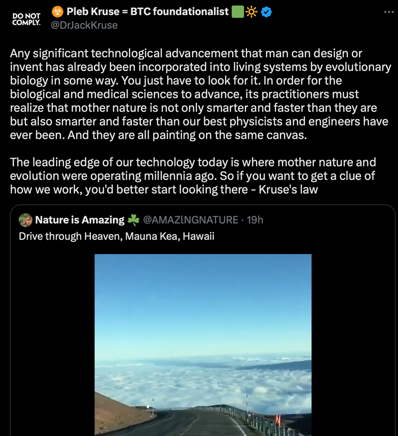
Post translational time stamping is present from cyanobacteria to mammals. All organisms have evolved timing mechanisms to adapt to environmental changes in order to optimize survival and improve fitness for an environment. To anticipate these regular daily electromagnetic cycles of light and dark, many organisms manifest near 24-h cell-autonomous oscillations that are sustained by transcription–translation-based or post-transcriptional negative feedback loops that control a wide range of biological processes. With an eye to identifying emerging common themes among cyanobacterial, fungal and animal clocks, some major recent developments in the understanding of the mechanisms that regulate these oscillators and their output need to be discussed. These include roles for antisense transcription, intrinsically disordered proteins, codon bias in clock genes, and a more focused discussion of post-transcriptional and translational regulation as a part of both the oscillator and output.
Circadian rhythms in every organism are cell autonomous, appear to have arisen only a few times in evolution, and can be driven by one of a few lineage-specific but otherwise highly conserved central oscillators. While oscillators driving bacterial and plant clocks are distinct from each other and from other known clocks, fungal and animal cells share circadian oscillators of conserved regulatory architecture: transcription-translation feedback loops (TTFLs) comprised of two parts.
Specifically, 1) a positive arm with a heterodimeric complex at its core that behaves as the activator of the system, promoting the transcription of 2) one or more components of the negative arm, which when translated inhibit the activity of the positive arm.
WHAT IS THE TTFL?
Transcription-translation feedback loop (TTFL) is THE cellular model for explaining circadian rhythms in behavior and physiology. It is widely conserved across species, and the TTFL is largely auto-regulatory with the assistance of the sun & moon and dark periods on Earth, in which transcription of clock genes is regulated by their own protein products. This implies that light and dark control genetic expression and not the other way around. The TTFL is a negative feedback loop, in which clock genes are regulated by their protein products. Generally, the TTFL involves 2 main arms: positive regulatory elements that promote transcription and protein products that suppress transcription. When a positive regulatory element binds to a clock gene promoter, transcription of DNA proceeds, resulting in the creation of an mRNA transcript, and then translation proceeds, resulting in a semiconductive protein product. There are characteristic delays between mRNA transcript accumulation, protein accumulation, and gene suppression due to translation dynamics, post-translational protein modification, protein dimerization, and intracellular travel to the nucleus. Across species, proteins involved in the TTFL contain common structural motifs such PAS domains, involved in protein-protein interactions, and bHLH domains, involved in DNA binding.
The two overarching areas characteristic of circadian systems in general: 1) the negative arm and its regulation of the core clock; 2) the control of output by the positive arm and its environment.
In ALL mammals the heterodimeric BMAL1-CLOCK complex positively regulates expression of negative arm component genes, the Periods and Cryptochromes(encoding PER1, PER2, PER3, CRY1 and CRY2), that combine with CK1 and several other proteins to make the repressive complex that depresses BMAL1-CLOCK activity and alters periodicity of the mammalian clock which alters its accuracy. Remember all circadian clocks are flow meters for entropy in a cell.
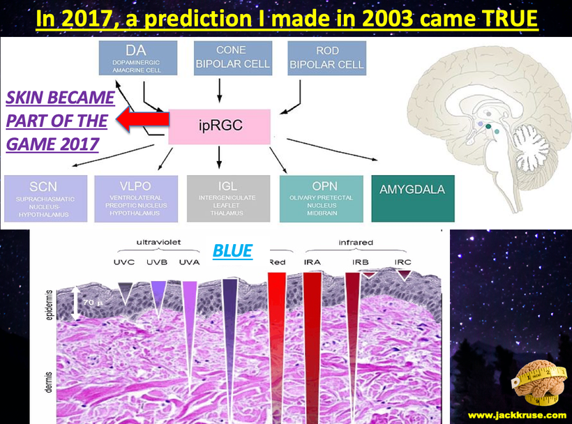
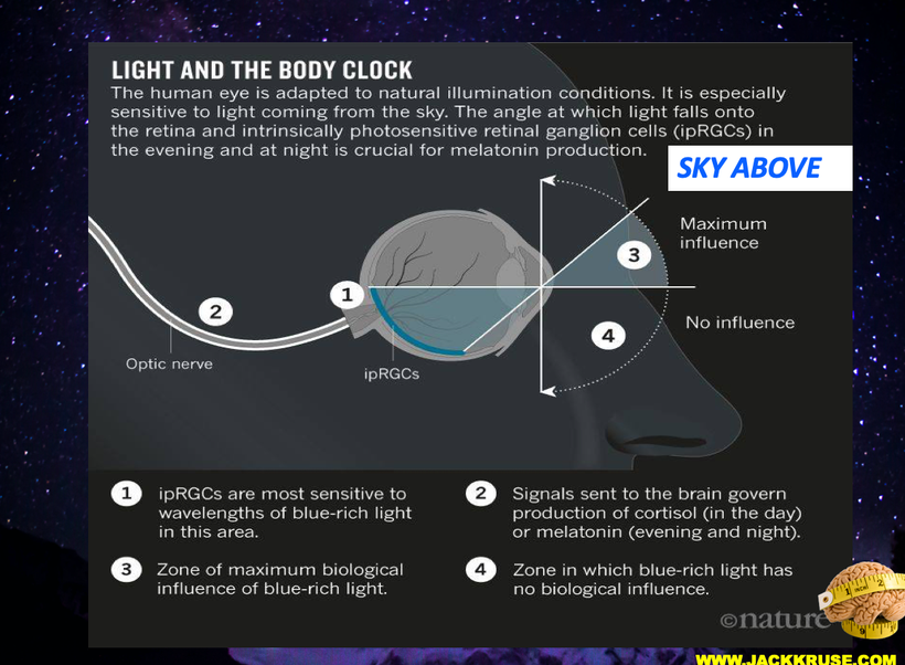
Solar light input into mammal TTFLs begins with dedicated non visual photoreceptors that elicit signaling that acts to induce (in fungi and mammals) or reduce (insects) the amounts of negative arm proteins mentioned above. In broad outline, Output occurs when the positive Arm heterodimer binds to DNA and activates expression of genes whose products do not impact the TTFL. The key take away is the clock gene actions are PROXIMAL to DNA translation and gene activation. This tells you that light inputs controls gene expression in mammals and it is not the other way around. Altering your genome will not improve your illness or disease if the light and dark environment is repair first.
HOW DOES TIME STAMPING WORK BY LIGHT AND DARK WORK?
Once enough modified protein products accumulate in the cytoplasm of a cell, they are transported into the nucleus where they inhibit the positive element from the promoter to stop transcription of clock genes. The clock gene is thus transcribed at low levels until its protein products are degraded, allowing for positive regulatory elements to bind to the promoter and restart DNA transcription. The negative feedback loop of the TTFL has multiple properties important for the cellular circadian clock. First, it results in daily rhythms in both gene transcription and protein abundance and size, caused by the delay between translation and negative regulation of the gene. The cycle’s period, or time required to complete one cycle, remains consistent in each individual and, barring mutation, is typically near 24 hours. This enables stable entrainment to the 24 hour light-dark cycle that Earth experiences from the sun & moon.
Additionally, the protein products of clock genes control downstream genes that are not part of the feedback loop, allowing clock genes to create daily rhythms in other processes, such as metabolism, within the organism. Light and dark cycles are the decentralized controllers of the TTFL.
THE TTFL USES MELANIN TO ELECTROCHEMICALLY TIME STAMP YOUR CELLS. This occurs in the retinohypothalamic pathways anterior to the SCN and it modifies what the SCN signals to all the other molecular clock genes it links to in tissues.
WHY IS MORNING LIGHT SO CRITICAL TO GET RIGHT?
CSP-1 (conidial separation 1) is a morning induced transcriptional repressor with a phosphorylation gated half-life is a key cog in driving EVENING gene expression in mammals. If you do not get AM sun your evening genomic expression will be AWRY. People have forgotten that leptin is released by fat cells and can only enter the hypothalamus under darkness after 4 hours. This should happen at night time. It cannot happen when CSP-1 is not created by AM light. These are the new recent insights into how circadian clocks in your eye and skin achieve phase-specific gene expression. This is how and why leptin resistance exists.
The negative element of the core circadian feedback loop is the frq or frequency gene. The frequency (frq) gene controls the morning-specific rhythmic transcription of a sense RNA encoding FRQ segment. As a result of this action, a long noncoding antisense RNA, qrf, is rhythmically transcribed in an evening-specific manner. It has been reported in the literature that the qrf rhythm relies on transcriptional interference with frq transcription and that complete suppression of qrf transcription impairs the circadian clock. The biological function of qrf transcription and its impact on the circadian clock are not understood in centralized science because centralized science has no light controls at night in labs.
CSP-1 expression is induced by light and glucose, and this finding suggests a rhythmic coordination of qrf transcription with metabolism. Because it is light and glucose we know POMC, ACTH, and melanin are the key to understanding CSP-1 biology. It also means that artificial light during the day or night is especially toxic when you know this is how the mechanism operates.
There are three type of melanins in humans and only one ACTH in humans. All three are used to time stamp the atomic lattice of cells to create an internal map of space time domains to be accurate measuring sticks for the flow of entropy inside of cells. This links melanin biology to Noether’s theorem directly. You have blogs on all these ideas now and it is time for you to link them all to comprend what I have been teaching your for 20 years. Light causes modern diseases.
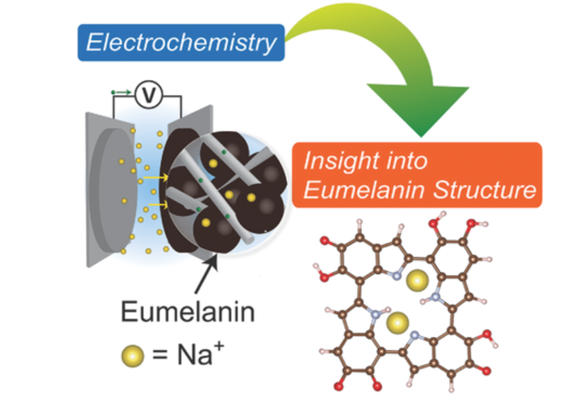
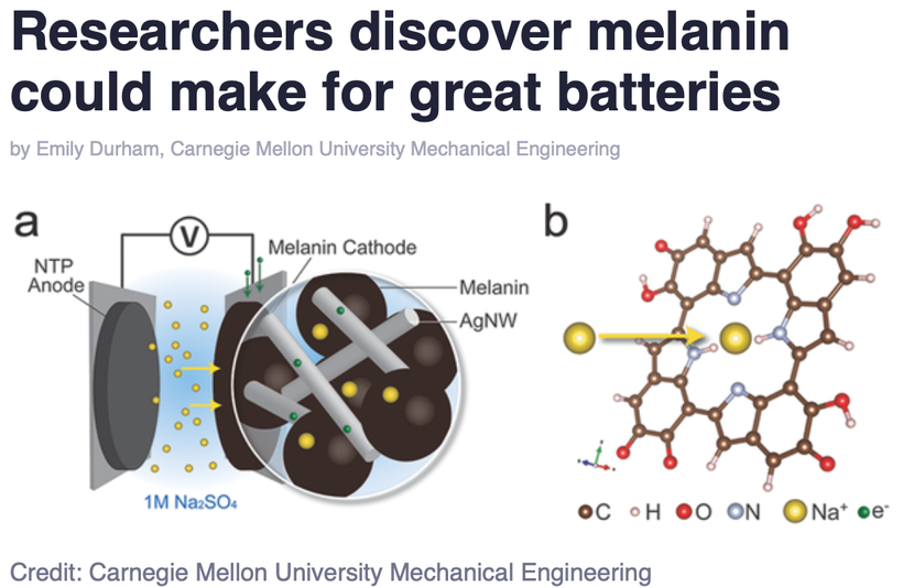
These 3 melanins are ALL extended heterogeneous biopolymers composed of molecular subunits with ambiguous macromolecular topology to modern centralized science. In the literature, an electrochemical fingerprinting technique has been described for melanin, which suggests that natural melanin pigments which contain indole-based tetramers seem to be always arranged into porphyrin-like domains to capture light and measure it in some way useful to the system.
Spectroscopy and density functional theory calculations suggest that sodium ions undergo occupancy-dependent stepwise insertion into the core of porphyrin-like tetramers in natural melanins at discrete potentials that time stamp the internal atomic lattice that allow it to act like a clock to measure the flow of entropy in the cellular system accurately just using light and dark as the feedback loops. It is fully decentralized because light and dark control this process. One is not more important than the other. A loss of melanin implies a loss of accurate time keeping inside the cell or tissue.
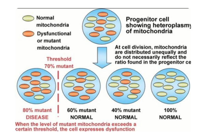
Lastly, in humans, the TTFL is a limit cycle, meaning that it is a closed loop that will return to its fixed trajectory even if it is disturbed by its environment, maintaining the oscillatory path on its fixed 24-hour period. It appears this is only true if the melanin structures it uses on surfaces and endogenously remain intact and are chronically replaced and renovated. If the endogenous electrochemical time stamping mechanism is damaged, the TTFL loses its periodicity and chronic modern disease results without any alteration to the DNA or RNA of a cell. These are diseases do not mimic genetic diseases like Tay Sach’s. These diseases are far more common than mutational diseases of DNA which are relatively rare. It means our circadian mechanism is open to the environment, and as such is not subject to calories measurement for metabolism because calories only is useful in closed thermodynamic loops.
CITES
1. Aronson BD, Johnson KA, Loros JJ, Dunlap JC. (1994) Negative feedback defining a circadian clock: autoregulation of the clock gene frequency. Science 263:1578-1584.
2. Belden WJ, Larrondo LF, Froehlich AC, Shi M, Chen CH, Loros JJ, Dunlap JC. (2007) The band mutation in Neurospora crassa is a dominant allele of ras-1 implicating RAS signaling in circadian output. Genes Dev 21:1494-1505.
3. Bell-Pedersen D, Shinohara ML, Loros JJ, Dunlap JC. (1996) Circadian clock-controlled genes isolated from Neurospora crassa are late night- to early morning-specific. Proc Natl Acad Sci U S A 93:13096-13101.
4. Cheng P, Yang Y, Heintzen C, Liu Y. (2001) Coiled-coil domain-mediated FRQ-FRQ interaction is essential for its circadian clock function in Neurospora. EMBO J 20:101-108.
6. Froehlich AC, Liu Y, Loros JJ, Dunlap JC. (2002) White Collar-1, a circadian blue light photoreceptor, binding to the frequency promoter. Science 297:815-819.
7. Gallego M, Virshup DM. (2007) Post-translational modifications regulate the ticking of the circadian clock. Nat Rev Mol Cell Biol 8:139-148.
8. Larrondo LF, Olivares-Yanez C, Baker CL, Loros JJ, Dunlap JC. (2015) Circadian rhythms. Decoupling circadian clock protein turnover from circadian period determination. Science347:1257277.
Xue Z, Ye Q, Anson SR, Yang J, Xiao G, Kowbel D, Glass NL, Crosthwaite SK, Liu Y.Nature. 2014 Oct 30;514(7524):650-3. doi: 10.1038/nature13671. Epub 2014 Aug 17.
Zhu Q, Belden WJ.J Mol Biol. 2020 May 29;432(12):3466-3482. doi: 10.1016/j.jmb.2020.01.009. Epub 2020 Jan 16.
12. Koike N, et al. Transcriptional architecture and chromatin landscape of the core circadian clock in mammals. Science. 2012;338(6105):349–354.
13. Li N, et al. The frequency natural antisense transcript first promotes, then represses, frequency gene expression via facultative heterochromatin. Proc Natl Acad Sci U S A. 2015;112(14):4357–4362.
14. Xue Z, et al. Transcriptional interference by antisense RNA is required for circadian clock function. Nature. 2014;514(7524):650–653.
POMC arose over 500 million years ago by an insertion of the melanocortin sequences into a prepro-endorphin gene. Evidence for this comes from structural identities with other opioid precursors in both the NH2- and COOH-terminal regions of POMC.
The phenomena of pro-opiomelanocortin (POMC) as a hormone precursor emerged gradually over time as observations slowly filled in pieces of the puzzle. Long before the concept of hormone precursors was realized, the bronzed skin color described by Addison in his patient with adrenal insufficiency (“melasma suprarenale”) gave perhaps the first hints of a connection between the hypothalamic, pituitary, adrenal (HPA) axis and skin color. A similar link between the pituitary and pigmentation came from the studies of Allen in 1916 and Smith in 1916 who both noted that immersing tadpoles in pituitary extract made their skins darker. In humans too, large doses of porcine pituitary extract also appeared to cause pigmentation in 1954, with this active extract of the pars intermedia of the pituitary henceforth termed “melanocyte stimulating hormone” or MSH.
In 1932, Cushing extended his clinical reports of a polyglandular syndrome caused by basophilic adenomas of the pituitary by linking this finding with adrenal hyperactivity. In the 1930s, work by Ingle and Kendall in the 1930s showed that administration of large amounts of “cortin,” a purified adrenal extract, produced atrophy of the adrenal cortex in rats. Importantly, they found that administration of the “adrenotropic principle” of the anterior pituitary was effective in preventing adrenal cortical regression following treatment with cortin. The first hints of a behavioral angle to POMC biology came from studies by Ferrari in the 1950s, when “stretching-yawning syndrome,” a bizarre crisis of muscular tone, occurred following central administration of MSH. Many other studies assessing the effects of central α-MSH on motivational processes followed, but it was not until 1976 that Panskepp observed for the first time that this peptide decreased food intake using light alone. What else might POMC do in a modern world run by artificial light now? Today, you get to see another perspective centralized science missed.
Viewed from the comfort and assured knowledge of the modern centralized molecular world, these observations and interventions could be considered overtly simplistic. However, I believe that these classic decentralized observations should be regarded as essential building core evolutionary building blocks, not only for our understanding of POMC peptide processing, but also for the work which subsequently tied together these seemingly diverse peptides.
Is this why did I had a ubiquitnation & mitochondrial series before the Quantum Engineering series on Patreon?
Yep.
In mammals, a master clock localized in the suprachiasmatic nucleus of the hypothalamus synchronizes cellular clocks in other central nervous and peripheral tissues with each other and with external time. At the molecular level, these clocks are based on an interregulatory network of clock genes, including the 3 Period genes (Per1–3), that control circadian rhythms (CRs) by rhythmic orchestration of 5–10% of the cellular transcriptome in a post translational tissue-specific manner. CRs time stamp genes after they are translated and cause diseases. I relayed this last month to the government of El Salvador in a speech I gave there.
Transcription-translation feedback loop (TTFL) is a cellular model for explaining circadian rhythms in behavior and physiology. Widely conserved across species, the TTFL is auto-regulatory, in which transcription of clock genes is regulated by their own protein products. Rev-erba is one and so is NF-kappa beta.
THIS IMPLIES TIME STAMPING CAUSES CHRONIC NEOLITHIC DISEASES.
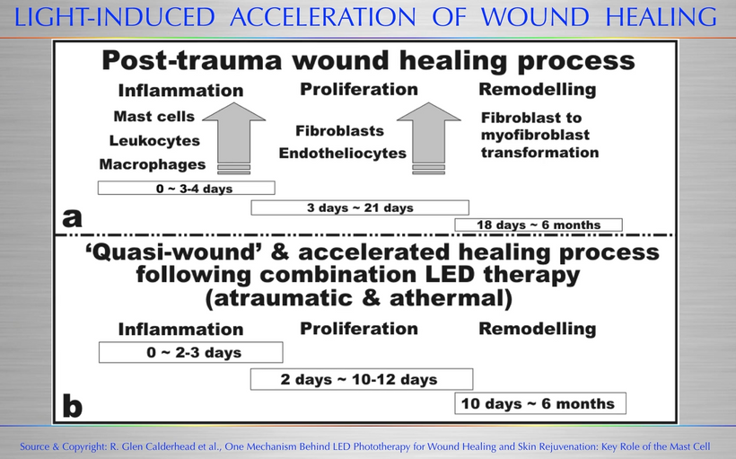
What can we use as an example to see if light stress on out largest organ causes diseases below?
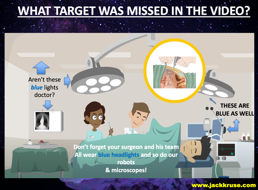
I posted the video above to show you how centralized edicational materials still miss the obvious elephant in the room. The lights used in surgery can cause the problem. the lights on laparascopic cameras are all high intensity blue lights, and it is already well know that other parts of the electromagnetic spectrum used in medicine can cause this problem without any previous surgery. Look at the picture below that I lifted from the video to show you how their own video on this topic misses the elephant in the room on adhesions. I even mentioned this to Huberman during my podcast when I told him everytime I open a skull I now worry about what I am doing because of my understanding.
Have you ever heard of adhesions of the gut?
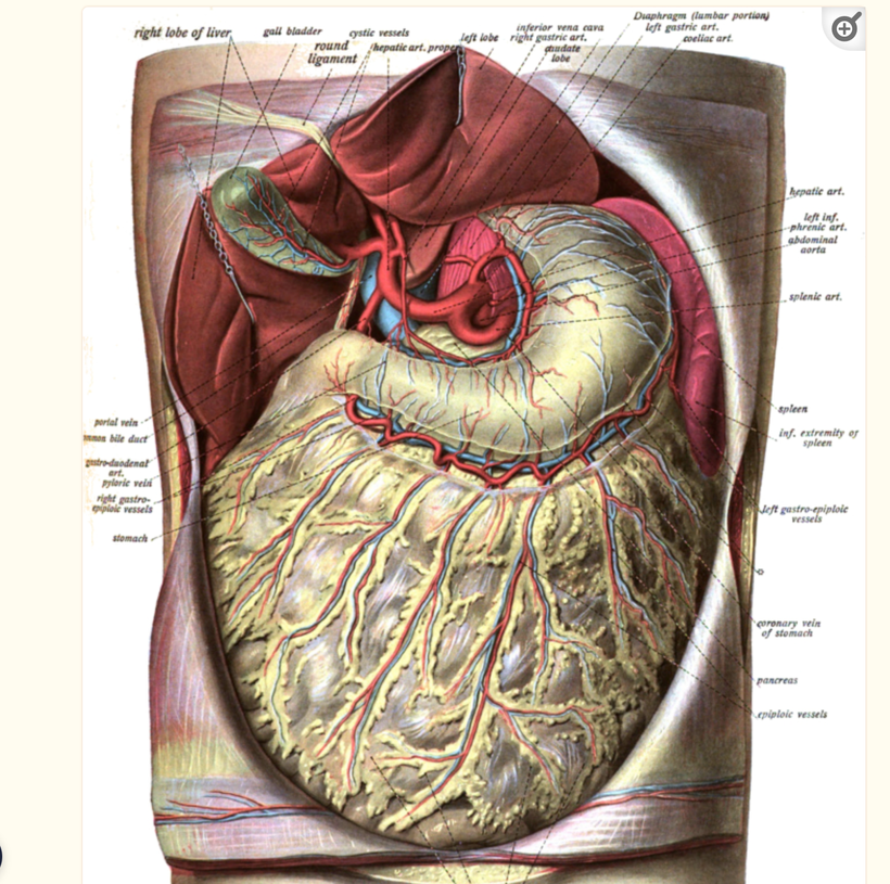
Abdominal adhesions commonly form after intra-abdominal surgery, radiation, and inflammatory processes in humans. In a subset of patients, adhesions lead to problematic symptoms such as abdominal pain, bloating, and bowel obstruction. The mechanisms of adhesiogenesis are not well understood by centralized science but are believed to involve mesothelial surface disruption with subsequent fibrinocoagulative and inflammatory signaling processes. I believe the reason centralized science has missed the boat on adhesions is because aberent light causes it. It is a biophysical disease at its core. Melanin in your omentum is a big deal to the gut clocks below. The melanin in the omentum links to the POMC genes in the skin of the rectus sheets on your abdomen.
WHAT ARE ADHESIONS?
Abdominal adhesions are fibrous bands that span two or more intra-abdominal organs and/or the inner abdominal wall (i.e. peritoneal membrane) which typically form after abdominal surgery. Adhesions may also form secondary to inflammatory conditions of the abdomen in the absence of prior abdominal surgery or as a sequela of abdomino-pelvic radiation. Although the majority of patients with intra-abdominal adhesions remain asymptomatic, a clinically significant subset of patients will develop “adhesive disease”, a symptomatic state ranging from mild and/or vague to highly distressing and even life-threatening symptoms.
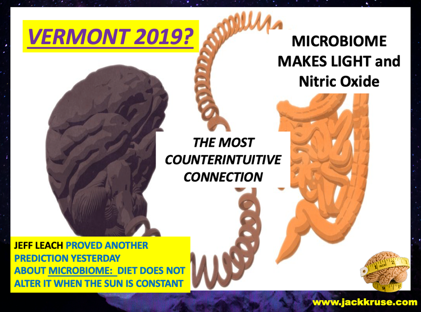
Considering the fact that adhesions have no characteristic laboratory features and are not readily visible by currently available imaging methods, many cases of adhesive disease will go undiagnosed for prolonged periods of time, causing medical providers to find themselves in a diagnostic and therapeutic quandary. Patients, consequently, after extensive non-diagnostic testing and empiric treatments, may not only experience protracted symptoms and adverse medical outcomes, but can also suffer from significant emotional distress or demoralization, which in turn may be misdiagnosed as depression, anxiety, or a functional bowel disorder. This topic was going to be the headliner in 2019 in Vermont. That never happened.
WHAT IS THE LIKELY DECENTRALIZED CAUSE OF ADHESIONS?
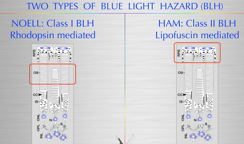
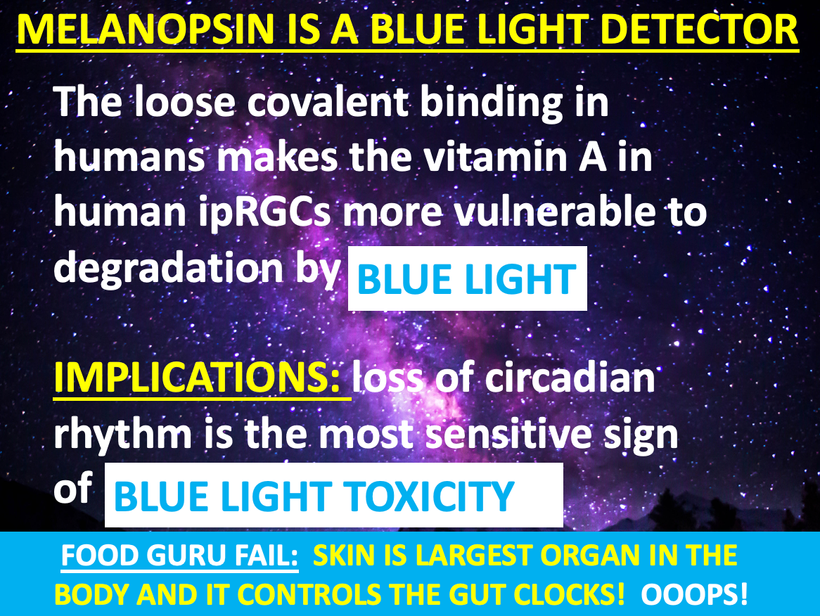
Irradiating human skin with blue light in frequency bands that causes both type of blue light toxicity is likely the cause of abdominal adhesions. People with autoimmune conditions are at increased risk of adhesions and keloid formation on their skin. The link between all these conditions is the chronic irradiation of human skin with artificial blue light devoid of IR- A and UV frequencies with blue light. One must remember that isolated blue frequencies activates the blue light hazard in mitochondria powered by melanopsin signaling that cause pseudohypoxia in mitochondria. When will centralized medicine learn how light stress not from sunlight ruins mitochondrial biology? Melanin is a key battery in the skin and deep inside of our systems.
This type of isolated light irradiation can set up adults for epithelial cancer, myopia, low dopamine diseases, diabetes, and skin cancer as they age and their heteroplasmy rates grow larger faster as they age into adults formats. If you destroy the battery you have no energy left for health and disease is the result. Could it also be the cause of adhesions? Might this mean adhesions in the gut are a warning signal that surgeons should be paying attention to in patients?
It is for me. When i see arachnoid webs in the brain I know what it means to my patients.
Women with keloids on their cesarean scar have been found in prospective studies to have increased adhesions between the uterus and the bladder and between the uterus and the abdominal wall. This certainly supports my theory and my ideas on this topic. This is cited below.
In mammals, a master clock localized in the suprachiasmatic nucleus of the hypothalamus synchronizes cellular clocks in other central nervous and peripheral tissues with each other and with external time. At the molecular level, these clocks are based on an interregulatory network of clock genes, including the 3 Period genes (Per1–3), that control circadian rhythms (CR) by rhythmic orchestration of 5–10% of the cellular transcriptome in a post translational tissue-specific manner. This tells us that most human disease is not DNA/RNA based, it is mitochondrial based because circadian disease phenotypes mimic those of chronic diseases in centralized healthcare. Circadian biology and oscillations time stamp genes after they are translated and cause modern chronic diseases. How does it happen in humans?
Transcription-translation feedback loop (TTFL) is a cellular model for explaining circadian rhythms in behavior and physiology. It is widely conserved across ALL species, & the TTFL is auto-regulatory, in which transcription of clock genes is regulated by their own protein products. Rev-erba is one and so is NF-kappa beta. I’ve written about both of these things in the past on my blogs.
Rev-erbα, a nuclear receptor and circadian clock component, is a mediator of microglial activation and neuroinflammation in the connection between the CNS and human gut.
I believe people who get adhesions (Crohn’s, UC, SIBO patients) all have destroyed Transcription-translation feedback loop destuction due to an altered circadian mechanism. I propose that such a mechanism can be found within the molecular control of circadian rhythms and “Clock” gene biology and a lack of melanin in some key places in the spinal cord that connect the gut to the CNS.
All melanin’s can create a toxin when they breakdown in human tissue. For example, neuromelanin can reversibly bind and interact with amine containing neurotoxins, (e.g., MPTP(1-methyl-4-phenyl-1,2,3,6-tetrahydropyridine is an organic compound. It is classified as a tetrahydropyridine.) MPTP augments their actions in the terminal, eventually leading to the instability and degeneration of melanin-containing neurons due to oxidative stress and mitochondrial dysfunction.
In particular, a lack of neuromelanin and POMC translation by a lack of UV light appears to confer susceptibility to chemical toxicity by providing a large sink of iron-bound, heme-like structures in a pi-conjugated system. This system seemingly allows for stabilizing interactions including pi-stacking, sodium inclusion, as well as ligand binding to iron in cells. Given the progressive accumulation of neuromelanin components with aging. it seems melanin degradation corresponds with an apparent decrease in dopamine synthetic pathways in man. This raises an immediate question of whether melanin can also create or is capable of binding dopamine, the primary functional monoamine utilized in gut enterocytes and its corresponding gut neurons, should be raised by centralized research. To date, it has not been.
A number of physiological processes demonstrating circadian variation have been shown to involve ‘Clock genes’ in the suprachiasmatic nucleus (SCN), which then entrains a similar set of Clock genes in peripheral tissues such as the heart, brain, spleen, lung, liver, skeletal muscle and kidney. The intrinsic time-keeping system influences activity, such as sleep, temperature regulation, rates of metabolism, immune responses, blood pressure and hormone secretion. The function and availability of mediators involved in the inflammatory response, fibrinolytic and anti-coagulation pathways are all under the tight control of the molecular Clock system. These include IL-6, PAI-1, fibrinogen, fibroblasts and TNF-α. I am making the educated guess that disruptions in the ‘Clock system’ are central to the causal pathway of adhesion formation here. I am also implying this symptom is a gateway disease to many other diseases linked to altered cellular time stamping in humans.
The transcriptomic analysis of hippocampus from Rev-erbα-/- mammals has revealed a predominant inflammatory phenotype results in mammals and it suggests it comes from a dysregulated NF-κB signaling due to circadian dysfunction at its core. When one considers that melanin derivatives are always found in pathologic specimens it is not hard to come up with this linkage.
DEEP DECENTRALIZED SCIENCE THEY ARE MISSING
The brain gut connection requires pristine circadian rhythm control to stay healthy. Loss of Rev-erbα in primary astrocytes of the spinal cord of mammals had no effect on basal activation of the neurons that innervate the gut but do potentiate the inflammatory response to lipopolysaccharide (LPS). In vivo, Rev-erbα-/- in a mammalian model has exhibited enhanced hippocampal neuroinflammatory responses to peripheral LPS injection, while pharmacologic activation of Rev-erbs with the small molecule agonist SR9009 suppressed LPS-induced hippocampal neuroinflammation. This explains to us how a loss of circadian control allows the LPS of the microbiome to lead to a disease phenotype just with a loss of circadian control post translationally of DNA. This means DNA alteration is not the cause of a majority of modern human disease. This is 180 degrees from the current centralized perspective.
Rev-erbα deletion due to a loss of clock gene control clearly influences gut enterocyte function and neuronal health in a coordinated fashion. When this link breaks it makes the formation of adhesions post surgically much more likely. Lab experiments done have shown when gut neurons are conditionally cultured on media from Rev-erbα-deficient primary gut-glial cultures, it exacerbates oxidative damage in cultured neurons that innervate the gut. Rev-erbα-/- mammals have not only shown gut dysfunction in these models but they also exhibited significantly altered cortical resting-state functional connectivity. This finding tells me that many central neurological diseases maybe masquarding in centralized medicine clinics as circadian diseases of theTranscription-translation feedback loop(TTFL).
One neurologic disease in particular intrigues me with its skin & gut connection is ALS. ALS patients tend not to show up in low latitudes and they tend to be quite pale because they lack UV exposure POMC requires for translation of melanin. In human ALS, abnormalities have been found in mitochondrial structure, mitochondrial respiratory chain enzymes, and mitochondrial cell death proteins indicative of some non-classical form of programmed cell death. In ALS patients this happens in the spinal cord, skin, and their gut in pulsatile fashion over timescales. This is a hint to me that it might be a disease of time stamping of the TTFL masquarading as a neurological disease.
The creation of MPTP, which is lipid-soluble, readily penetrates the blood—brain barrier and enters the brain cells. Because it is amphiphilic, it is captured into acidic organelles, mostly lysosomes, of astrocytes. MPTP itself does not appear to be toxic, but its oxidized product, 1-methyl-4-phenylpyridinium (MPP+), is toxic. This has been linked in Parkinson’s Disease but I think it might link to ALS because the toxic MPP+ could affect defective mitochondria in neurons in the anterior motor horn from circadian rhythm damage in the gut. These malfunctioning mitochondria might contribute to neuronal death in ALS through the biophysical entity called the mitochondrial permeability pore (mPTP) activation.
The major protein components of the mPTP are enriched in mammalian motor neurons which also have abundant POMC genes. Early in the course of disease in ALS humans seem to present with mutant superoxide dismutase-1 enzymes in their mitochondria. The mitochondria in motor neurons undergo “trafficking abnormalities” which results in dramatic remodeling of mitochondria in ALS patients that results in the formation of mega-mitochondria (larger size) and coinciding with increased protein carbonyl formation and nitration of mPTP components. Anything that enlarges is a thermodynamic sign to me. In fact, lab experiments in nocturnal mammals have shown that genetic deletion of a major mPTP component called cyclophilin D which has robust effects in ALS by delaying disease onset & phenotype and extending survival of the anterior motor neurons.
When this process is disrupted the MPP+ action it appears to destroy the mPTP in a cascading manner in the spinal cord mitochondria and spreads like an infection would in the anterior motor neurons. I think the circadian disruption in the anterior motor neurons causes a paralysis like effect that we normally see in REM sleep. In ALS, the circadian disruption shows up when we are awake and it has gone unrecognized as a completely disrupted rhthym disorder because of how we perceive the disease and we have not looked at the data carefully enough. I happen to have one person with this disease who is doing something no one else has done with this diagnosis. He is improving his solar redox and improving mtDNA function by living in the tropics adjacent to a flat beach. So far he is doing a lot better than other ALS patients who aren’t paying attention to their light stamping problem. In fact, I believe some of these patients diseases are made worse when they travel outside of time zones. That seems to make the diseases more aggressive.
I think this model in mammals explains why anterior motor neurons can be destroyed in isolated fashion in the spinal cord when the circadian rhythms are disrupted in the nerves that innervate the gut but link back to their embryologic origins. The inflammatory cascade could start out in the splanchnic nerves below but as the spinal cord anterior motor neurons loses REV-erb alpha it could create an inflammatory cascade of LPS induced damage to “infect” other adjacent motor neurons by destroying their clock gene TTFL’s. This is how ALS might be progressing in these patients.
The Rev-erbα, mPTP in mitochondria, and TTFL should be thought of as as a key biophysical link between the circadian clock control and neuroinflammation in the CNS. People forget that the spinal cord is part of the CNS and so is their skin. Nerves link the gut to both.
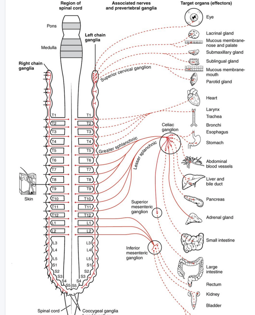
Look more carefully at the picture above then you did in QE #47. Note the skin is on the left of the picture and it is our largest organ and it has a somatotopic organization to the ENTIRE spinal cord and to our internal organs by way of nerves.
The Quantum Enginnering #47 blog has the key metric of why the eye, skin, PVN, and DLF of the brain stem sleep and pain areas give the input into celiac plexus to cause gut problems via the microbiome. The implications of this connection are massive in many other diseases besides bipolar disorder. I told you that there and I am doubling down on it here.
WHAT DID I SAY IN QE #47 AGAIN ABOUT THIS PROCESS?
I said, “The dorsal longitudinal fasciculus (fasciculus of Schutz) is a periaqueductal (area around ventricular system Aq above) ascending and descending fiber system arising from the hypothalamus and terminating to the autonomic nuclei of the pons and the medulla, conveying autonomic fibers from the brain to the gut in humans. It also conveys pain messages and is important in the sleep pathways of humans. These are usually altered in bipolar patients as well because of a lack of melanin in these areas.
The dorsal longitudinal fasciculus is found within the dorsal brainstem tegmentum. It passes through the periaqueductal gray matter and contains both ascending and descending fibers. The ascending fibers pass from the reticular formation (sleep region/insomnia) passing to the hypothalamus thus transmitting information related to the viscera in the thorax and abdomen. THIS IS HOW THEY PASS THE ANTERIOR MOTOR HORN CELLS AND THE GUT VISCERA!
People who travel a lot across time zones or people who use a lot of blue light or nnEMF at night or day in their cities are going to have massive sleep disturbances because they lack charge density in this spot. They mimic people with mental disorders like bipolar patients because they have broken the same rules of Nature. I view insomnia as a mental disorder in my decentralized medicine framework.
This topologic neuronal surface is a critical barrier to the brain health of humans. This is the pathway where metabolic syndrome, poor sleep, and fatty liver all come from fundamentally. This pathway could be in many blogs but I left it out this detail. Why? You already thought this stuff was too hard. Why add another level of complexity with neuroanatomy?
Many of these same findings are found in diabetics, bipolar disorder, and those with sleep disorders like insomnia. Patients with bipolar disorder tend to have all these symptoms at times that vary based on how defective their FM antennae are in their eyes & brains.”
DOES ANYONE SEE THOSE LINKS NOW IN THIS BLOG IN THE SERIES?
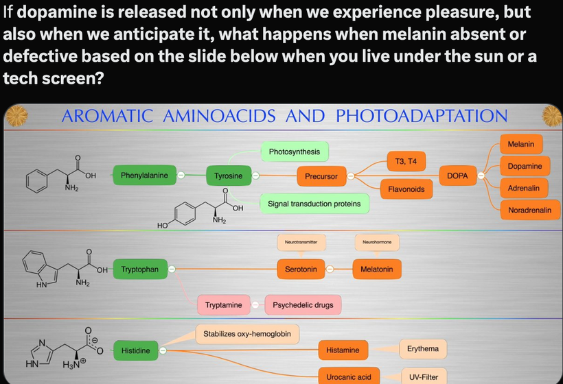
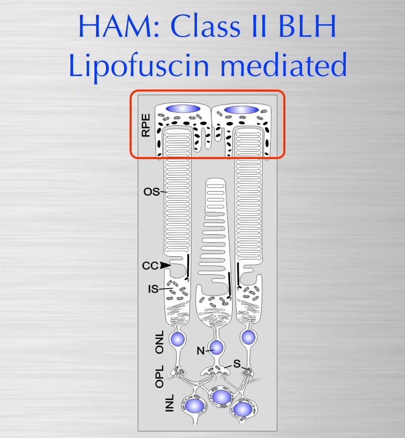
Now, go back and look at the lipofuscin blog in this series. That chemical comes from melanin degradation and is always associated with aging and light stress. Dopamine is made in the eye several ways by sunlight and it can be made in the gut by your microbiome due to the link to melanin. People forget that bacteria release 5000 times more light than eukaryotic cells and this light is capable of making dopamine from the aromatic amino acids in the gut. This is why serotonin and phenylalanine and tyrosine are stored in the enterochromaffin system in our gut.
The more and more you look into its biology, I feel more confident in saying that light is the most powerful drug for living systems. The light release is more important than the fuel in the diet for our health outcomes because food has light information built into photosynthesis. This stimulus leads to light release to start the optical signal cascade in the gut using aromatic amino acids as the mover in the GI tract. When you are blue light/nnEMF toxic you cannot repair the gut or sleep issues at all. This is why many people get gut adhesions without having had any surgery. The same thing will be true for pain thresholds and opiate use. I can tell you this. Your gastrointestinal system will never function optimally in a circadian mismatch (melanopsin/encephalopsin dysfunction) and that will bypass however good a diet is.
The study linked below in cite 6 is a longitudinal study on TBI following college football athletes across a sports season.
•Nanopore 16S rRNA sequencing of gut microbiome reveals changes after head injury.
•Serum biomarker GFAP increased during the competitive period of the season.
•S100β and SAA blood levels were positively correlated with Eubacterium rectale.
•Gut microbiota is suggested as a future biomarker for diagnosis following head & neck injury.
The blue light hazard/nnEMF links the gut to the brain, heart, and vascular damage leading to neurodegeneration that causes protein folding issues in blue light-damaged tissues as cites 4 and 5 show. Few are making these links in diseases like adhesions and ALS.
But I am.
CITES
Van Goor H. Consequences and complications of peritoneal adhesions. Colorectal Dis. 2007 Oct;2(9 Suppl):25–34.
Menzies D., Ellis H. Intestinal obstruction from adhesions; how big is the problem? Ann. R. Coll. Surg. Engl. 1990;72:60–63.
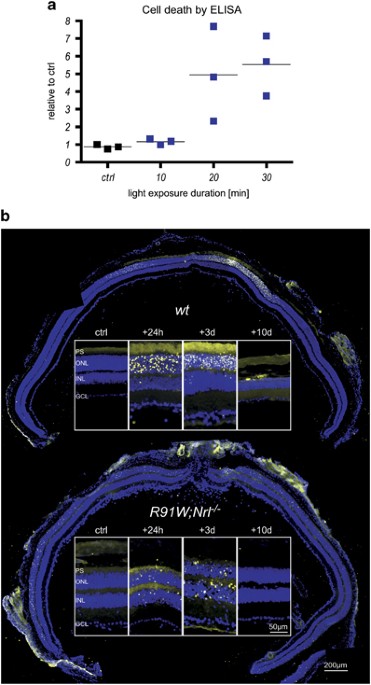
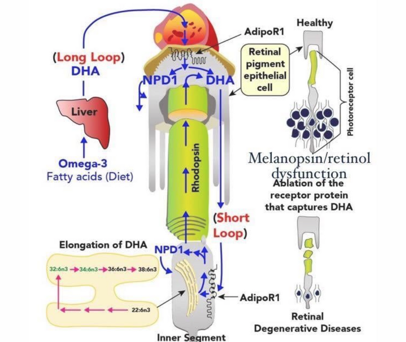
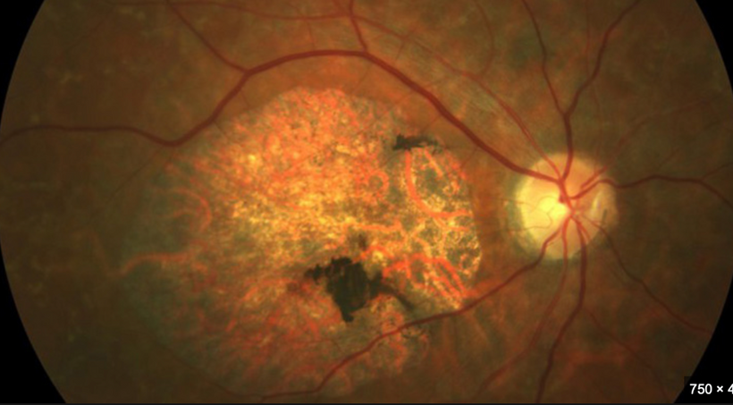
Little is known about the mechanisms underlying macular degenerations, mainly for the scarcity of adequate experimental models to investigate cone cell death. Recently, we generated R91W;Nrl−/− double-mutant mice, which display a well-ordered all-cone retina with normal retinal vasculature and a…
Concussions, both single and repetitive, cause brain and body alterations in athletes during contact sports. The role of the brain-gut connection and …
Allen BM. Extirpation experiments in Rana pipiens larvae. Science 44: 755–757, 1916. doi: 10.1126/science.44.1143.755.
Smith PE. Experimental Ablation of the Hypophysis in the Frog Embryo. Science 44: 280–282, 1916. doi: 10.1126/science.44.1130.280.
Lerner AB, Shizume K, Bunding I. The mechanism of endocrine control of melanin pigmentation. J Clin Endocrinol Metab 14: 1463–1490, 1954. doi: 10.1210/jcem-14-12-1463
.Ingle DJ, Kendall EC. Atrophy of the Adrenal Cortex of the Rat Produced by the Administration of Large Amounts of Cortin. Science 86: 245, 1937. doi: 10.1126/science.86.2228.245.
Panskepp J, Reilly P, Bishop P, Meeker RB, Vilberg TR, Kastin AJ. Effects of alpha-MSH on motivation, vigilance and brain respiration. Pharmacol Biochem Behav 5, Suppl 1: 59–64, 1976. doi: 10.1016/0091-3057(76)90329-4.
The auditory nerve transmits auditory information up a series of nuclei to the cortex where perception occurs. These nuclei include 1) cochlear nucleus, 2) superior olivary nuclei, 3) lateral lemniscus, 4) inferior colliculus, and 5) medial geniculate nuclei.
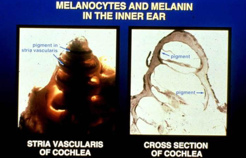
Melanin pigment is normally present in the outermost layer of the retina of the eye, the inner ear adjacent to capillaries in stria vascularis near hair cells, and vestibular organs. In the skin, it is in the basal layers. In the gut, it is found in the enterochromaffin cells. Significant reduction in melanin pigment in mammals is associated with embryonic miswiring and disruption of visual and auditory functions. This has implications for human diseases like autism which usually affects all sensory inputs to the thalamus. In this way, it mimics what I wrote about Autism in this series in QE#45. The consequences for the visual system include abnormal development of the retina and misrouting of optic pathways into the brain impairing visual acuity, eye movement, and stereovision. Lack of melanin pigment in the inner ear is associated with greater susceptibility to noise damage and poorer localization of sound in space.
Auditory sense in mammals is unique in another way and may be critical to why melanin exists where it does. Unlike other cells within the brain, hair cells within the Organ of Corti of the cochlea do not have axons. Neurons within the spinal ganglion have peripheral axons that synapse at the base of the hair cell soma. These axons make up the auditory nerve. Most (90%) of auditory nerve fibers receive their input from the inner hair cells. Thus, the inner hair cells facilitate a majority of auditory processing. My bet is this arrangment is how sound waves are converted to light waves that the auditory nerve can process.
The melanin sheets in the ear are proximal to the auditory nerves. When melanin is hypoxic it can break down into noradrenaline and dopamine. The mammalian auditory brain stem, and in particular the medial nucleus of the trapezoid body (MNTB) (Wynne and Robertson 1996), receives extensive adrenergic input. This tells me that the sheet in the cochlea is creating neurotransmitters by melanin degradation in the auditory pathways as the slide below shows.
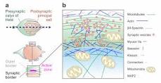
The mammalian auditory brain stem receives profuse adrenergic innervation, whose function is currently poorly understood by centralized science. Noradrenaline increases high-frequency firing at the Calyx of Held synapse during development by inhibiting glutamate release.
The calyx of Held synapse plays an important role in the auditory system, relaying information about sound localization via fast and precise synaptic transmission, which is achieved by its specialized structure and giant size. During development, the calyx of Held undergoes anatomical, morphological, and physiological changes necessary for performing its functions. The large dimensions of the calyx of Held nerve terminal are well suited for direct electrophysiological recording of many presynaptic events that are difficult, if not impossible to record at small conventional synapses. This unique accessibility has been used to investigate presynaptic ion channels, transmitter release, and short-term plasticity, providing invaluable information about basic presynaptic mechanisms of transmission at a central synapse.
DECENTRALIZATION ANSWER to why all senses have melanin sheets between them and the environment?
Why does the spiral cochlea have this huge melanin sheet? Melanin shows up in mammalian tissue because of the melanin concentration hormone. Melanin-concentrating hormone (MCH) was originally isolated from salmon pituitaries (think of Huberman fish gaff on melanopsin now) where it induces the aggregation of melanin granules in melanophores, which results in pale skin color. MCH sequence is conserved in ALL MAMMALS analyzed to date, including mice, rat, rabbits, and humans. In mammals, the neurons that synthesize and release MCH are present mainly in the hypothalamus and nearby areas. Is the cochlea near the hypothalamus? No. But the melanin stimulatory hormone is very prominent in the brainstem and the cochlear is very close to that. Lack of melanin pigment in the inner ear is associated with greater susceptibility to noise damage and poorer localization of sound in space.
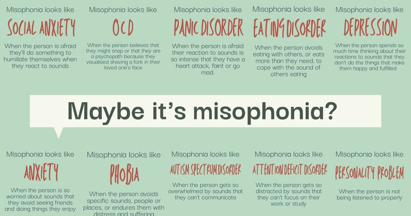
I think a relative lack of melanin in different areas of the cochlea is the cause of misophonia. Misophonia is a disorder in which certain sounds trigger emotional or physiological responses that some might perceive as unreasonable given the circumstances. Those who have misophonia might describe it as when a sound “drives you crazy.” Their reactions can range from anger and annoyance to panic and the need to flee. If your centralized health professional doesn’t know about misophonia, they may take all that information and try to fit it with something they do know about. Here are some diagnoses you might hear before a health professional finally acknowledges your misophonia:
Melanocytes are distributed in the stria vascularis and spiral ligament regions and recent evidence illustrates the distribution of melanin in the human cochlea with the lower relative amount of melanin observed in the basilar turns compared to apical turns. Sensorineural hearing loss caused by presbycusis (old age heteroplasmy), maternal-fetal rubella infection, aminoglycosides, and noise exposure disproportionally impacts the basal turn with variable losses of spiral ganglion and hair cells reflected in a down-sloping audiogram. Melanin is sparse in those regions. The location of melanin and its known function as a free radical scavenger may explain some patterns of sensorineural hearing loss and highlight that melanin has special abilities in the sensory pathways of mammals.
Might it have to do with the binary code of biology and the fidelity of sound that I raised in the Kruse for Dummies lecture? I believe it does. Look at how many areas sound inputs are sent. If we are built to semiconductor sound waves this implies we need a big equalizer in our ear no?
Since this synapse is massive in all mammals it has been studied. What do we know? Researchers have studied multiple mammal species and to the extent that a comparison is possible, the time course for development embryologically matches earlier experiments performed in rats, cats, mice, or gerbils, suggesting that the development of this synapse is highly conserved across all mammalian species. There is another surprise. The essential steps to build hearing occur largely before the onset of hearing, supporting the view that sensory activity does not play a major role in the formation of this synapse. So the sound stimulus is unnecessary to morphology. This is a clue that morphology likely links back to the binary code in morphogenesis.
Do you know what the Calyx of the Head is in humans? It is the big equalizer mentioned above.
Function. The calyx of Held is a part of the auditory system, connecting the globular bushy cells (GBCs) of the anteroventral cochlear nucleus to the principal neurons of the medial nucleus of the trapezoid body (MNTB).
Most of the MCH-positive fibers in mammals have been detected throughout the brainstem and the cochlea is very close to it in mammals. Interestingly, there was a profuse MCH innervation of brainstem areas that are involved in the control of REM sleep.
The calyx is a giant glutamatergic terminal formed by the main axon of globular bushy cells (Fig. 2, GBC). These cells have their cell body in the cochlear nucleus contralateral to the MNTB and receive large axosomatic terminals from the auditory nerve (endbulbs of Held).
The calyx of Held is probably the largest synaptic terminal in the brain, forming a unique one-to-one connection in the auditory ventral brainstem. During early development, calyces have many collaterals, whose function is unknown.
During embryonic and postnatal development, the calyx of Held undergoes a significant transformation in order to possess morphological and functional properties necessary for performing its major role in relaying acoustic information from the environment to our brain.
SUMMARY
The implication of this blog are that hearing, tinnitus, and acoustic changes can all be changed by solar exposure on the skin. If this is true is there evidence that heavily melanated human skin leads to less acoustic disease?
There is. There is a ton of data I provided you below.
Many studies have shown that blacks have a 40%–70% lower prevalence of hearing loss than do whites.
In a previous cross-sectional study, Lin et al. (below in cites) demonstrated an association between certain Fitzpatrick skin phototypes and lower odds of hearing loss among Hispanics, which they suggested might be due to differences in melanocytes between darker-skinned and lighter-skinned individuals. Melanocytes are known to be present in the inner ear, and results from evolutionary studies have suggested that darker-colored individuals tend to have more internal (nonskin) melanin. Furthermore, studies in humans have demonstrated a positive association between number of melanocytes in the skin and in the inner ear.
Melanocytes have antioxidant functions that might serve to protect against reactive oxygen species that are associated with the death of inner ear hair cells in noise-induced hearing loss . Furthermore, cochlear melanocytes serve as intermediate cells in the stria vascularis and are important in the generation of the endocochlear potential. All this tells me that getting a tan is a great way of maintaining your acoustic system and lowering your risk for both acoustic diseases like tinnitus and misophonia, ocular diseases like retinopathy and thalamic diseases like autism. Get your skin in the game. Decentralize your biology to get back what you’ve lost.
J Kil, GH Kageyama, MN Semple, LM Kitzes, Development of ventral cochlear nucleus projections to the superior olivary complex in gerbil. J Comp Neurol353, 317–340 (1995).
K Kandler, E Friauf, Pre- and postnatal development of efferent connections of the cochlear nucleus in the rat. J Comp Neurol328, 161–184 (1993).
BK Hoffpauir, JL Grimes, PH Mathers, GA Spirou, Synaptogenesis of the calyx of Held: Rapid onset of function and one-to-one morphological innervation. J Neurosci26, 5511–5523 (2006).
VC Wimmer, H Horstmann, A Groh, T Kuner, Donut-like topology of synaptic vesicles with a central cluster of mitochondria wrapped into membrane protrusions: A novel structure-function module of the adult calyx of Held. J Neurosci26, 109–116 (2006).
DK Morest, The growth of synaptic endings in the mammalian brain: A study of the calyces of the trapezoid body. Zeitschrift Anat Entwicklungs127, 201–220 (1968).
Lin FR, Maas P, Chien W, et al.. Association of skin color, race/ethnicity, and hearing loss among adults in the USA. J Assoc Res Otolaryngol. 2012;13(1):109–117.
Lin CS, Zak FG. Studies on melanocytes. VI. Melanocytes in the middle ear. Arch Otolaryngol. 1982;108(8):489–490.
Dubey S, Roulin A. Evolutionary and biomedical consequences of internal melanins. Pigment Cell Melanoma Res. 2014;27(3):327–338.
Wolff D. Melanin in the inner ear. Arch Otolaryngol Head Neck Surg. 1931;14(2):195–211.
LaFerriere KA, Arenberg IK, Hawkins JE Jr, et al.. Melanocytes of the vestibular labyrinth and their relationship to the microvasculature. Ann Otol Rhinol Laryngol. 1974;83(5):685–694.
Nofsinger JB, Liu Y, Simon JD. Aggregation of eumelanin mitigates photogeneration of reactive oxygen species. Free Radic Biol Med. 2002;32(8):720–730.
Meyskens FL Jr, Farmer P, Fruehauf JP. Redox regulation in human melanocytes and melanoma. Pigment Cell Res. 2001;14(3):148–154.
Henderson D, Bielefeld EC, Harris KC, et al.. The role of oxidative stress in noise-induced hearing loss. Ear Hear2006;27(1):1–19.
Takeuchi S, Ando M. Inwardly rectifying K+ currents in intermediate cells in the cochlea of gerbils: a possible contribution to the endocochlear potential. Neurosci Lett. 1998;247(2-3):175–178.
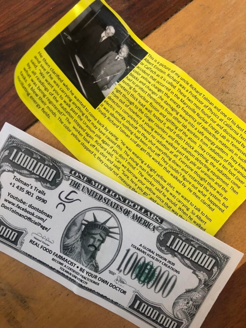
For a long time, there has been a guy running around the world advocating wearing yellow clothing to block radiation. Is this pseudoscience or might there be something to this? You might even run across this pamphlet from time to time.
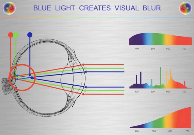
What is a possible mechanism for this observation in Nature? Many people do not know when I look into the eye of a patient with an ophthalmoscope one of the things I am looking at is the macula lutea. The macula contains the fovea where color vision is the sharpest. “Lutea” is yellow in Latin. This area collects yellow carotenoids to decrease the visual blur that is induced by blue light because blue light is the high frequency of light and it is the frequency that bends the most for the macula to handle. Yellow is the complementary color of blue.
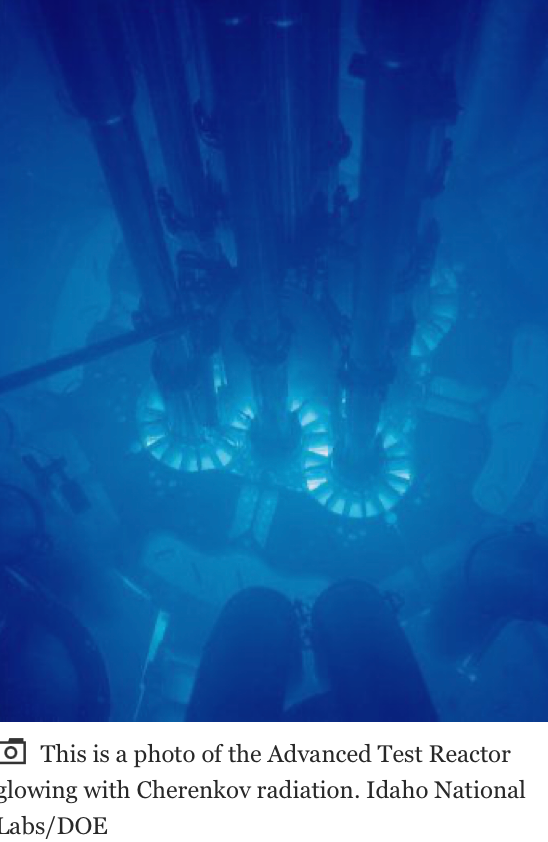
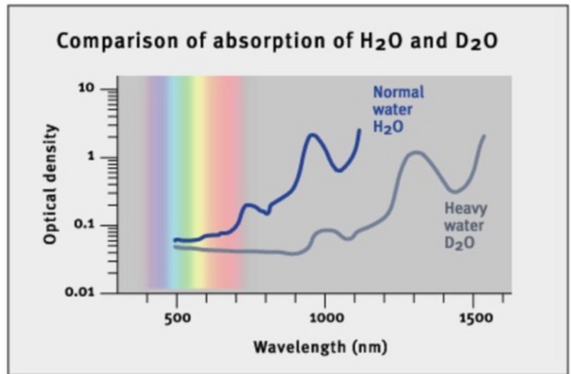
In nuclear reactors, the heavy water that contains spent fuels emits Cherenkov radiation. The frequency spectrum of Cherenkov radiation by a particle is given by the Frank–Tamm formula. Unlike fluorescence or emission spectra that have characteristic spectral peaks, Cherenkov radiation is continuous. Around the visible spectrum, the relative intensity per unit frequency is approximately proportional to the frequency. That is, higher frequencies (shorter wavelengths) are more intense in Cherenkov radiation. This is why visible Cherenkov radiation is observed to be brilliant blue. Cherenkov radiation is a form of energy that we can perceive as a blue glow emitted when the electrically charged particles that compose atoms (i.e. electrons and protons) are moving at speeds faster than that of light in a specific medium. Ironically, most Cherenkov radiation is in the ultraviolet spectrum—it is only with sufficiently accelerated charges that it even becomes visible to our retina; the sensitivity of the human eye peaks at green, and is very low in the violet portion of the spectrum. This helps explain this scientifically. Anyone who calls it pseudoscience without examining it for themselves should not be listened to.
HERE IS WHAT THE RADIATION LOOKS LIKE INSIDE A NUCLEAR REACTOR
DOES SCIENCE PROVIDE A POSSIBLE ANSWER FOR THIS OBSERVATION IN NATURE?
Can Gold be used as nnEMF radiation shield for melanin protection?
Now for more science related to this idea.
In 1988, this was published in the NY Times and I remembered reading it. Remember gold emits a yellow color because of Einstein’s relativity equations.
What was in the article I read in 1988 in New York City?
I was dental school in 1988 and I was learning about class 5 gold foil dental reconstructions and came across this article back then.
GOLD, one of the heaviest chemical elements, is the basis of a new lightweight plastic foam under development as a radiation shield.
Scientists at Texas A&M University have found a way to intersperse gold atoms with other atoms in the long molecular chains that make up polymers. By bubbling gas through the gold polymer, it can be expanded into a light foam that reportedly shows great promise as a shielding agent against neutrons and other types of radiation.
Dr. John Fackler, director of the program, said the new polymer combines gold with triphenylphosphine, a compound of carbon, hydrogen, and phosphorus, in a form that may be suitable for making anti-radiation garments.
The polymer is 11 percent gold by weight, and the gold atoms in the substance efficiently scatter or absorb most forms of radiation, including X-rays. Chemically incorporated into a polymer, gold is less poisonous than other heavy metals that also block radiation.
Metallic gold is not poisonous, but when incorporated into compounds it may be. Dr. Fackler said that because gold is chemically very stable, it tends to revert to its native state from some of the compounds it forms. ”We often have trouble with shiny, yellow metallic gold precipitating out of liquid compounds – just what refiners want, but the opposite of what we want,” he said.
The gold polymer the Texas group has developed looks like plain white plastic, Dr. Fackler said, but it emits a yellowish glow when exposed to ultraviolet light.
SUMMARY
Many might find this shocking but there is a lot of science behind these claims about the color yellow and the atom of gold. One of my former members in the mining industry could not believe this when I share it with him during a consult I did on him in 2015 about his melanoma risk.
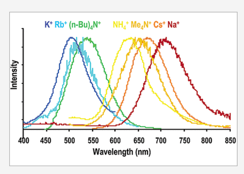
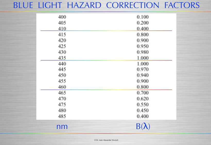
Above you see a series of bis(thiocyanato)gold(I) complexes with Au−Au interactions show luminescence in the range from 500 to 670 nm. These frequencies include the blue light frequencies that cause the blue light hazard in the human retina. This picture below from Alexander Wunsch, MD show this effect.
The series of salts correlates emission energy with the reciprocal of the Au−Au distance. As the Au−Au distance increases, the emission energy decreases. The ligand system provides no framework for the Au−Au interaction. The emission energy seems totally determined by the Au−Au distance.
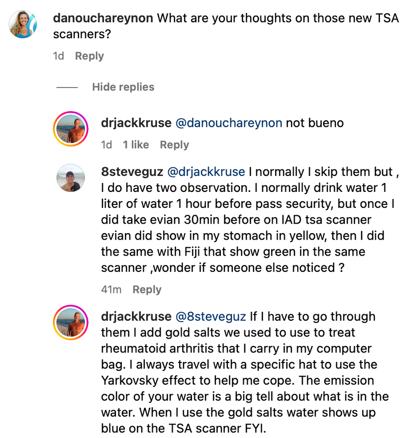
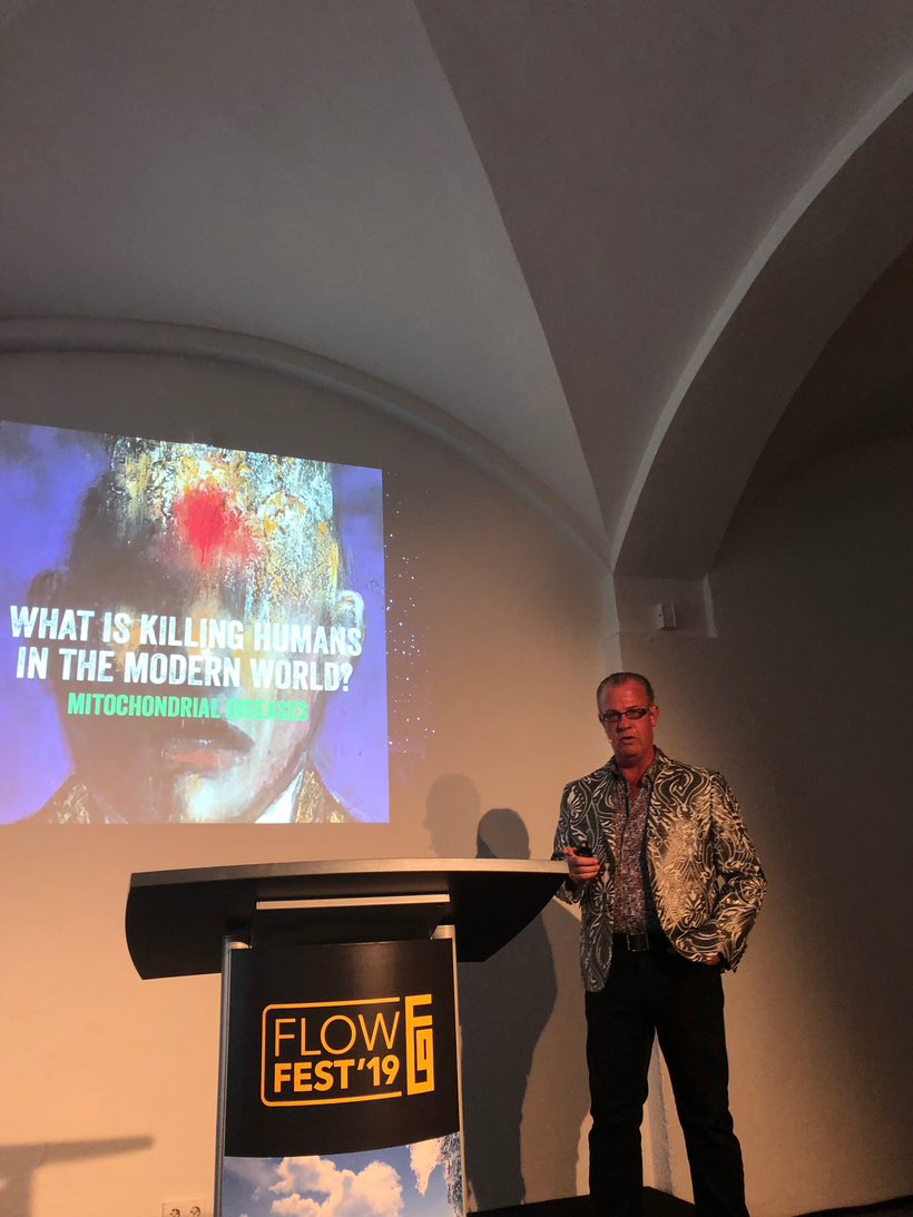
So when I am in a country where I cannot opt out of the new TSA scanners I reach into my computer bag to protect melanin in body. I usually take it 30 minutes before flight. Last time I had to do this was in Germany when I went to speak at Flowfest. I posted about this hack on Instagram.
Gold therapy, while not commonly used in the treatment of RA today, is still available in oral capsule form as Ridaura (auranofin). The injectable forms—Myochrysine (aurothiomalate) and Solganal (aurothioglucose)—are no longer manufactured but they can be compounded by a pharmacy. Below is my travel jacket I wore to Europe the last time I went there. The jacket is definitely not a fashion statment.
Jason Bowden Smith, a former entrepeneur member who was a mining industry executive, was with me in Mexico 4 months before the Germany talk and I told him about the jacket hacks and the gold hack and why I was doing it to go to Europe. Here is the picture taken that morning at 8:34 AM on January 4, 2018. He just did not believe what I shared with him. That maybe explains why reality unfolded as it did.
The gold salts and clothing are expensive hacks to protect melanin, but I think my health is worth the expense. I first mentioned this effect to Jason Bowden Smith in a consult in 2016. He was from Australia and had a very challenging medical history. We did a few consults when he was alive & during a consult I told him about these effects for long flights to mitigate TSA scanners, cosmic/gamma radiation exposure, and WiFi toxicity from passenger use around him.
Gold therapy can induce an internal Yarkovsky effect (below) for melanin protection. People who have had melanoma and who still are afraid of the sun should be told about this mitohack. Very few of my clients needed this. Unfortunately he did need the advice and he got it. UInfortunately, he believed a lot of the propaganda that is floating around in Australia even today about the sun.
Jason took a lot of circumpolar flights in his life. I thought this was linked to his REAL melanoma & thyroid risk and I told him about POMC and melanin. I told him a lot of what you’re learning now in this blog series a long time ago. In fact, Jason was with me in a meeting the night before my clinic opened in New Orleans in December 2018 to talk about a business opportunity about this very topic since he had deep connections with gold miners in Australia. My nurse, Chantal was present at this meeting.
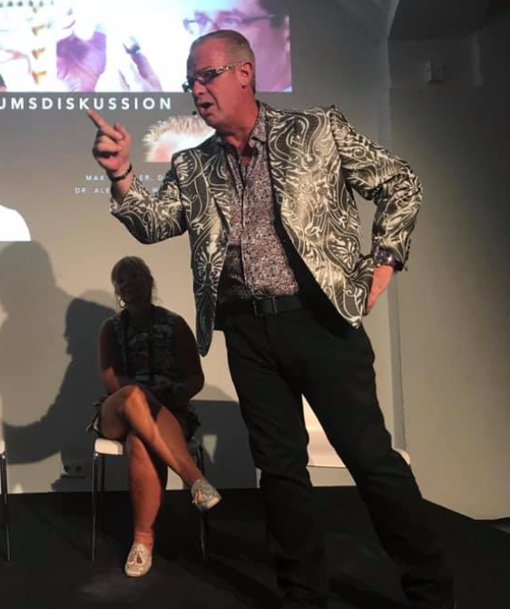
Anyone who was with me in Germany knows I did not feel well when I was in Germany because they had recently turned on 5G in the city I was speaking in. I distinctly remember Jason mocking me before my talk when I took my clothes off and laid in the sun to get ready to speak. He kind of pissed off my nurse who was with me at the event. He just did not believe that something on the surface could have that big an effect on our mitochondria below. I told him that the gold/yellow can protect quantum coherent process happening with 1/2 nuclei spin magnetic moments in our colony of mitochondria to maintain quantum coherence. I told him everything that is important in quantum biology happens on the surface before it changes the matter inside of us. I told him that in June of 2018 I was goning to unleash this in Vermont. I did. Here is the slide from that event.
I spoke in Germany right after the Vermont event on July 6th, 2019. Something else happened at that talk that others can verify. The meeting organizer subtracted 45 minutes from my talk and only gave me 15 minutes to speak. Matt Maruca and Jason were both there also as a speaker and they saw this go down. He made me a bet that there was no way I could do a nnEMF talk for the German biohacking community in the 15 minutes. I went and powered up in the sun, drank one liter of DDW, and laid shirtless for two hours before my talk and missed many talks doing so. Matt then got a red LED stop clock and put it in his lap to show me my time to see if I could do it. Notice the first slide in the talk.
I finished the talk at 14:59 and everyone who was at the event was astounded I got the talk done in such a short time frame. The talk was so good that the Q & A lasted two hours outside the event when I was ushered off the property because the event was closing. Jason Bowden Smith spent that two hours in the sandpit of the event with me trying to ask me more about this science. It was the last time we met live in person and we never spoke about it again. He died soon after it. I feel that he never called me back to speak about things because he felt foolish that he had an answer from 2016 and thought it was a joke. Generally I don’t joke around on consults. I am all business.
Below this was me in Germany live on the stage during the Q&A before it spilled outside. That jacket was tailored to be infused with gold microfiber threads hidden in the silver etchings, hence the reason I bought it and wore it. I still have this jacket today.
The Yarkovsky effect is defined as a thermal radiation force that causes objects to undergo semi-major axis drift as a function of their size, orbit, and the COLOR of the material properties in the matter that makes up the object in question.
Melanin has a massive Yarkovsky effect inside of cells because of its color. so anything you can do to reflect and deflect incoming radiation should help protecrt you melanin sheets.
The Yarkovsky effect is hard to predict, though. Scientists have to know the asteroid’s mass, size, and shape. They also need to know the patterns of light and dark on its surface, because lighter colors reflect more sunlight, while darker colors absorb more.
The best measurement of the Yarkovsky effect so far is on an asteroid named Bennu. The effect has altered Bennu’s position by about a hundred miles in just a dozen years. So when you hear something that sounds ludicrous. Remember it is the height of ignorance to say it is pseudoscientific without looking deeper at the issue before making a decision.
Just because something’s logical, doesn’t mean it’s not wise. Something can make sense in and of itself, but when tested against reality yield an undesirable or inefficient outcome. Being seduced by impractical logic is antithetical to wisdom, you just feel smart despite being wrong. Sometimes chosing comfort over wisdom might kill you.
2. A version of this article appears in print on Sept. 6, 1988, Section C, Page 11 of the National edition with the headline: SCIENCE WATCH; Gold as Radiation
Supraventricular tachycardia SVT is a broad term for a number of tachyarrhythmias that originate above the ventricular electrical conduction system (Purkinje fibers). read the link below to learn about SVT. This rhythm is usually the first symptom in melanin degradation of the cardiac plexus. I showed you this picture in Quantum Engineering #47 for the first time.
In ACLS codes we use adenosine via IV push to rid the heart of this detrimental rhythm when the patient is not symptomatic. SVT can lead to rapid cardiac standstill. We are seeing this more commonly now due to the spike protein inflammation of the heart by mRNA vaccines. I believe this was behind the demise of Demar Hamlin and JJ Watt.
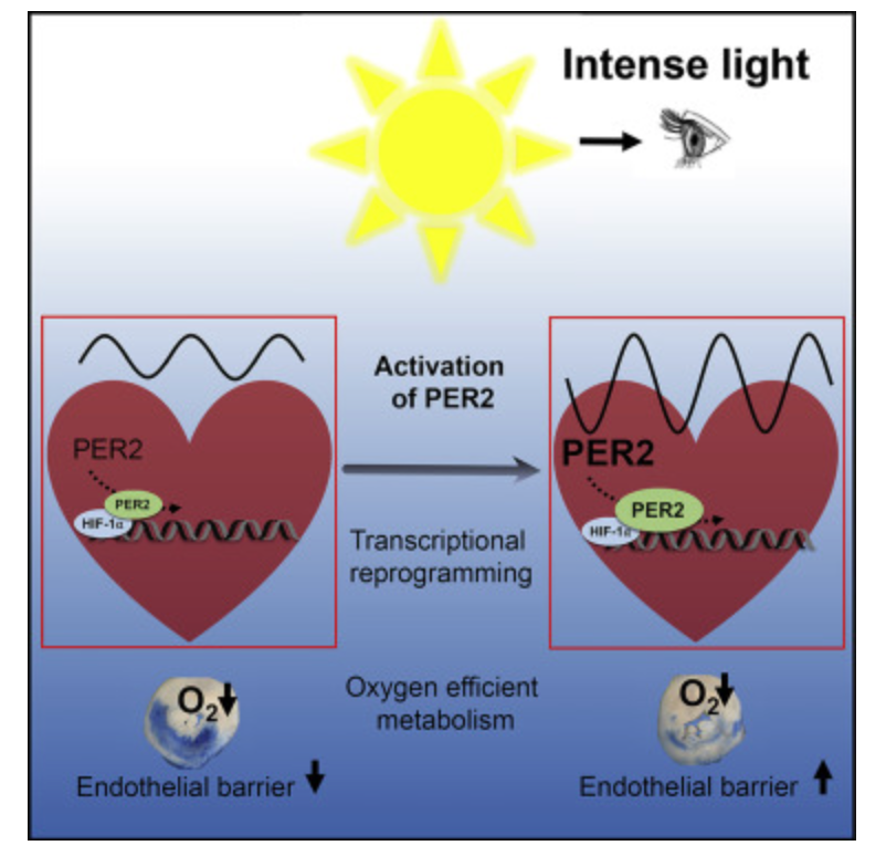
IS THERE NOW EVIDENCE THAT INTENSE RED LIGHT can do THE same thing using the PER circadian gene? Does this imply that red light is a drug equivalent?
YES, it does. The picture above shows you this.
Is there more to this circadian story you need to know? YES.
You know my answer and you should know what Dr. Tina Kuru says about this………..Red light is a drug equivalent to adenosine use. This is a message Big Pharma wants buried from centralized MDs.
In this paper below in an effort to find out why red intense light can do this, researchers developed a photonic strategy using optogenetics to protect the heart using intense light to target and manipulate the function of the PER2 gene which is expressed in a circadian pattern in the part of the brain that controls circadian rhythms. (Sounds like something Dr. Kruse would suggest no?)
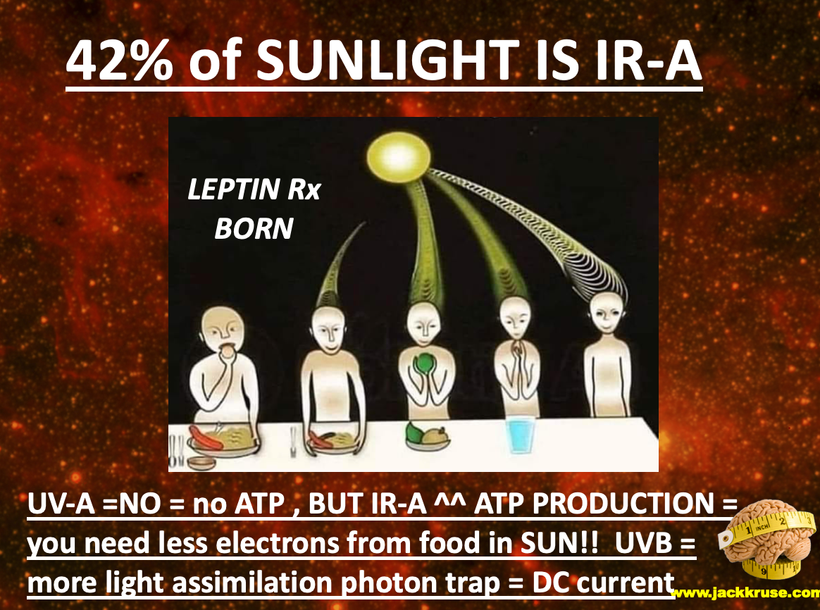
You do know that sunlight is made of 42% intense IR-A light huh?
By amplifying this gene to improve the periodicity of the cardiac clocks, the researchers were just using LIGHT PHOTONS to do this job; they found that it protected cardiovascular tissues against LOW OXYGEN conditions like myocardial ischemia, caused by reduced oxygen flow to the heart. Dr. Kruse called low oxygen situations pseudohypoxia (low NAD+). These are all associated with low NAD+ levels in cytochrome 1 and leptin resistance with low delta psi on the inner mitochondrial membrane = low redox power.
They also discovered that the light increased cardiac ADENOSINE, a chemical that plays a role in blood flow regulation. Hey didn’t Dr. Kruse just do a massive post on ADENOSINE last week on his page?
Hey, isn’t leptin resistance a synonym for melanopsin dysfunction? Isn’t melanopsin dysfunction a synonym for a melanin problem? Yes, it is. Tell me again how that works Dr. Kruse?
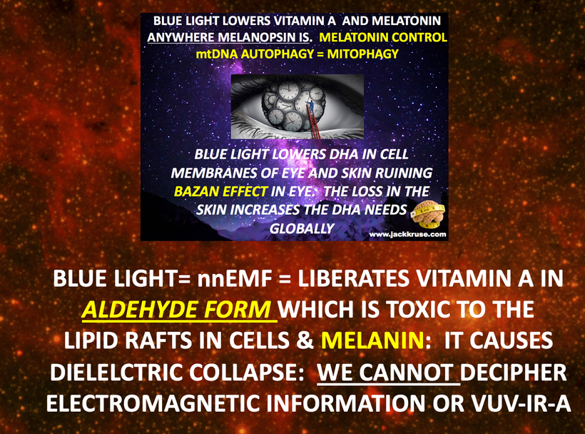
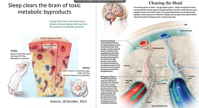
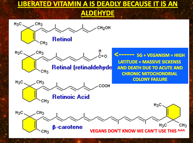
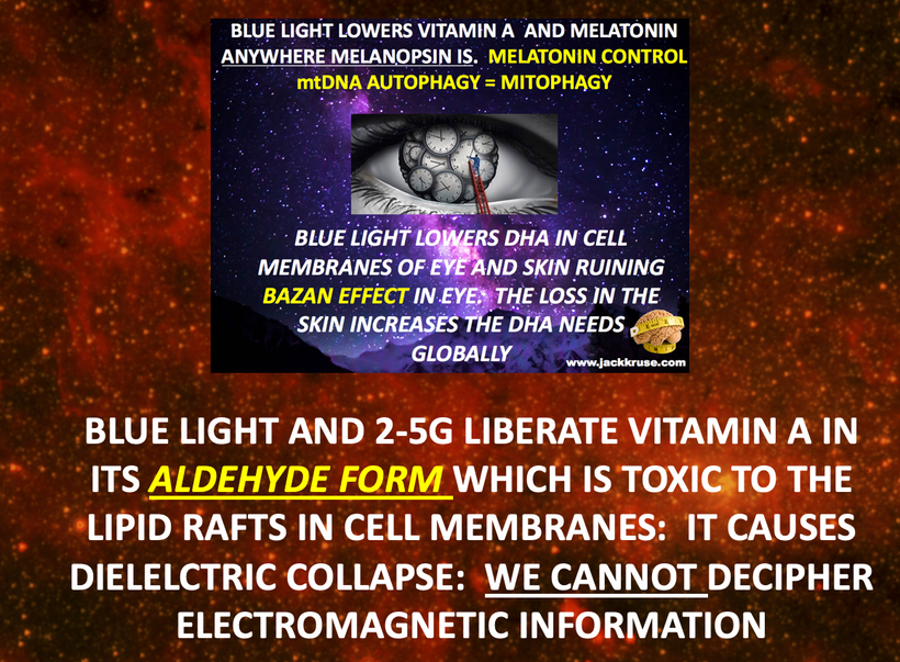
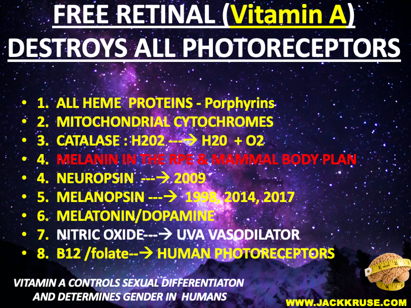
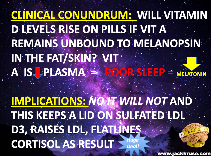
Dr. Kruse Response: Blue light and nnEMF liberate Vitamin A from our cells and cell membranes to raise its presence in the blood plasma and this lowers plasma levels of Vitamin C and Vitamin D. This destroys melanin in the cardiac plexus and it degrades into adrenaline and noradrenaline that begins to stimulate the heart adrenergically. It also has effects on the glylympaphatic system in the brain and on slow wave sleep. When Vitamin A is liberated by non terrestrial light or trauma, we are inducing a light stress and this causes Vitamin A to becomes an aldehyde that becomes a wrecking ball for many non visual photoreceptors like melatonin, dopamine, NO, adrenaline, noradrenalin, L-Dopa, P-450 enzymes, RBCs, cytochromes, and melanin.
This wrecking ball action destroys the small molecule modulators of the mammalian circadian mechanism. PER1 and PER2 are gears in that eye clock mechanism. Once the molecular clock in the eye and peripheral clocks goes awry the implications for many neolithic diseases spiral out of control. What are some of the Vitamin A proteins involved in this downward spiral?
They are called retinoic acid receptor-related orphan nuclear receptors or RORs for short. The RORs have several isoforms too called RORα-γ. These proteins are also under the transcriptional control of CLOCK/BMAL1 heterodimers. CLOCK and BMAL1 are positive regulators of circadian gene expression, and PER and CRY are the NEGATIVE FEEDBACK LOOP regulators that operate under day and night cycles.
These are the positive and negative feedback arms of the circadian mechanism. They must be coupled properly to terrestrial light to operate well and control all growth and metabolism, protein synthesis and hormone production and release. It also controls receptor biology. It controls EVERYTHING. These are post transcriptional explosions that blow up the whole system of optical signaling and require the cell and mitochondrial to rebuild them all while our power plants are experiencing a brown out. This is not ideal.
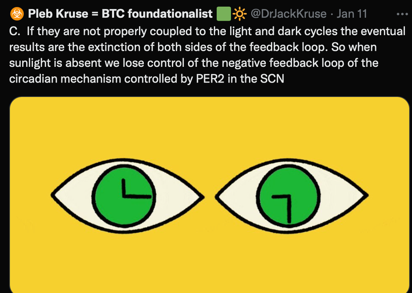
If they are not properly coupled to the light and dark cycles the eventual results is the extinction of both sides of the feedback loop. That is how all human disease begins. It is circadian biology that couples all the molecular clock genes in humans and the SCN of the eye drives the program and the major timekeeper.
Remember this lesson from the past:
Classic Paroxysmal SVT has a narrow QRS complex & has a very regular rhythm. … A rapid heart rate will significantly reduce the time which the ventricles have to fill. This is why degradation of melanin to adrenalin and noradrenalin is a real problem. Carefully look at how melanin degrades on the top line during periods of hypoxia. The chemistry in hypoxia goes from right to left.
The heart fills during diastole, and diastole is normally 2/3 the cardiac cycle. A rapid heart rate will significantly reduce the time which the ventricles have to fill and as a result this lowers cardiac output and oxygen delivery = hypoxia. As a result of this the heart tries to beat faster to improve global hypoxia and this worsens the situation. It fast forwards on itself. The reduced filling time results in a smaller amount of blood ejected from the heart during systole. The end result is a drop in cardiac output & hypotension.
With the drop in cardiac output, a patient may experience the following symptoms. These symptoms occur more frequently with a heart rate >150 beats per minute as the ECG strip shows:
Shortness of air (S)
Palpitation feeling in the chest (S)
Ongoing chest pain (U)
Dizziness (S)
Rapid breathing (S)
Loss of consciousness (U)
Numbness of body parts (S)
The pathway of choice for SVT in the tachycardia algorithm is based on whether the patient is stable or unstable clinically.
The symptoms listed above that would indicate the patient is unstable are noted with the letter (U) in a code situation. This can present outside a code situation when someone has a very low redox state because of a very poor environment linked to blue light and nnEMF toxicity. This mimics adrenal fatigue and brainstem pathology, sleep disorders, and eating disorders I wrote about in the QE#47 blog. Stable but serious symptoms are indicated with the letter (S) above.
Insert any 3G-5G city or environment = an acute or chronic adenosine problem. What fucks up sleep ultimately at the dorsal longitudinal fasiculus? Problems with adenosine at the brain stem level. This is why ACLS algorithms for advanced caridiac life support uses adenosine to treat acute mitochondrial failure in the heart that results in SVT cardiac rhythms……..guess what drug is used for cardiac tachycardias in ACLS?
ADENOSINE via IV push = a DEFECT IN PER 1 or PER2. SOUND FAMILIAR?
How do you like me now…………?
Do you still think tech use and abuse is safe and has no side negative effects for the heart of your blood vessels? That damage can be found early in your brainstem by the DLF where glylymphatic drainage occurs. How about your eye clock or the molecular clocks in your skin. These are all places where melanin, melatonin, melanopsin and leptin are.
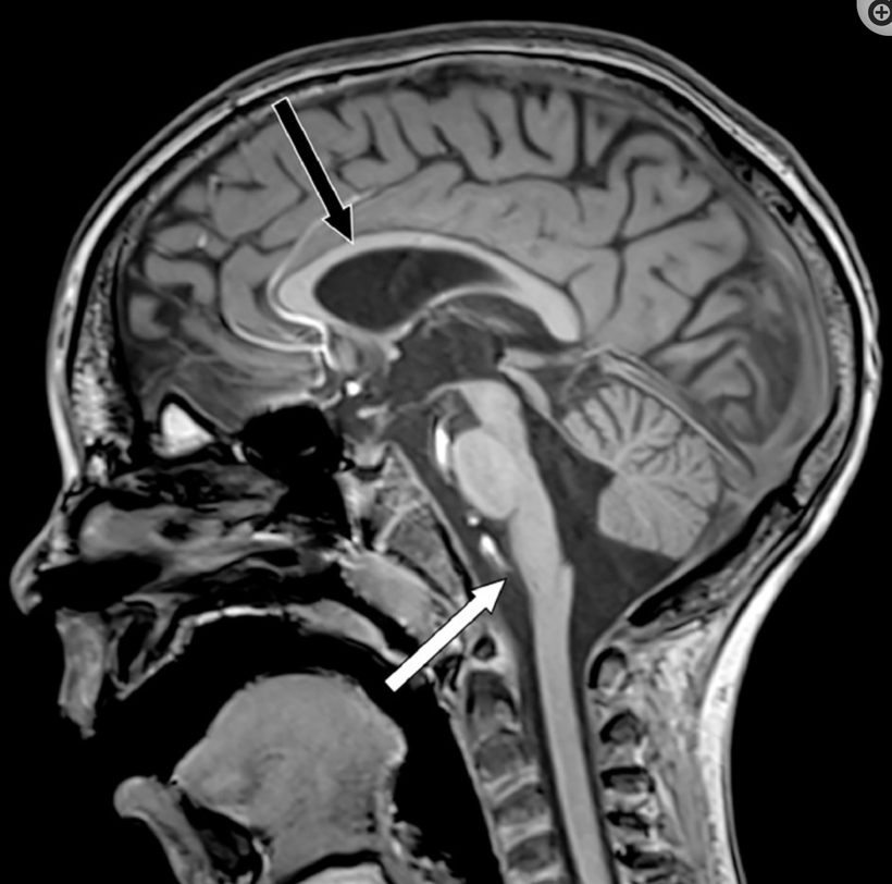
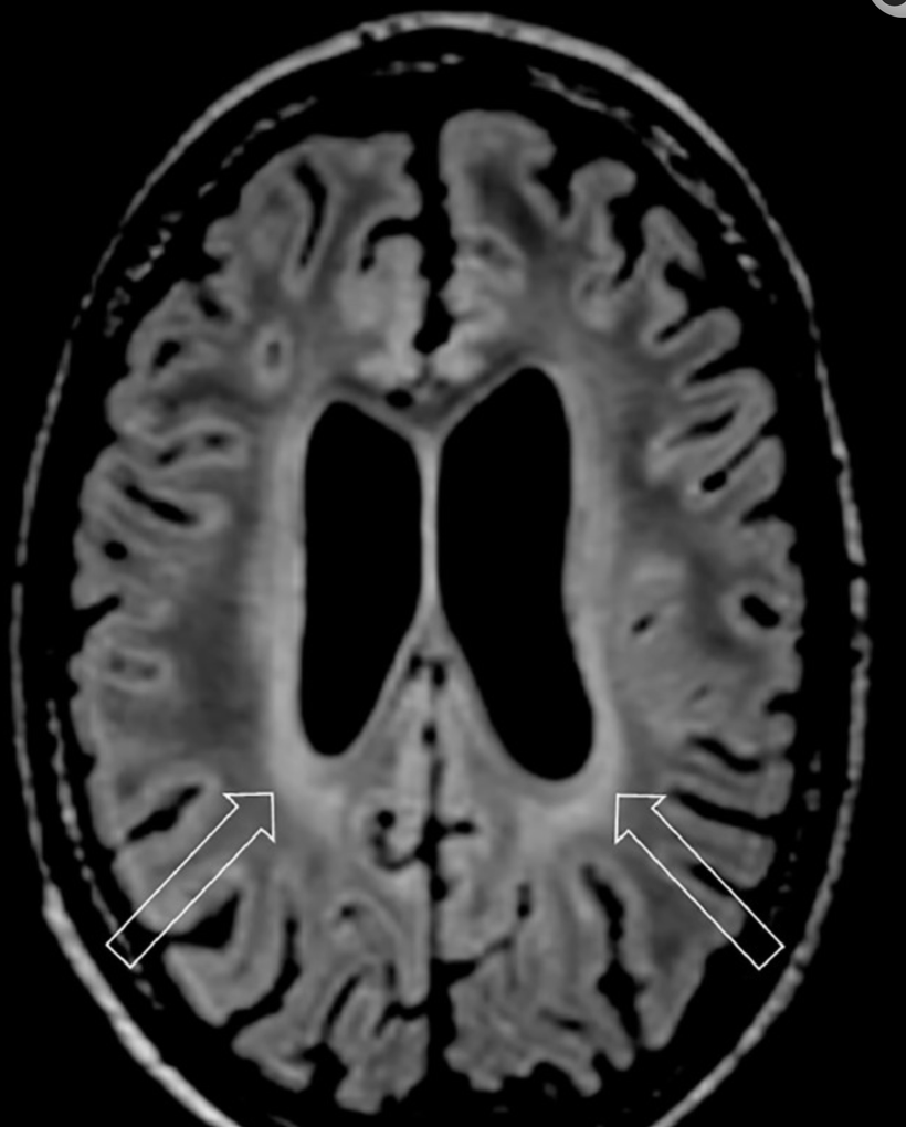
Adenosine building up occurs normally during the day when red light is powerful as we live and is one thing that drives us to feel sleepy during the night. Then while you sleep, the adenosine is cleared out by the glylymphatic system in the brainstem at the perivascular spaces, leaving you refreshed and ready to go in the morning… for most people. This does not happen in those with sleep apnea. Those with OSA have circadian mediated glylymphatic failure of AQA 4 in the brain stem due to lowered periodicity of PER1 & PER2 (above pic). I can see the widened perivascular spaces on 3 Tesla MRI images at the DLF. This is how decentralized medicine is practiced. Today, most people that have a version of EHS that affects adenosine at the brainstem level which causes their sleep destruction and hypoxia via this mechanism. This mimics adrenal problems (PVN) and causes wild symptoms doctors cannot explain.
Who else can get these problems sans a technology addiction?
if you follow your genetics from 23 and me…
There is a genetic variant in the adenosine deaminase (ADA) gene that decreases the clearance rate of adenosine. People who carry the variant may get more slow-wave, deep sleep at night, but they also may need to sleep a little longer o feel refreshed the next morning. These people are at high risk of neurodegeneration and for normal pressure hydrocephalus and glylymphatic failure.
What is the decentralized diagnosis that your centralized sleep MDs will never resolve for you?
Check your genetic data for rs73598374 (23andMe v4, v5; AncestryDNA):
C/C: normal clearance of adenosine
C/T: reduced clearance of adenosine, more deep sleep but may feel sleepy when waking up
T/T: reduce clearance of adenosine, more deep sleep but may feel sleepy when waking up.
How do you like me now?
Intense RED LIGHT via PBM/LLLT is the first step I like I use at Kruse Longevity Center for melanin renovation of the cardiac plexus that I wrote about in QE #47. It is always cardioprotective. The red light of the sun is better than my fake red light panels. Shocking huh?
It shocks only those who are not on the path of becoming a decentralized Black Swan Mitochondriac.
If you do not listen to me you might wind up needing a shock from a defribrillator sometime in your life from tech abuse or from excessive spike protein build up in and around these areas in your body. See my blog on Demar Hamlin or JJ Watt for more detail.
Today you get to see see what melanin looks like on MRIs of some of my patients.
Melanocytes are normal, neural crest-derived cells present in the human leptomeninges (pia and arachnoid membranes) primarily at the base of the brain, the posterior fossa, and around the upper cervical spinal cord. They are very close to cells with POMC expressed to some degree.
Key point: When these tumors are found in the trigeminal cave, then they are associated with a nevus of the dermatomes corresponding to the trigeminal nerve on the face. The benign dermal melanocytic nevus usually involves the ophthalmic (Va) and maxillary (Vb) divisions of the trigeminal nerve (CN V). This again shows the neural create connections and the neuroplasticity pathways one should expect in melanogenesis from these movements in mammals.
Primary melanocytic tumors of the CNS can manifest as solid masses or as diffuse dissemination within the subarachnoid space. They range in histologic grade from benign to malignant, differentiating between the following entities:
Melanocytomas are more common in women (mean age 45-50 years old). A prolonged evolution of clinical signs of myelopathy or radiculopathy prior to surgical resection (from 5 to 10 years) has been documented.
I believe this is true because women tend to wear more clothing over their bodies due to cultural and societal reasons.
Approximately 100 cases of melanocytomas have been reported in the CNS (brain and spinal cord) since Limas and Tio coined the term “meningeal melanocytoma” in 1972. In the spinal cord, most cases of melanocytomas are found in the extramedullary intradural compartment, at the cervical and thoracic spinal levels. An intramedullary location as our case depicts is extremely rare, with only 24 cases reported before.
The unique paramagnetic properties of melanin result in a relatively specific MRI pattern for melanocytoma, consisting of iso- or hyperintensity on T1WI and iso- or hypointensity on T2WI, and with homogeneous enhancement. The differences in MRI signal intensities relate to a variable degree of tumor melanization.
Paramagnetic means it is drawn to magnetic fields. This is a clue why the tumor was in this location.
Definitive diagnosis is based solely on histopathological and immunohistochemical examination.
The distinction between melanocytoma and melanoma rests on the identification of cytologic atypia, mitotic activity, necrosis, and neural parenchymal invasion.
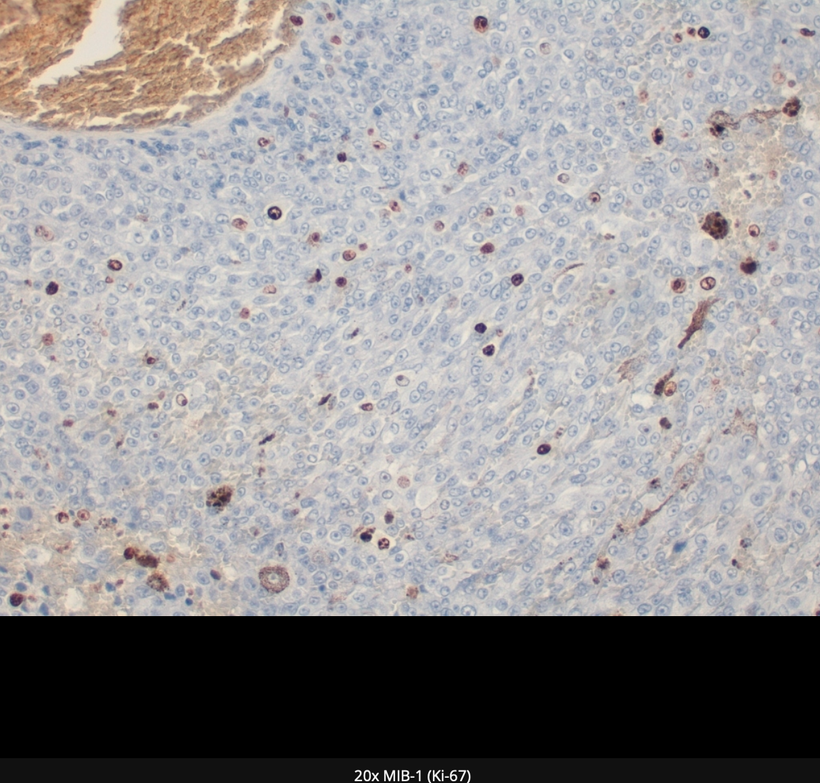
MIB-1 (Ki-67) labeling index seen in melanocytomas is low (0%-2%) while in primary melanomas is higher (2%-15%). Based on a proposition by Brat et al. the WHO classification assigns an intermediate grade to melanocytomas with increased mitotic activity and infiltrative growth that fail to meet all characteristics of malignant melanoma. In this case, MIB-1 (Ki-67) proliferation index was 5% and a definitive diagnosis of intramedullary Intermediate-grade melanocytoma of the thoracic spine was made.
CURRENT CENTRALIZED MEDICINE BELIEFS
Although classified as benign, meningeal melanocytomas may behave aggressively and a limited number may transform into malignant melanomas. Complete excision is the treatment of choice, however, this is often not possible as intra-operative hemorrhage may be severe. Furthermore, local recurrence has been reported even after gross total removal. Due to the risk of tumor recurrence even after complete excision, adjuvant radiation therapy is advised in cases of both complete and incomplete resection. I no longer believe this.
EPIDEMIOLOGY
Peak presentation is in the fourth and fifth decades, although these tumors have been diagnosed in all age groups. Occurrence in children is very rare!
RADIOLOGY IMAGING
The patient presented after breast cancer resection with a 2-year history of progressive paraparesis, paresthesia, and dysesthesia in the left lower limb. Neurological examination demonstrated generalized hyperreflexia and clonus of the left foot.
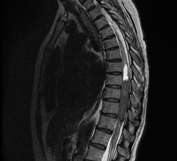
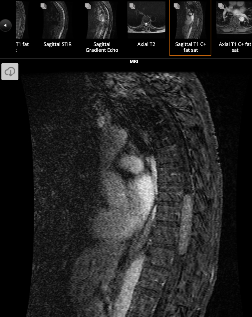
The above MRI is done on a 1.5 Tesla magnetic and shows a solitary well-defined fusiform intramedullary lesion involving the spinal cord at the T8-T11 level, hypointense on T2WI, moderate hyperintense on T1WI, and presenting homogeneous enhancement of its solid component after gadolinium administration.
Lesion associates a cystic component in its cranial pole, and presents susceptibility artifacts in the T2WI-GRE corresponding to its melanin component and the presence of degraded blood products.
T2WI hyperintense per focal tumoral edema at the T5-T7 levels and conus medullaris is noted, suggestive of myelopathy.
OTHER IMAGES:
3 case questions are available
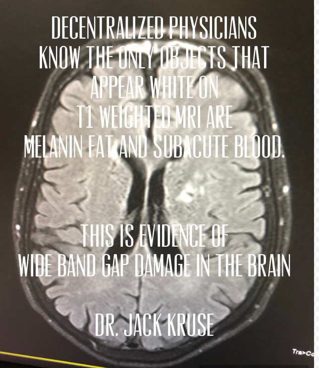
Q: The presence of hyperintensity on T1WI can be an important clue leading to a specific diagnosis, as happens in this case above. Which are the causes of T1 hyperintensity?
A: Melanin. Gadolinium. Fat. Blood forming proteinaceous substance. Some paramagneticstages of blood. Mineralization. Slowly-flowing blood. Calcium. (Pic below)
Q: What’s the most frequent T1WI appearance of meningeal melanocytomas?
A: Isointense or hyperintense depending on the amount of melanin content present.
Q: What’s the most frequent T2WI appearance of meningeal melanocytomas?
A: Isointense or hypointense depending on the amount of melanin content present.
The slide below reminds us that when we are looking for melanin loss we should see it missing in T1 weighted MRI images.
Radiological differential diagnosis includes:
primary or metastatic malignant melanoma
melanotic schwannoma
melanotic meningioma
melanoblastosis
cavernous malformation
astrocytoma
ependymoma
Operation report:
Tumor resection was performed through osteoplastic laminectomy and under electrophysiologic intraoperative monitoring. Tumor fragments were soft, brownish, and hemorrhagic. Melanin tumors are always bloody because they are always associated with brisk blood flow due to their need for massive oxygen consumption. This mimics what we see in retinal bleeds around the RPE and Bruch’s membrane.
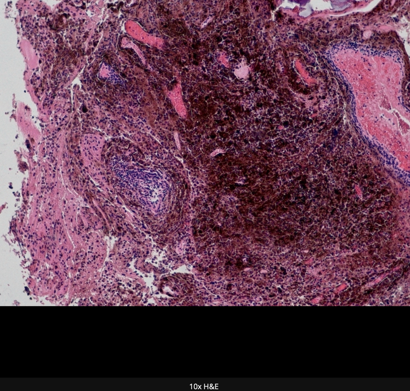
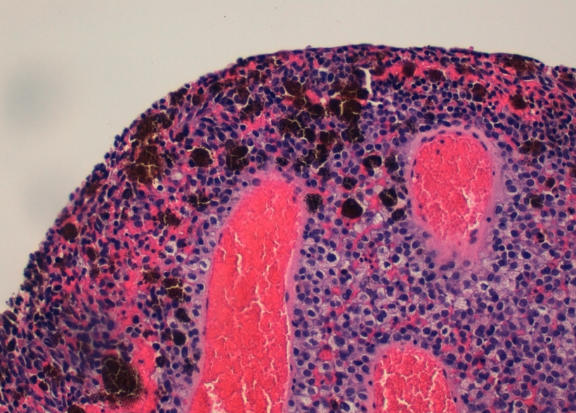
PATHOLOGY SLIDES
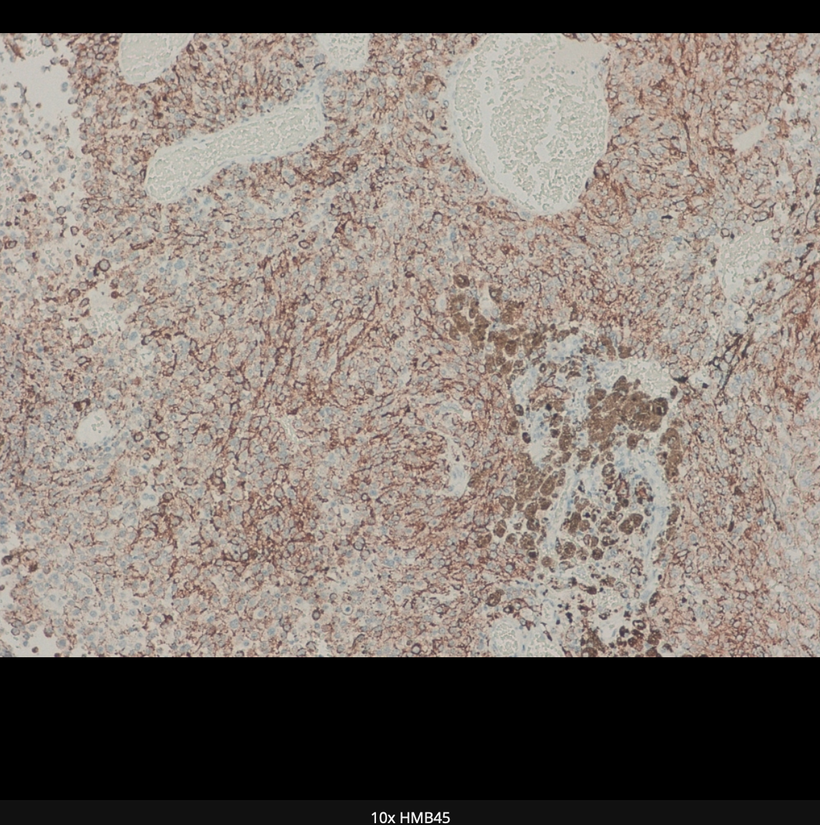
H&E stained slides above show in sequence sheets and nests of cells with mild nuclear pleomorphism and prominent nucleoli; note the marked melanin pigment deposition. An immunohistochemistry study revealed positivity for melanocytic markers Melan A and HMB45. MIB-1 (Ki-67) proliferative mitotic index of 5%.
KEY BLOG POINT: This index clued me in that her previous breast cancer was likely related to this mass and its location. She had a WiFi router under her bed in her apartment at the level of her mid-thoracic spine.
Meningeal melanocytomas are most commonly found in the cervical and thoracic regions (intrathecal-extramedullary). Within the spine, melanocytomas present as intradural masses, and maybe intradural extramedullary or rarely intramedullary. They are most commonly found in the upper cervical region, as melanocytes are most concentrated at this location. I believe the real reason is that this area is usually covered and not illuminated by sunlight. Even more interesting is they are less common in the intracranial compartment because rarely do we lose all VUV-IR-A inside the brain. We would most commonly see this in glioblastoma multiformans cases instead.
CITES
1. Louis DN, Ohgaki H, Wiestler OD, Cavenee WK, Burger PC, Jouvet A, Scheithauer BW, Kleihues P. The 2007 WHO classification of tumors of the central nervous system. (2007) Acta neuropathologica. 114 (2): 97-109. doi:10.1007/s00401-007-0243-4 – Pubmed
2. Karikari IO, Powers CJ, Bagley CA, Cummings TJ, Radhakrishnan S, Friedman AH. Primary intramedullary melanocytoma of the spinal cord: a case report. (2009) Neurosurgery. 64 (4): E777-8; discussion E778. doi:10.1227/01.NEU.0000341516.22126.AA – Pubmed
3. Liubinas SV, Maartens N, Drummond KJ. Primary melanocytic neoplasms of the central nervous system. (2010) Journal of clinical neuroscience: official journal of the Neurosurgical Society of Australasia. 17 (10): 1227-32. doi:10.1016/j.jocn.2010.01.017 – Pubmed
4. Czarnecki EJ, Silbergleit R, Gutierrez JA. MR of spinal meningeal melanocytoma. (1997) AJNR. American journal of neuroradiology. 18 (1): 180-2. Pubmed
5. G.Q. Hou, J.C. Sun, X.J. Zhang, B.X. Shen, X.J. Zhu, L. Liang, X.L. Zhang. MR Imaging Findings of the Intraspinal Meningeal Melanocytoma: Correlation with Histopathologic Findings. (2012) American Journal of Neuroradiology. 33 (8): 1525. doi:10.3174/ajnr.A2987 – Pubmed
6. Wagner F, Berezowska S, Wiest R, Gralla J, Beck J, Verma RK, Huber A. Primary intramedullary melanocytoma in the cervical spinal cord: Case report and literature review. (2015) Radiology case reports. 10 (1): 1010. doi:10.2484/rcr.v10i1.1010 – Pubmed
7. Brat DJ, Giannini C, Scheithauer BW, Burger PC. Primary melanocytic neoplasms of the central nervous systems. (1999) The American journal of surgical pathology. 23 (7): 745-54. Pubmed
Energy is a physical concept but is not really explained well in biology. In physics, it is well explained by Noether’s theorem. That equation says that any symmetry, either local or global, implies there must be a conservation of some physical quality in reference to energy transformation to keep the system functioning.
Mammals break time symmetry by conserving POMC biology. Most people have no idea that just about everything I post falls directly in line with Noether’s idea in biology. Until you understand it clearly, you will keep making mistakes in judgments on how you live and in your health.
This is exactly what mammals do with melanin and Wide Band Gapped semiconductors within their cells. It is the focus of my current Patreon series. Time symmetry (circadian biology) can be broken, but to do it chronically, and still maintain your health, you have to conserve one thing to keep the system from falling apart = The one thing mammals conserve to satisfy Noether’s axiom is their melanin content. The KT event imprinted Noether’s theorem in every mammal cell on this planet to reserve time for this subfamily of living creatures.
What did her theorem tell us? Noether taught us that with respect to energy and momentum in the universe, energy “informs” space and time how to curve. This solves many problems in physics but has yet to have the same in biology because most biologist still has no idea how life is controlled by non-linear optical arrangements of semiconductive proteins inside cells. How did this woman give Einstein’s theories a major boost after his miracle year?
HOW DOES EMMY NOETHER THEOREM TOUCH BIOLOGY? LET’S US CANCER AS AN EXAMPLE
Most people have the centralized perception that cancer shortens our time on Earth but in the June 2016 webinar, I uncovered principles that cancer may be an old solution that nature was looking for again when we lost a key feature in cells, to allow us to operate well in our new modern world. This is regressive atavism making a reappearance in the series.
The decentralized perspective inserts POMC biology and a lack of UV light as the missing link in most cancerous tissues and this disease really develops this idea fully. Either way, no matter your perspective we are talking about “time” when it comes to cancer.
In fact, the higher the grade of the tumor the less time you have to live. This is the current belief of most patients and of allopathic centralized medicine. Becker’s work actually refutes this idea.
Have you ever thought about why time and energy are linked fundamentally and really explain cancer well?
Amalie Emmy Noether was born on 23rd March 1882 in a world in which women were not always appreciated for their intellect. In the German city in which her family lived, the local university—the University of Erlangen entirely prohibited women from being accepted as students. Despite this, Noether managed to gain special permission to sit in on lectures. This exception presumably had something to do with her father, who was a professor of mathematics at the school. Seven years later, she became the first woman to earn a doctorate degree from the university.
In 1915, David Hilbert and Felix Klein invited her to work at the University of Göttingen, a world-renowned mathematical institute, to help solve a key problem in the field of general relativity (Einstein), which treats gravity as a bending of space caused by mass and energy.
The theory seemed to have serious cancer at its core: Energy caused the bending of space, but gravity itself was energy. Thus, it would seem that the energy of bending space made yet more energy. Presumably, this would bend space more, resulting in more energy. It seemed like the theory could cascade in this manner to the point that energy would grow forever. And because this didn’t happen in reality, there needed to be a solution to the problem. Emmy’s solution, which has come to be called Noether’s theorem, was worked out in the same year she arrived and had far-ranging implications in physics.
Her theorem has yet to hit biology because biology still doesn’t realize that all biochemistry is quantized by light via non-linear optics in cells. Light tells cells how space-time how to bends inside our cells at the electronic level in us too. It turns out MELANIN is key to this information transfer. Melanin is a 100% story tied to Noether’s theorem.
HOW?
Noether’s theorem defines how time stamping in cells remains accurate. This ensures the accuracy and periodicity of the circadian mechanism in cells. All mammals have to do is keep their skin in the game where melanin is located in ALL mammals.
Energy is a physical concept but is not really explained well in biology. In physics, it is well explained by Noether’s theorem. That equation says that any symmetry, either local or global, implies there must be a conservation of some physical quality in reference to energy transformation. Modern cancers in mammals reflect a disease state of a lack of energy and time. Might cancer just be a maladaptive state to the light we live under when we look at Norther’s theorem as it relates to POMC biology? I think so. So for the concept of time, what is really conserved in cells so they become able to avoid cancerous transitions in their organs?
KEY BLOG MOMENT: It turns out, the answer is that light energy their surfaces sense itself, must be conserved at the electronic state in cells for time to manifest in living things. When UV light is conserved in biology melanin and mitochondrial biology become optimized to maximize energy conservation at the electronic level of our cells. Energy can only be conserved if the information quanta in sunlight are brought to mitochondria to help maximize autophagy and apoptosis.
Cancer is not really the disease it appears to be. It is a maladapted state that mammals took advantage of 65 million years ago. Today, mammals are being redacted from it because they chronically live under the light that is devoid of UV light exposure. Cancer is a state where space-time is deformed in a cell because UV light is missing at the electronic level of cell organization. This is why cells move in our body even today. They are looking for a source of UV light. Without movement, we call metastatsis occurs. This movement has a negative connotation in centralized medicine today but in mammals, but it really is not a defect in us. In fact, mammalian metastasis is one of our superpowers. Without diapedesis our immune system would fail. Without it, neural migration of melanin fails. Without it, embryological construction of the mammalian body plan fails. This is one of the most counterintuitive consequences of Noether’s Theorem.
As a decentralized physician, your job is to ask a lot of questions about light abuse in every cancer patient you see to get to the root cause of where their mitochondrial and melanin biology went awry. POMC loss begins where the primary cancer began. Once you find the primary cancerous lesion you can guess where the melanin loss is if you are good at remembering where that tissue came from on an embryological basis.
Is your life lived the same way every day as we go around the sun? Will you do it this way for 75-85 years and just call it a life? If you know I am speaking to you, pay deep attention to this blog. I want you to make one small change today. I want you to stop settling for easy and begin to embrace living your days with an edgy rawness that must be found in SUNRISE.
RAW: When you wirelessly connect to others from here on out do it in a raw primal fashion. Make them feel “your rawness”. Never let them feel totally comfortable around you again. Let them know you have depths they have not been invited to. Make them want to visit these levels. Become like the masterpiece in DaVinci’s collections he never finished. Become like a painting in his studio that wasn’t dry yet because of the heat in the room. Push people to their edge with your heat. One hard nudge from you and you become able to smear paint all over their painted facades. You’ll begin to change your life and you’ll certainly change everything about them without their consent.
Nature is the way of seeing the truth. Only Nature penetrates human habit, pride, passion, and intelligence to reveal wisdom. Nature erects a boundary around us that tethers us to her. Sunrise and sunset are two such boundaries. If we stray too far from the laws in either, we pay a deep toll. The relationship between what we see and what we know is never settled. Each morning we see the sunrise you are beginning the needed melanin renovation to keep you alive and away from diseases like cancer.
We know that the earth is turning toward the sun to create the picture we see. Yet the knowledge, the explanation, never quite fits the vision we experience and we never really understand how it renews us. That is due to a lack of awareness/dopamine that creates our need for transformation. This demand is built in by seasonal light changes. The demand is not great at the equator but rises to extremes at higher latitudes. This is why life is sparse above 60 latitudes.
Transformational change is a state of bold total annihilation or what we were to something we envision ourselves to be. It can not simply be a better version of the business-as-usual approach. Transformational learning is a shift in our context of reality and our point of view about ourselves.
The next slide blows the entire centralized paradigm of science up. You are not defined by your nuclear genome. You are defined by your ability to adapt to your environment. The light in that environment is the key source of energy and this is why MELANIN is the most critical semiconductor in your body. Without it, your circadian mechanism cannot operate. This explains why circadian gene regulation occurs at a post transcriptional level in mammals. If Darwin was right it would happen at a PRE transcriptional level, but it does not.
SUMMARY
Melatonin and melanin are biogenic amines linked to sunlight’s local effect on cells. POMC was created to observe these local effects in cells on the central energy pathway in mammals, namely the leptin-melanocortin pathway. Let us expand this idea to the laws of physics to see how biology follows those rules. Melatonin recycling changes local symmetry in tissues, therefore it affects the space/time dimension possible in tissues and the timing mechanisms in cells. Symmetry is often thought of globally, but it turns out Einstein’s general relativity, put local symmetry front and center in physics in 1905.
It appears melatonin and melanin control the local symmetry of light collection for cells too. It is a proxy for how effectively we collect light locally within cells and this is shared with the entire organism via leptin biology. This explains why leptin has an absorption spectra of 220nm. This wavelength is below terrestrial sunlight so the brain can be informed about the state of VUV creation from melanin sheets inside of our tissues.
Einstein’s idea was unorthodox in physics in 1905, so my ideas in this series may seem queer at first until you see its beautiful simplicity in design. Moreover, that simplicity appears over and over again in how the melanin/melatonin/POMC cycle uses Einstein’s ideas locally in cells to maintain our longevity. These things are both related to how light can flow in cells and tissues locally.
Only UV light is capable of non-linear optical effects, so it makes a lot of sense why cells are built around this part of the spectrum of visible light. Look at the SECOND picture above in this blog and carefully re-examine it now that you have read what Noether’s Theorem means to mammals.
I said this in Reality #11, “most modern technology works electronically by making electrons do the things we want them to do on semiconductors. All electronic semiconductor circuits work on the basic idea that any given electron can influence the control of other electrons and holes adjacent to those electrons. What programs electrons in cells? UV light does.
Physics since Einstein and Noether has taught us that energy and momentum in light energy tell space and time how to curve. As light falls to us it gains energy and momentum, so in this way, on a relative basis UV light photons have a weight or inertia associated with it. That weight and inertia affect our surfaces where POMC is located to record that measurement to make things like melanin, Vitamin D3, and matrix water as a biochemical signal of the photons at the electronic state in cells.
Gravitational lensing is tied to energy flows by weight and momentum changes in light. UV light offers massive changes in momentum because it has the smallest and most powerful energy in the terrestrial spectrum of solar light.
Neother’s theorem is also found in the story of grey hair. Why? Grey hair is a story of melanin and circadian biology gone awry.
This story hyperlinked here shows the IRF4 gene to gray hair by interfering with the production of melanin.
What did centralized science forget in this story above? Local control of symmetry in the melatonin cycles: the development of grey hair and melanoma are linked by melanosomes and melatonin cycles locally in cells. One thing the study in 2016 did get right is this: The mammalian story of melanin and POMC clearly shows we are NOT at the mercy of our genes.
That idea is the belief of centralized science today. I reject it as a decentralized physician now. The current study on mammalian hair genes found that environmental factors controlled about 70% of cases of hair graying. Isn’t that being explained in this series and really in those blogs now? To the centralized mindset, genes appear responsible for about 30%, at least in the Latin American cohort. Might that be a facade because Latin America is inside the tropics where UV light is very stable from season to season? I think so.
They said, “The study confirms that (going gray) is at least a mix of genes and environment”
I chuckled when I read it.
The genes in both diseases are not important in this process. The light environment that turns it on and off is what is important. Light is critical in that environmental switch on or off. Losing the local symmetry of melanin/melatonin cycles from poor environmental light signals is the key to why IRF4 turns hair grey and what causes melanoma and all cancers in mammals. Melatonin inhibits Electron Chain Transport to allow cells to recapture apoptotic efficiency to avoid cancer. This is the decentralized position on what really causes cancers.
Conservation of energy is a law of physics, and Noether’s theorem says that the laws of physics come from symmetry. Specifically, Noether’s theorem says that every symmetry implies a conservation law. Mammals must conserve melanin to remain at a dissipative state at their electronic level. Noether’s theorem clearly lays out why centralized science is a joke. She deserved a Nobel for her work, but the centralized machine does value women like Ms. Noether.
The neural retina is considered to be an extension of the central nervous system and has several features in common with the brain. The neural retina is similarly sensitive to degenerative processes, in which the closely adjacent retinal pigment epithelium (RPE) which contains melanin plays a key role in normal and abnormal physiology. There are various forms of degenerative processes, including one of the leading causes of visual impairment, age-related macular degeneration (AMD). The molecular mechanisms of AMD are continuously being elucidated, with a focus on oxidative stress due to the lack of melanin in tissues. Natural melanin is known to protect the skin from ultraviolet (UV) irradiation. This is in large part attributed to its surface quinone residues that can efficiently scavenge all free radicals. Mitochondrial oxidative stress is believed to be a contributing factor in the etiology of numerous neuronal disorders. However, the precise mechanism(s) by which mitochondrial reactive oxygen species (ROS) modify cellular targets to induce the death of neurons remains unknown.
Melanin is known to be the highest-quality scavenger of ROS and RNS in humans. Melanin is also paramagnetic and this makes it sensitive to the magnetic fields created by the spinning F0 head of the ATPase.
When we lose RPE tissue from donors with Acute Macular Degeneration we find that they all exhibit REDUCED mitochondrial redox power and glycolytic function compared with healthy donors. Recall that mitochondria make the majority of melatonin used to regenerate photoreceptors. Dopamine is the other chemical used in photoreceptor regeneration. It can be made directly from melanin. (See pic below) One should look at the top row in the slide below and realize that the eye is to the left of the aromatic amino acids and the brain side of the flow chart to the right side. Dopamine can be made directly from DOPA or melanin in the retina/brain when hypoxia is present. The topline of this slide below can go from left to right with higher oxygen tensions (UV light present) and from right to left when pseudohypoxia (low NAD+/high lactate/ low thiamine or pyruvate) is present in the mitochondria of the tissue in question.
The mitochondria are a major source of ROS where an estimated 0.4–1% of total oxygen consumed in this vital organelle is reduced to O2 . − (superoxide radical)
From a pool of RPEs from AMD donors, their tissues were found to be MORE resistant to oxidative inactivation of the (TCA/glycolysis) two energy-producing pathways and were less susceptible to oxidation-induced cell death compared with cells from healthy donors. Investigation of the potential mechanism responsible for differences in bioenergetics and resistance to oxidative stress showed RPE from AMD donors had increased PGC1α protein expression as well as differential expression of multiple genes in response to an oxidative challenge.
These findings seem like a paradox to most of the beliefs flying around neurodegeneration and retinal pathology. It raises the question what is oxidative inactivation?
In eukaryotes, two isozymes of aconitase exist; one localized to the matrix of the mitochondria and the other in the cytosol (also known as iron regulatory protein 1). M-aconitase catalyzes the reversible isomerization of citrate and isocitrate via its intermediate form, cis-aconitate, in the tricarboxylic acid (TCA) cycle. Because of m-aconitase’s unique [4Fe-4S]2+cluster which contains a labile iron atom, and its proximity to mitochondrially generated ROS, it is an ideal candidate for oxidative inactivation. Indeed several neurodegenerative diseases in which oxidative stress has been implicated, as well as in vivo and in vitro models of these disorders collectively demonstrate decreased aconitase activity
Retinal Pigment Epithelium (RPE) is fully loaded with melanin and located under photoreceptors’ outer segments and plays an important role in the maintenance of photoreceptors by completing the visual cycle and phagocytosis of shed photoreceptor outer segments. Lipofuscin is a NATURAL byproduct of the visual cycle, and it is currently believed to be a nondegradable compound that accumulates in the RPE cells and eventually damages the RPE cells and inevitably causes photoreceptor degeneration. Vitamin A has an absorption spectrum of 328 nm. This is UVB light. Vitamin A is the only Vitamin known to emit light. The emission of light is called fluorescence when absorbed light of a short wavelength and emitting light has a longer wavelength.
Lipofuscin is the major cause of fundus fluorescence that can be detected by Fundus Autofluorescent (FAF) imaging systems.
The neural retina is the light-sensitive tissue of the eye. It consists of several layers of neurons interconnected by synapses. The primary light-sensing cells in the retina are photoreceptor cells, rods, and cones. The RPE is the pigmented single-cell layer located right behind the retina, firmly attached to the underlying choroid and in close contact with photoreceptor cells. The RPE (below) has several crucial functions for vision, namely, scattered light absorption, epithelial transport, spatial ion buffering, visual cycle, phagocytosis of outer segment photoreceptor membranes, secretion, and immune modulation.
With exposure to light, during rhodopsin photolysis, toxic retinoid side products (Vitamin A) can be produced in photoreceptor cells. Biogenesis of these products occurs when two molecules of all-trans-retinal condense with one molecule of phosphatidylethanolamine in the photoreceptor membrane.
Evolution has developed a powerful mechanism that prevents the accumulation of retinoid side products in terminally differentiated photoreceptor cells. Throughout life, the debris of the photoreceptor outer segment (POS) apical part is phagocytized and digested by RPE cells (above), while new photoreceptor discs with rhodopsin molecules are synthesized by the photoreceptor inner segments. However, the lysosomal enzyme system of the RPE cell is not effective in the degrading of POS debris, because the latter is supposed to contain modified retinoid side products of rhodopsin photolysis, as well as modified lipids and proteins.
In other words, the lysosomal enzyme system of the RPE cell cannot recognize such modified molecules and does not digest them. As a consequence, lipofuscin granules (LGs), containing retinoid derivatives, are formed and accumulated in RPE cells with age. They have been long believed to be just a cell metabolism by-product. However, it has been established that LGs are one of the sources of reactive oxygen species (ROS) in RPE cells. This explains why melanin is the most prominent protein in the RPE because it handles the ROS/RNS from the LGs with ease. If melanin is not present or degraded then lipofuscin becomes a problem. Visible light exposure induces ROS formation in LGs, initiating oxidative stress in RPE cells. Oxidative stress is believed to be central to the development of AMD. The quantum biologic reality is quite different. If melanin is not present then and only then is ROS/RNS/ and lipofuscin a problem because it also consumes surrounding DHA that is needed in the repair process.Its absence is characterized by an increased level of ROS resulting in damage or modification of cellular proteins, lipids, and DNA, impairing their physiological functions. Therefore, the development of AMD is associated with the progressive accumulation of lipofuscin.
Lipofuscin is a pigmented, heterogenous byproduct of failed intracellular catabolism conventionally found within lysosomes or the cytosol of aging post-mitotic cells. Numerous studies indicate that the formation of lipofuscin is due to the oxidative alteration of macromolecules by oxygen-derived free radicals generated in reactions catalyzed by redox-active iron of low molecular weight. This protein usually is found in places where POMC is not turned on by endogenous UV light stimulus and or where melanin is absent and is supposed to be. Melanin is an excellent absorber of free radicals to limit oxidative damage. This changes the optics inside the cells. Melanin is the semiconductive protein that is designed to chelate the iron complexes to clear them. When melanin is degraded or absent this cannot occur.
The nature and structure of lipofuscin complexes seem to vary among tissues and show temporal heterogeneity in the composition of oxidized proteins (30–70%), lipids (20–50%), metals cations (Fe3+, Fe2+, Cu2+, Zn2+, Al3+, Mn2+, Ca2+) (2%), and sugar residues (Benavides et al., 2002; Double et al., 2008).
Looking at the retina is the best way to predict the topographic risk of neuronal ceroid lipofuscinosis. Below you can see the lipid deposition in the macula in AMD. This lipid deposition mimics what we see in atherosclerotic plaque formations when there is melanopsin damage, lack of melanin, and/or a lack of ferroelectric currents on the endothelium. In the eye, a lack of the ferroelectric current can occur at multiple locations to cause various diseases associated with lipid accumulation.
High lipofuscin levels in the RPE have been associated with retinal degeneration and blindness in Stargardt disease patients and animal models. Currently, centralized science believes there is no treatment to prevent and/or revert lipofuscin-driven retinal degenerative changes in humans. This slide and paper below show that with the blue light hazard injury or nnEMF damage to brain tissue LED light can really make a difference.
That belief is present because none of them realize a combination of UVA-IR-A light can stimulate POMC to create melanin to clear the iron complexes that are common in this disease.A lack of ATP is a big factor in this disease.
Most modern ophthalmologists are fully unaware that IR-A light can create ATP even when the cellular damage is overwhelming. I found this out in my clinic three years ago with some patients with AMD and bone injuries. I did not know about this issue until that time because I do not treat AMD.
There is also a belief in ophthalmology that there is no way to recover melanin creation in the RPE once humans are born. The use of high intensity IR-A light needs to be considered. This belief also needs to be revisited because it is clear that there is neuroplasticity present in the layers of the human eye that contain melanin & DHA in concert.
Stargardt disease is usually caused by changes in a gene called ABCA4. This gene affects how your body uses vitamin A. Recall when Vitamin A is liberated from opsins in the retina it is freed by incoming light. Vitamin A is normally recycled in the eye by the rhodopsin system to prevent accumulation.
The body uses vitamin A to make cells in the retina (the light-sensitive layer of tissue at the back of the eye). Then the ABCA4 gene makes a protein to clean up the fatty material that accumulates after the photooxidation of light that’s left over. In Stargardt disease, this gene doesn’t work — so the fatty material builds up in yellowish clumps on the macula. Over time, this fatty material kills the light-sensitive cells and destroys central vision.
Stargardt disease is an inherited genetic disease, which means it gets passed down from parents to children. This disease mimics what we see in the peripheral retina of people with type two blue light hazards.
When lipofuscin is seen in the inferior peripheral retina it is the first sign of coming neurodegeneration in the brain in the frontal and temporal lobes. This is where Alzheimer’s disease and frontotemporal dysplasia form.
The key feature for most neurodegenerative disorders is the accumulation of misfolded protein aggregates, framing them within the classification concept of proteinopathies or “protein conformational disorders”. However, it is important to underscore that not all neurodegenerative diseases are considered protein-conformational disorders. Proteins are semiconductive materials in cells that need constant care and rejuvenation. Melanin performs this vital function in tissues. If the redox power in the cell is suboptimal those semiconductive protein components will accumulate in the extracellular matrix of tissues ruing optical signaling.
Although the molecular underpinnings of neurodegeneration are still not completely understood by centralized medicine, if you are following my thesis, it should be obvious where the problem lies. As melanin is degraded or lost we see the loss of the RPE microvilli on the basal side of the RPE. This infolding by the microvilli increases the surface area of the RPE over a millionfold. The loss of microvilli is the first clinical sign of a POMC problem in the eye. This is the clinical sign one can see on OCT retinal scans that melanin needs renovation.
As melanin is lost at the tissue level so is mitochondrial function, so is the electric potential on lipid rafts, and melatonin levels drops from mitochondrial dysfunction. Recall from the picture above, that dopamine can be made DIRECTLY from L-DOPA when tissue level hypoxia is present. Dopamine can be created from melanin in mammals, but in the face of tissue level mitochondrial dysfunction but this ability is lost as well. This destroys many photoreceptor regeneration pathways (below).
Every thing listed above is destroyed and can lead to a myriad of diseases that afflict modern humans.
As a result, the ROS made in the eye cannot be absorbed by melanin. This increases the oxidative stress associated with the eye and into the brain via tracts that are topographically linked to the RPE and the rest of the human cortex. It also empties the retina of DHA because DHA breakdown products act to limit inflammation by stimulating autophagy. Without autophagy, we get apoptosis (apoptosis = cell loss = tissue/organ failure) with resultant necrosis. This can happen in any organ.
When it happens in the retina and AMD begins. Type two blue light hazard destroys the photoreceptors at the junction of where they meet melanin sheets in the RPE as seen below.
DHA in mammalian tissue is broken down into elovanoids and resolvins. Elovanoids and resolvins enhance the expression of pro-survival proteins in cells undergoing uncompensated oxidative stress. DHA helps stimulate autophagy to recycle photoreceptors in mammals. We have the most DHA in our tissues as a mammal for a reason; our brain is unique in the mammalian family. As we age heteroplasmy rises and so does melanin degradation. Thus, aging is associated with increasing levels of pro-oxidant factors (reactive oxygen species, ROS/RNS)
Neuronal Ceroid Lipofuscinosis (NCL)
This disease shows the effect of lipofuscin accumulation which rapidly destroys optical signaling in the brain by destroying white matter in the brain. Clinically, NCLs are associated with variable, progressive symptoms, including dementia, visual loss, seizures, and cerebral atrophy. One of the most striking morphologic changes in neurons during normal aging is the accumulation of lipofuscin (LF) aggregates, iron complexes, as well as, neuromelanin pigments. LF is an autofluorescent lipo-pigment formed by lipids, metals, and misfolded proteins, which is especially abundant in nerve cells, cardiac muscle cells, and skin. This disease shows the effect of rapid damage to the non visual photoreceptor system. Below you see the thinning or the corpus callosum (black arrow) which connects both halves the brain. You also see the thinning of the brainstem where the sleep centers are (white arrow). Even the cerebellum has prominent folia present below.
Within the Central Nervous System (CNS), LF accumulates as aggregates, delineating a specific senescence pattern in both physiological and pathological states, altering neuronal cytoskeleton and cellular trafficking and metabolism, and is associated with neuronal loss, and glial proliferation and activation. Traditionally, the accumulation of LF in the CNS has been considered a secondary consequence of the aging process, being a mere bystander of the pathological buildup associated with different neurodegenerative disorders. This will be seen in an enalarging ventricular system as the matter of the brain becomes atrophic. This is visualized by the two arrows in the MRI below. The real offender is a lack of melanin and DHA in our tissues due to nnEMF and blue light destruction liberating Vitamin A.
The enlarged state of the ventricles above destroys the FM radio station you learned about in QE #47 & 48. All of these things show you where melanin is being destroyed with mitochondria as the non visual photoreceptor system is being destroyed.
I believe self-organizing criticality theory (SOC) needs to critically look at the work of Ilya Prigogine on how dissipative systems operate in mammals. I believe this is why SOC is stuck in the mud with modern centralized neuroscientists. To understand this idea you need to understand how the liberation of Vitamin A from our non-visual photoreceptors begins the process of diseases by destroying all of our photoreceptors.
People in centralized science seem to forget that all of the cytochrome proteins in our mitochondria are also heme-based photoreceptors that are linked to this damage cascade. This is found in every human disease I have studied the last 20 years, from Autism & Alzheimer’s diseases all the way to zoonosis. This fully explains how our environmental choices lead to mitochondrial destruction in a few steps. This is wholly an idea in decentralized science that needs to be understood.
Why do cells organize by changing their size and shape in mitochondria before diseases or phenotypes begin to vary Uncle Jack?
Efficiency of maintenance of metastability in the tissue is the short answer. The science of how heat interacts with matter is called thermodynamics and this science can be boiled down to 4 laws. Most people talk about the main three laws, but the fourth one shows us how energy flows in the universe by a microscopic reversible process. The 4th law is “The Onsager reciprocity relationship.”
This law shows how symmetrical coupling of processes (circadian mechanisms in cells) can arise naturally in a system under energy flow that moves from an areas with a lot of energy to one with a small amount of energy. The area around a mitochondria where melanin is unusually prominent in humans and cell membranes of the skin is loaded with energy because of proteins that act like a capacitor. This is what melanin does for cells. It drives the Onsager reciprocity to operate in cells. A capacitor is a battery holds a ton of charge. This is what melanin is doing in cells. Lipofuscin is evidence of a destroyed battery.
In nature, Lars Onsager showed that energy moves from high potential to lower potential naturally. He is often given credit for this unofficial 4th law of thermodynamics. All these laws of thermodynamics are subject to one main law of how energy relates to matter in the universe. That law is mass equivalence, or E=mc^2.
All things in biology, boil down to size and shape, not genetics as the current centralized paradigm professes. This is why size is a signal for ongoing thermodynamic changes in the Vitamin A cycle of the POMC neurons in the suprachiasmatic nucleus before gender tracts in the hypothalamus are laid down in the embryo. This is evidence of light changing the signal for mitochondrial autophagy and apoptosis programs related to gender issues in decentralized fashion. We see the changes in the tissue at a macroscopic level.
SUMMARY
The blue light hazard creates lipofuscin in tissues by Vitamin A liberation. It is not a consequence of any other process linked to DHA as some of the ignorant have spewed on social media.
Vitamin A liberation always is associated with sleep disturbance.
DHA content in your cell membranes INCREASES your ability to engage in autophagy because it breaks down into chemicals involved in the visual and non visual photoreceptor system that resolve inflammation via wound healing. Vitamin A destruction caused by blue light impedes this effect by lowering resolvins and elovanoids in the photoreception repair process in humans. This is what leads to increases in lipofuscin in tissues. It is an early marker of organ failure. https://www.sciencedirect.com/science/article/abs/pii/S0531556514000722
Autophagy is critical in growing and maintaining the neocortex size of humans. This is especially true in cases of neurodegeneration and autism when you are trying to repair defects from Vitamin A liberation in any mammalian tissue. This turns Vitamin A into an aldehyde that acts to destroy all visual and non-visual photoreceptors that impede tissue regeneration in humans. This leads to increases in lipofuscin. It is a marker for Vitamin A liberation.
Anyone who thinks this is due to too much DHA needs to be placed on ignore. it is 180 degrees opposite this opinion.
The quality of their sleep (also linked to the Vitamin A cycle) is also critical because it maximizes autophagy and does not deplete our stem cells in the recycling process. The more efficient autophagy is the more you protect your stem cell supply and the longer you can live or regenerate.
Autophagic efficiency is linked directly to energy-mass equivalence (Einstein). The more electrons you have in your brain the more autographic efficiency you will have to absorb exogenous or endogenous light production. All cells created in the young human’s brain are newly minted post-mitotic cells (think Autism as an example). Broken autophagy at a tissue level almost always tells us there is a problem with the sleep centers in dorsal longitudinal fasiculus mentioned in recent blogs. Many diseases have this effect built into pathogenesis.
Autophagy augments longevity in all post-mitotic cells in 5 ways:
The 5 cytoprotective effects of autophagy:
1. Reduced accumulation of toxic protein aggregates or misfiled proteins via Vitamin A damage
2. Destroying bad mitochondria via mitophagy and sensed via size and shape changes in mitochondria
3. Reduced apoptosis to prevent stem cell depletion = organ failure
4. Reduced necrosis to stop organ failure.
5. Improved hormesis by increasing the redox stickiness of semiconductors made of carbon and water like melanin & collagen.
Why is blue light bad for mitochondria and why does it cause us to eat more than we should? All foods are linked directly and indirectly to photosynthetic webs. Most modern humans are divorced from evolutionary theory and from mitochondrial science. Blue light/nnEMF spreads out the respiratory proteins on cytochromes and destroys the cytochromes via Vitamin A liberation and this DECREASES our natural coupling ability in our mitochondria DNA was given to us by our maternal haplotype. https://www.ncbi.nlm.nih.gov/pmc/articles/PMC4684129/
People at war with their species’ directives from Nature will always cause collateral damage in the lives of those around them. It begs the question, how smart are we really today? Humans now write books to collect their ideas. Books are really harbors for these beliefs. This book below might hint that our current beliefs around technology might be the death of human intelligence. Humans are now creating books with the idea that we can “do better” than nature. Might this be a situation where our ‘best invention’ or our final act on the planet? Our assumptions are based on the lenses we use to view the world. Are we now at the time where we have to put some Windex on our glass eyes? I believe it is the job of ‘mitochondriacs’ not to be on the same side as the executioners.
2. Is time a risk for us all?
Without risk, life is worthless; without risk, biology does not work. Evolutionary biology is based on the random risk of natural selection and the conditions of existence. Evolutionary biology has used time as its canvas of innovation. Darwin coined both of these terms but only explained the first well. The second idea was his better one, but he simply could not explain what conditions of existence meant. The reason was simple. He did not understand quantum field physics.
Of the two, he said he knew, the latter was the most powerful force in biology. He was dead right, but since his time evolutionary biology and my critics have believed natural selection is the most important. It is not true. Today, we know “conditions of existence” meant epigenetics.
Darwin spent the rest of his life looking for ‘epigenetics’ in an idea labeled “Pangenes”. He believed that Pangenes were responsible for translated environmental influences that have now become known as epigenetics today. Today we know that small frequency EMFs called the Schumann resonance imprints water and create a life where there was none by using the power of quantum coherence of water.
Evolution is in large part, about how these electromagnetic epigenetic signals are transferred from living organism to living organism by bioelectrical cellular communication; it’s comparing quantum resonance notes and frequency notes and copying and pasting new epigenetic signals on RNA and DNA to make new genetic recipes to create a survival solution from life’s current condition of chaos. Water hides the mechanisms of quantum field theory from our senses. The miracle of your brain isn’t that you can see the world as it is. It’s that you can see the world as it isn’t to understand how you fit in Nature.
When we understand how that dish was created, we gain deep insight into how to keep that dish tasting fresh every time a generation is replicated. Today trans-generational epigenetics tells us something is badly off in our species. How we use light determines the time we experience.
3. What is time to you?
Time is among the very few things that once lost can never be recovered. Think about it: the average life of a person is just a reservoir of 2.4 billion seconds or 75 years in first-world nations, where life expectancy is quite high. The situation is worse in third-world nations. This is how we know in reality time is relative. So, with each passing second our reservoir sheds time, just like in an hourglass.
Time is always in motion. It does not stop for anyone, rich or poor, famous or ordinary, Muslim or Christian, man or woman. Time, like light, never stops moving.
Moving light amongst atoms creates the sensation of time in cells.
4. If time is light, what light are you using to spend your time?
Even with the best light of the sun, time depreciates in quantity, but appreciates in value as time goes by. As we age, we realize that we are not invincible and that death is always around the corner. It becomes crystal clear that time is a great asset that we have wasted away chasing after things that have no importance at all. Darwin never linked biology to light and light to timing. This is why his theories remain ONLY operational back to the Cambrian explosion. At that moment in Earth’s history, his theory becomes impotent.
The Cambrian period, part of the Paleozoic era, produced the most intense burst of evolution ever known out of the blue 650 million years ago. The Cambrian Explosion saw an incredible diversity of life emerge, including many major animal groups alive today.
The major animal body plans that appeared in the Cambrian Explosion did not include the appearance of modern animal groups such as: starfish, crabs, insects, fish, lizards, birds and mammals. These animal groups all appeared at various times much later in the fossil record. Cephalopods however were one of the first animals to emerge and their body plan was the rough draft of what would become the homo sapien brain 650 million years later.
The forms that appeared in the Cambrian Explosion were more primitive than these later groups, and many of them were soft-bodied organisms. However, they did include the basic features that define the major branches of the tree of life to which later life forms belong. For example, vertebrates are part of the Chordata group. The chordates are characterized by a nerve cord, gill pouches and a support rod called the notochord. In the Cambrian fauna, we first see fossils of soft-bodied creatures with these characteristics. However, the living groups of vertebrates appeared much later. It is also important to realize that many of the Cambrian organisms, although likely near the base of major branches of the tree of Life, did not possess all of the defining characteristics of modern animal body plans. These defining characteristics appeared progressively over a much longer period of time.
5. Was time the canvas of Darwin’s theory?
Darwin lived in a world where the church made everyone believe for 2000 years that the Earth was no older than 6000 years old. Lyell’s observations in geology had a tremendous impact on Darwin before he got on “The Beagle”. It made Darwin realize just how important timing was to biology. This idea was to think of time are grand scales = BIG TIME.
Life however uses time at small scales and this is why cells were built. Cells are like the states inside the borders of the USA. Some states share a time zone and others do not. This is why California and Florida are so foreign to many people. They are small little rooms in us where atoms must be specifically arranged to operate with amazing fidelity Tissues are comprised of small states called cells. And within the cell, there are municipalities and counties. All of those local zip codes in cells are under the direction of the light of our sun.
Today, it remains noteworthy in centralized science taught to physicians that it is exactly this timing principle, that is foundational to the cumulative power of incremental change over millions and billions of years and thousands of generations. Darwin realized time was critical to life’s formation. Both of his ideas, natural selection and conditions of existence, are subject to a deep understanding of true space-time in a palindromic fashion.
Mitochondria are dissipative structures in cells, but not the only ones. They transform energy and create order from the disorder in light energy they use to operate. The water mitochondria create via the metabolism of food is probably the single most important dissipative structure that life is based upon in cells. According to Ilya Prigogine, determinism loses its explanatory power in the face of irreversibility and instability in dissipative systems. This is a major departure from the approach of Newton, Einstein, and Schrödinger, all of whom expressed their theories in terms of deterministic equations.
What does all this scientific mumbo jumbo above imply with respect to time?
Time is the most critical issue to the dissipative system. While most current thermodynamical analyses used in biology completely ignore space-time structure, the “thermodynamics of organized complexity” applying to living systems depends WHOLLY on space-time heterogeneity, which allows a ‘free’ variation of microscopic states within macroscopic constraints. THIS DEFINES WHAT A MITOCHONDRIA WAS DESIGNED TO DO AND TO BE IN A VARYING EMF FIELD CREATED ON THIS PLANET by the sun.
Darwin knew conditions of existence (environment) were the most critical part of his theory, but he could not figure out how natural selection and conditions of existence operated in unison to create evolutionary change. The neo-Darwinist who came after him totally destroyed his theory because they focused on nuclear DNA as the change agent. It is not. Genes only code for part of the semiconductor life uses to change life.
This illustrates the point that when the centralized paradigms in science do not know what they do not know and when what you do not know turns out to be the most important thing in how evolution unfolds, epic mistakes in understanding are the result. This is how unintended consequences happen. We are looking at the wrong genome to understand life.
This missing link in his work almost sunk his theory back in his epoch. They both lie at the heart of his evolutionary opus if you read it. I have read it many times. Just like an individual basalt flow, an asteroid strike, or the tiny amount of sedimentary rock, that can be carried away annually by a waterway, the change within species due to natural selection is quite small if we just consider it over its life span of some years or decades. Darwin was able to grasp how far it could take life when given the “proper geological time” to work with. That time was able to apply consistent pressure for a few tens or hundreds of thousands of years, and those small changes start to begin to add up to massive alterations to the organism = Big time is geological time scales. Evolution happens at the attosecond, not geologic time.
Darwin proposed his theory at a time when Newton’s perspectives about time drove thinking. Back in Darwin’s epoch, time was not relative, it was absolute because Newton’s ideas dominated physics. Darwin never knew Newton was wrong about time so this never affected Darwin’s theory. Time being relative has big implications for biology. It means inside cell compartments, time zones, and zip codes exist because the light is moving at relative speeds depending on the matter that it interacts with within a cell.
These interactions create new spectral densities that control how change happens in a cell. In fact, it is these light frequencies that turn on and off genes coded for in DNA. What Darwin did not know is that our mtDNA is the source of that coherent biophoton light. The gene is not the key but the light that turns them on or off is the key to the mystery.
Cells capture sunlight using melanin and ater semiconduction and transform that sunlight in cells into biological biophotons. That transformation creates the light that sculpts all life. That is the hammer of evolution. In a weird twist of fate, Darwin and Genesis have a lot in common and neither one of them ever realized it. Biological bio-photons switch on the body’s processes like an orchestra conductor bringing each individual instrument into the collective sound. At different frequencies, they perform different functions.
This is why Darwin’s theory has remained unfinished for 170 years. We do not understand how light is being used in cells. If the majority of light around us is unseen, what is it capable of doing to cells? What are the implications for time? Is this what drives evolution? Is this what makes disease show up immediately without any change to nuclear DNA? Of course, that is what I now believe.
6. Does the perspective we have of time color our beliefs in science?
Time at small scales is why Nature innovated cells.
In my writings, I am pointing out the very same problem that modern chemistry and biology have today, because of how they fail to include molecular timing as a vital ingredient into the recipe of how life forms and evolves from the chaos the world it finds itself in presently. We know light controls timing. What we are finding out now is how light does it. Moreover, we are seeing diseases in modern man that illuminate these ideas. Decentralized clinicians are now asking “how does modern life affects the evolutionary flux right now?” The answer is called transgenerational epigenetics. This should be called the new name for “conditions of existence”.
Today, modern science has no idea that the smallest changes in “molecular timing” can cause massive changes in the cellular response in one hundred years to cause massive neolithic disease at very short time scales of human life. My ideas and Darwin’s idea are cut from the very same cloth as Darwin, but my clock is radically different than Darwin. Because time is relative I can say Darwin was wrong because his theory was based on an innacurate viewpoint of how biology uses time. What is the difference and why am I provocative in saying Darwin was wrong? Because he had a Newtonian understanding of time and I have an Einsteinian understanding that everything is relative including time and this is a game changer in understanding the big themes in biology.
Absolute time of Newton means there is cause and effect. When time is relative there can be no cause and effect because everything is based on a probability. This is a huge problem for modern medicine who thinks the cause and effect in RCT is the gold standard. It is a centralized fallacy.
Darwin used “big time”, to figure out his evolutionary opus, while I am using “femto and atto-time scales” to figure out the riddle’s buried in Nature. This idea was hinted in the Rubin/Huberman podcast when I talked about Dr. Gazi Yasargil. He innovated Cushing and Dandy’s world of neurosurgery by bring the operating microscope to surgery so neurosurgery lost its macroscopic perspective of neurologic disease and got it at the microlevel. Today, I am doing the same with quantum biology. I am making biology and medicine leave the paradigm of biochemistry and thermodynamics to head to the quantum level of understanding. I am shrinking down our field of vision to make sense of the macroscopic world of disease. Because of this perspective, it turns out, timing at the smallest scale in cells can cause disease to show up out of the blue. Pun intended.
Einstein’s science is based in quantum field theory and his Theory of Relativity also uses “time” as a key variable. Noether’s thereom is tied to Einstein’s theory of time. The analogy is startling, when you consider it. Modern science has lost the perspective of nanoscopic time on the biochemistry of life because it works on quantum coherence and not in the world of classic physics that modern science sees today. The bioenergenic stoichiometry of ATP hydrolysis can not explain real time enzyme fluxes and kinetics when they are measured in living tissues. I can, using quantum field theory.
To see how blind biology really is, they gave Peter Mitchell a Noble Prize for this idea in 1978. Because of this alone, I do not expect them to adapt fast. Embarrassment is hard to overcome in centralized science. Every organic chemist on this planet believes Mitchell’s is right about membranes and chemiosmosis and if you continue to follow those organic chemists, you will die faster than you should. This is a bold statement, but it is based on the universality of quantum thermodynamics in cells.
Once I realized disordered quantum time was the etiology of our demise, I sensed that nothing so makes us desire to spend out time wisely than to reflect on vita brevis; the brevity of life. Disordered time increases entropy production in cells. It does this by moving atoms around incorrectly and the light becomes impotent inside of cells to carry on the business of living properly.
7. When you see time as relative and not absolute what is the result?
Based upon what I know now about time, I believe we need to think differently than we ever have heretofore about how light sculpts time in our cells. When you fully understand the implications of Einstein’s ideas on biology you will begin to see what unfolds in a doctor’s office and on social media in a different light.
Ever since Einstein papers in 1905, scientists have also been scratching their heads about how to make sense of space and time. Before then, almost everybody thought Isaac Newton had figured it all out. Time “flows equably without relation to anything external,” he declared. Absolute space is also its own thing, “always similar and immovable.” Nothing to see there. Events of physical reality performed independently on a neutral stage where actors strutted and fretted without influencing the rest of the theater of life.
But Einstein’s theories turned Newton’s absolute space and time into a relativistic mash-up — his equations suggested a merged spacetime, a new sort of arena in which the players altered the space of the playing field. It was a physics game changer. No longer did space and time provide a featureless backdrop for matter and energy as it flowed. Formerly independent and uniform, space and time became inseparable and variable. And as Einstein showed in his general theory of relativity, matter and energy warped the spacetime surrounding it.
Cells create their own space-time effects using sunlight.
Melanin captures sunlight and buries it at the electronic level in cells with the help of water created by the metabolism of food. This gives cells the ability to bend space-time differently in different compartments of a cell’s zipcode. Each zipcode is like and instrument in the orchestra. To make the music of life, each zipcode needs to be conducted to create a harmony. Circadian biology is the conductor of all the zipcodes. Centralized modern medicine can not fix this mess by itself, but we can teach people how to slow time down for patients to give them a chance to collectively solve the issue for themselves. Once manufactured EMFs are created by energy transformation they can not be destroyed, but the law of energy conservation says they can be transformed. Every EMF ever created outside the Schumann resonance is like a 45 caliber bullet shot into the human SCN that goes on forever bouncing off the ground and hitting the ionosphere.
As the hyperlink above shows, extraterrestrial nnEMF just being on the outside of the protection of our magnetosphere carries risk to cells.
8. Does the simple variation of the tilt of Earth change how cells experience times at the smallest scales?
If you focus on the fuel & the timing of how food is made photosynthetically, while neglecting the fidelity of the matrix engine, you’ll never understand how biology operates electronically. The tilt of the axis of the Earth is the source of all the quirkiness in circadian biology. This non-linear relationship should astound you. When you think about it, and I mean really think about it, as I have done for 20 years you begin to see how the process links the environment to cellular processes.
Circadian rhythms provide a selective advantage by anticipating organismal cyclic needs and guaranteeing optimal metabolic capacity during active hours when the sun is out. It is wholly dependent on the invisible parts of the terrestrial spectrum of the sun. If those hours are in the sun the clocks work differently if they are in the darkness and nocturnal. Impairment of circadian rhythms is associated with an increased risk of type 2 diabetes and emerging evidence suggests that metabolic diseases are linked to perturbed clock machinery and not the fuel source at cytochrome 1 (NADH/NAD+). I told you a long time ago tryptophan was a time crystal and most of you laughed. NAD+ is made from tryptophan. I am laughing at you now for mocking me when I knew better. NAD+ informs the circadian system how to tick with a better periodicity. The more periodicity in the clock mechanism the better the clock is in controlling the flow of entropy in all the zipcodes in cells. That is how we are organized below the level of the cell. 100% physics. Biology is not a foundational science. It is a complex of non linear physics and condesed matter physics.
It appears that certain fuels operate the matrix better when cytochrome 1 is “time defective” in recycling NAD+ to move protons in mitochondria. That is it should not be carte blanche to vilify foods, as most LCHF people do. We have to stop blaming foods for what improper light causes. Bad light causes bad timing to occur. Why it happens is very counterintuitive, but I hope you are begining to see how Uncle Jack sees the world now.
In the universe, photons do not experience time. They are packets of energy that have no mass and must constantly move. Photons ONLY interact with electrons in matter in a cell. All parts of the electromagnetic spectrum interact with matter differently = this is where time relativity comes from.
Look at the picture below. The atoms in the center have different electrons, therefore living cells experience time differently.
All food webs are linked directly back to the sun via photosynthesis. It should be the reason you learn more about the quantized cytochrome engines that are defective because of a broken circadian mechanism that happens due to your choices made of light used in your environment. When you realize this is all tied to our planet’s tilt, you just fall back into a chair and are stunned. This is foundational to the Black Swan mitochondriac perspective. The circadian clock regulates many transcriptional–translational processes influencing whole-cell metabolism and particularly mitochondrial activity. It uses light variations to change the charge density of things DNA codes for to get the job done. Below is how light creates the zipcodes in organelles in cells.
9. How does time link to life?
Life is built around the complexity of how light powers electrons. Life is all about ionization. Ionization is the process by which an atom or a molecule in a cell acquires a negative or positive charge by gaining or losing electrons. This often in conjunction with other chemical changes from the action of electron movements and alterations in their charge. Light excites and powers electrons & protons to do some unusual things that cells depend upon. (pic above)
In the simplest Quantum Field Theory that describes our reality, the quantum electrodynamics of Julian Schwinger, Shinichiro Tomonaga and Richard Feynman, there are only two quantum fields: the electromagnetic field and the electron field.
The inception of QFT is usually dated 1927 with Dirac’s famous paper on “The quantum theory of the emission and absorption of radiation” (Dirac 1927). Here Dirac coined the name quantum electrodynamics (QED) which is the part of QFT that has been developed first.
The Heisenberg uncertainty relation means that a quantum field cannot sit still. Instead, it froths and boils, a bubbling soup of particles and anti-particles, constantly created and destroyed. This complexity is what makes quantum field theory hard to comprehend. Even nothingness is difficult to understand in quantum field theory.
QFT is used in particle physics to construct physical models of subatomic particles and in condensed matter physics to construct models of quasiparticles. Life uses many quasiparticles to comminicate via non linear optics framework. QFT treats particles as excited states (also called quanta) of their underlying quantum fields, which are more fundamental than the particles.
Quantum Field Theory (QFT) is, at least in its origin, the result of trying to work with both quantum mechanics and special relativity. Loosely speaking, the uncertainty principle tells us that we can violate energy conservation by ∆E as long as it is for a small ∆t.
“Small time” is how cells experience time at the atto or femto/atto time scales.
Few know that the uncertainty principle doesn’t just refer to an observer not being able to know simultaneous position or velocity of particles and waves in Nature, uncertainity also tells us there is no determinacy between time and energy.
It also means there is no cause & effect. There is only a probability that something can happen. This is a big deal for modern science who still believe in cause and effect in how science is done in centralized medicine via RCTs.
I think they know it, but they hope you do not realize it. Why? If you did fully understand it, they’d have no business. when you see my perspective clearly you begin to view the PEER literature as fake news. This will offend most PhDs. It should. You are accomplices in this theft of time.
FAKE NEWS IS A BUSINESS PLAN now.. FAKE CENTRALIZED SCIENCE has become the most PROFITABLE version of manufactured truth for the profiteers.
I think Satoshi had to have an interest in mathematics and physics at some point in his life. The way he built BTC code is that proof. He seemed to know that the uncertainty principle connecting energy & time could be violated, so time scarcity had to be BTC base case in its code.
The principle of indeterminacy applied to time and energy is critical in decentralized structures like cells.
Deep implications of this perspective of time I’ve developed post Darwin?
Spacetime & gravity has to emerge from something else. Why? Because of the incongruity present in relativity and quantum mechanics. It’s impossible to understand how Einstein’s gravity & the math of quantum mechanics can reconcile their longstanding incompatibility.
Einstein’s view of gravity as the manifestation of spacetime geometry has been enormously successful in science. But quantum mechanics has also shared this success. QM describes the machinations of matter and energy on the atomic scale with unerring accuracy.
It has been tested thousands of times and QM is undefeated. Attempts to find coherent math that accommodates quantum weirdness with geometric gravity, have met formidable technical and conceptual roadblocks. I believe both emerge from light, the electromagnetic force. We just have not realized it yet.
Quantum entanglement doesn’t happen in spacetime as most believe.
Entanglement actually creates spacetime.
Light creates entanglement and time.
Entanglement arises from the connection between particles.
Life takes full advantage of all of these connections via the partnership of melanin and water in cells.
10. What is time on a psychologic level for humans?
On a psychological level, time is quite malleable to our perception of time. When life speeds up, time slows down, such as at moments of great threat like trauma that require a slower arena for the will to act. If you are about to be hit by a car, time should slow down for you, allowing you perhaps to jump or duck (if there is time of course). Anyone with panic disorder will tell you that their 10-15 minute long panic attacks actually feel eternal. Of course things are not that simple. Time goes slowly when one lacks meaning in their life as well. Think back to the old clock on the wall of your last class of the week back in high-school. Children experience time differently than adults do as well. It seems to them Christmas takes ten years to get here every year.
SUMMARY
The point here is to make you aware that time is relative in how we experience and perceive it and today, no one is realizing that how cells experience time tells the story of modern diseases. That is what I want you to realize today.
Darwin was able to think what might happen while he slowed down time in his own mind. I am presenting the other side of the coin for you to consider. What happens to biochemistry when timing at femtoseconds is altered because our electric or magnetic fields are altered as life evolves over a short period of time? Essentially, how do alterations in light change quantum time in your cells to speed up epigenetic de-evolution to cause disease?
I can not compete with your current beliefs about time, nor do I want to. When you read my work, I need you to suspend your beliefs around time, then allow me to show you what I have found about how time really is below the cell level.
Let the science of Einstein and Darwin lead you as it lead me for 20 years. I had to erase my beliefs to get to a new understanding of what nature is doing at the smallest scales in mitochondria. I was a victim of poor thinking because of my centralized education. I decided to unlearn to relearn and then became a survivor. I have chosen to become a warrior for these new ideas.
I spent an entire year and half of my life realizing that all I was taught had to be rethought with this new perspective on time. I was trained to believe all the lessons in physics, chemistry, and biology textbooks were accurate. Then, I had the realization being wrong for 40 years really has no feeling unless I ascribe one to it.
Just because something’s logical, doesn’t mean it’s wise. Something can make sense in and of itself, but when tested against reality yield an undesirable or inefficient outcome. Being seduced by impractical logic is antithetical to wisdom, you just feel smart despite being wrong. I am addicted to be correct. This is why I could care less what others think about me or my methods. I am vicious and relentless for the truth.
If you know you’re wrong, and feel like your on solid ground you might feel like you are correct. This is where most of your centralized thought leaders are right now in science and politics. It was my Dunning Kruger moment close to 20 years ago.
I was in that place in 2005. I got stuck in the box of thinking I was correct, when I was dead wrong. Today, I am asking you to think deeply about the time paradox and the possibility that quantum field theory may explain every perplexing thing you see in a doctors office today.
The variables we are facing today in our modern electromagnetic environment are going to help us understand the results and the diseases we face in modern healthcare.
Darwin used the fossils in the ground to gauge his idea of “deep time” in millions of years. He realized small changes over” big time” could lead to massive changes in geology and for life. Lyell’s ideas in his book gave him that insight.
Darwin and Einstein gave me the insight to figure out why everyone in my clinic had inflammation in their MRIs and trashed labs when I began to look closely at them. When cells timing is off = small quantum spacetime has to be altered at some level. This is axiomatic because we know time is relative. This implies molecular chaos can result when light changes inside of cells and disease can happen on quicker timescales than we all believe possible. When we cannot hold the charge density light brings to our tissues we experience time differently. We get diseases faster and we die sooner than we should.
This is why Alzheimer disease can happen in 40 year olds today. This is why 3 year olds can go through puberty, and 10 year olds get carotid stenosis now and develop heart disease by 20 today. This is why autism incidence and prevalence has exploded in three decades. It is also why myocarditis can show up and kill people after weeks to months of getting a jab. Diseases of aging are now diseases of youth because our environment has changed dramatically on Earth with respect to light use.
Altered quantum time under the influence of manufactured light is the MAIN reason why this is happening today.
Humanity can’t manage time because we can not stretch a minute or stop a clock, yet. Time goes on and we can make a better use of time, by managing ourself better using our choices and priorities around light. This is the new knowledge I’ve been sharing for close to two decades now. We manage time best by managing the light around ourselves better. Decentralized MDs of the future need to become expert metronomes.
With every minute that passes from here on in, realize we are trading our lives for something. Let’s ensure that the trade is worth it and that it’s not squandered.
We ALL have the same twenty-fours a day to enjoy and use to the best of our ability that any successful person who has ever lived did or does. That’s 86,400 seconds a day, 168 hours a week, 24 hours a day for 7 days a week to fulfill our mission. Soon you will realize every person mission on this planet should be aligned to making sure our cells are using sunlight because we are all entangled to its power.
How do you see time now?
I see time in the first cite on Tweet number 11-12.
Linear optics includes most applications of lenses, mirrors, waveplates, diffraction gratings, and many other common optical components and systems. Cells use some of these things but were limited for a long time because the two domains of life had no way to use this sunlight maximally because primative cells were destroyed by UV light. This kept life simple. The development of photosynthetic crystals 30 million years before the Cambrian explosion changed the possibilities of cells. They began to be able to use non-linear optics to change their interiors.
Nonlinear optical (NLO) crystals serves the goal for generating coherent laser in the ultraviolet (UV) and deep-ultraviolet (DUV) frequency range through second-harmonic generation (SHG).
RBCs and chlorophyll are porphyrins. They absorb UV-IR light but they are able to split water which has a huge ionization energy of 12.06 eV. Nature decided to use NLO crystals to create an ocean of freed electrons from water to move in a semiconductive circuit of carbon. Among the different classes of nonlinear materials, prophyrin compounda are a kind of macrocyclic conjugated organic molecule which have an extensive system of delocalized π-electrons, constitute a major organic molecules suitable for NLO device applications. It seems nature got in this game long ago.
Materials with large birefringence (Δn) are highly needed by fiber-optic isolators, whereas crystals showing strong second-order harmonic generation (SHG) are the key component for all-solid-state laser devices.
Fiber-optic isolators are passive devices that reduce back reflections in optical fibers and backscattering of light to improve the signal to noise ratio.
Nonlinear frequency conversion provides essential tools for cells in generating new colors/frequencies as well as unique quantum states of light. Nonlinear optical processes in solid-state materials are widely used for generating quantum light, including single photons, entangled-photon pairs, and quadrature-squeezed states.
The monolithic integration of optical elements onto a wideband gapped semiconductor chip provides several advantages. These include suppression of phase fluctuations and other sources of noise and decoherence, the compactness required to build complex quantum photonic networks that would be impossibly large with traditional table-top optics, and an increase in system-level efficiency that ultimately impacts information processing and communication rates. Cells settled on specific atoms on a hydrated carbon base to build their optical network.
Cells have been using optical communication for 3.7 billion years but the tech industry just began using atomic sources of single and entangled photons for 55 years. The tech industry recently began to use spontaneous parametric down-conversion (SPDC). This is a χ(2)nonlinear process in which a pump photon is destroyed to create two correlated photons traditionally called the signal and idler.
It was not until 1995 that a high-quality, intense source of polarization-entangled photon pairs became available with table-top type-II spontaneous parametric down-conversion (SPDC), which enabled the production of all four EPR-Bell states
DNA/RNA only codes for proteins. On Earth, we only use 20. The 20 naturally occurring amino acids have the ability to make 75,000 different proteins because each difference in the amino acid sequence is a different protein. One change of an amino acid from the sequence is considered to be a new protein. It is also accepted if the protein has a repeating unit of one amino acid.
But Nature stepped up its game at the Cambrian explosion when more UV light fell to Earth and the two domains of life merged to form eukaryotes which protected DNA from UV degradation. How did this happen? I believe the Cambrian explosion is when melanin was created due to the elevated UV reaching the surface and MSH-like genes were innovated from viral parts in the sea and they eventually were added to the interior of early eukaryotic cells. To this day most of the integument cells have POMC located in them. Because melanin can absorb all frequencies of electromagnetic radiation the excess boost of UV became useful and not detrimental to the new domain of life. Very rapidly these cells were changed because cells could use more powerful frequencies of light in which to harvest solar power. That power was used to build complexity. This changed how cells could communicate.
The essence of what we are talking about here is how can terrestrial sunlight be boosted to take advantage of the chemistry of water and the 20 amino acids coded for in DNA.
In my podcast with Dr. Huberman told me about his fascination with cephalopods and how he uses them in his research. He seemed quite shocked and I told him our cells are doing the same thing in our brains except we cannot see the light as we do in squid because they have a simpler design. I asked him if he understood where the light was coming from and what it represented. He really had no idea. He has always been fascinated by these animals and how they take light in through their eyes and reflect it on their integument to emit the light to the environment to communicate. I explained to him what he sees in the tanks every day in his labs is evidence of semiconduction in cells and the cells using second-order electro-optical signals to create birefringence.Birefringence is the optical property of a material having a refractive index that depends on the polarization and propagation direction of light. Sunlight is unpolarized. Water surface is a typical polarizer in nature. Water becomes polarized by sunlight.
Electro-optics are part of non-linear optical communication.
Applying a voltage to a crystal changes its refractive indices and introduces birefringence. This is what a mitochondrion does inside human cells. We change the voltages on our inner mitochondrial membrane and this changes the colors a mitochondrion can emit. All those colors stimulate different chromophores inside the structure of a mitochondrion to run the metabolism of oxidative phosphorylation and create water, CO2, and heat to reverse the process of photosynthesis. The light show cephalopods are putting on is the optical representation of their metabolism in real-time. It doubles as a communication skill because they can vary their metabolism in the compartments of their cells. They emit these colors to communicate. It replaces their ability to speak. Humans have had 560 million years to upgrade and change their communication skills.
I told Dr. Huberman I liked cephalopods because as a neurosurgeon they allow me to look back and see the earliest organizations of a neural network. Many people do not know that Cephalopods evolved during the Cambrian period (∼530 Ma) very close to when this UV expansion fell to Earth. This also explains how we went from bacteria and archaea so fast into very complex cephalopod animals. The earliest melanin chemicals were critical to this rapid evolution. These animals came from a monoplacophoran-like mollusk in which the conical, external shell was modified into a chambered buoyancy apparatus. During the mid-Palaeozoic (∼416 Ma) cephalopods diverged into nautiloids and the presently dominant coleoids.
NON-LINEAR OPTICS is the base communication system inside of cells. Nonlinear optics allows us to change the color of a light beam, to change its shape in space and time, and to create the shortest events ever made by humans. Nature has been at this game a lot longer than Silicon Valley. They just began the game in 1961.
Nonlinear optical phenomena are the basis of many components of optical communications systems and optical sensing. NLO(Nonlinear) Crystals, means crystals that can generate nonlinear optical effects from a laser beam or electricity, a magnetic field, a semiconductor LED, or a strain field. As we age we lose the ability to make metabolic water and most of the biomolecules that use NLO are hydrated. As we become hydrated many people find that the addition of plant chemical psychedelics can replace the missing water to amplify NLO functions in cells. There is a famous quote attributed to Tesla about harnessing the magnetic flux of the sun to make it useful. Blood is one such crystal that allows mitochondria to keep a constant communication network operating between our sun and our colony of mitochondria. NLO crystal in us does this for living systems. Below is an NLO system in your standard laser pointer.
Why do cells value wide band gap semiconduction?
WBG semicondutors make VUV light and cells intern use this light to craft UV biophotons for signaling.
The real answer is they need it to create order out of the chaos in sunlight and to ramp up power generation to create the negative entropy state in a cell. This creates a dissipative structure in a cell that invites the complexity of biological cycles to begin to form and gain order and be controlled by light emission from within the cell. This requires atomic-level precision.
Semiconductor quantum wells have very large optical nonlinearity at low levels of excitation and give larger absolute changes in absorption and refractive index. This allows for the massive signal amplification from visible light we see in all living neural networks. Optical nonlinearity from free carrier absorption or free carrier refraction is important at wavelengths of 10 μm or longer.
Optical signal processing in cells for the last 560 million years is the future for human technology when they figure out how to use semiconductors as cells do with a carbon lattice that is hydrated. When Silicon Valley figures this out this will offer them an optical generation of electrons and holes to control the absorption and refractive index of their semiconductors. Cells already do this. They have been perfecting this for 3.8 billion years on Earth.
The nonlinear refractive index of water can be as large as 7 × 10^–10 cm2 W^–1 in the THz frequency range — a million times larger than the value in the visible and near-infrared, according to Russian researchers. The finding confirms earlier theoretical predictions that ionic vibrations in water generate large THz nonlinearities. https://opg.optica.org/oe/fulltext.cfm?uri=oe-27-8-10419&id=408122
What are the most important nonlinear effects of optical fiber communication?
Two important nonlinear effects in optical fibers fall into this category; both of them are related to the vibrational excitation modes of silica. These phenomena, known as stimulated Raman scattering (SRS) and stimulated Brillouin scattering (SBS), were among the first nonlinear effects studied in optical fibers.
Stimulated Raman scattering (SRS)
Stimulated Raman scattering (SRS) is an inelastic process where a photon of the incident optical signal (pump) stimulates molecular vibration of the material and loses part of its energy. Because of the energy loss, the photon reemits in a lower frequency (Smith, 1972). This is how we make light weaker than the incident light that is absorbed.
The interactions in SRS are due to molecular vibrations rather than acoustic ones. Scattered light can appear in both the forward and backward directions when this is used in cells.
STIMULATED BRILLOUIN SCATTERING
Stimulated Brillouin scattering (SBS) is similar to SRS in that energy is transferred from an optical pump beam to longer wavelengths through interaction with the glass medium, except that acoustic phonons are involved in the actions, and hence the frequency shift is small, about 11 GHz, and the bandwidth very small, typically 50 MHz.
The fundamental difference is that, the optical phonons participate in SRS while SBS is through acoustic phonons. As a result of this difference, SBS occurs only in one direction
Phasematching conditions for SBS result in unidirectional gain, i.e., in the backward direction relative to the pump, however, the gain is polarization-independent.
Because of the narrow bandwidth, the gain efficiency is large (e.g., 6 dB/mW in a typical fiber), and the threshold pump power low (e.g., 1 mW), however, long interaction lengths are required to achieve large gain, the noise figure is poor, and the saturated output power low (e.g., 1 mW).
Like SRS, SBS can limit the power that can propagate in an optical fiber without unwanted loss or onset of lasing, but is generally only a problem for sources with narrow linewidth, i.e., comparable to the Brillouin gain linewidth.
SUMMARY
Cells use visible light in many queer ways. Here youre being introduced to how wide band semicondutors are used to create light to signal and use light to create time & sound signals in cells to allow life to happen.
Light wakes us up but it also has the power to illuminate our mind to create things from the mist of ideas. Facts alone, cannot illuminate nature’s truths, only wisdom can. Remaining coherent with truth will always illuminate the path of another soul’s journey.
Our life seems to be a series of events and accidents. Yet when most of us look back we do see a pattern to our life. One day in your life really is a microcosm of what your life has been up until that point. With reflection, you realize everything you can imagine has its own new reality if you begin to live it and give it life. There is no greater agony than carrying thoughts of an untold story inside you. What light is doing inside of you should astound you.
When you begin to sense your real heart emotions in life, you begin to sense it is never too late to be what you might have been. This introspection makes you see your heart has always had problems, which your mind could never understand. The difficulty is trying to rebuild yourself with this insight, piece by piece, with no instruction manual, and no idea as to where all the important parts of your life fit. It’s the possibility of changing who you are with your imagination that ultimately, makes life interesting.
Time is valuable in life. Use it well and you can achieve greatness. Less excuses, more results. Less distraction, more focus. Less me, more we.