CPC #78: NAZI MKULTRA PROGRAM & CHARITY HOSPITAL BOXES

You might find placement of this blog in the middle of our endogenous melanin lessons shocking. When I am done with your brain today, you will see it was a masterful decision.

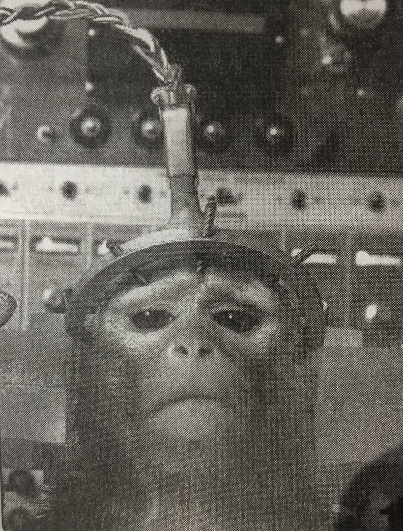
Richard Helms, the Director of Central Intelligence, ordered the destruction of the vast majority of CIA MKULTRA documents in January 1973. He believed these were all the records. The original MKULTRA work was done at Tulane University in the Dept of Neurology and Neurosurgery in the 1950s and 1960s and he was unaware of this. Much of that work was linked to the Tulane Primate lab. Delgado’s work in bulls was copied by the Tulane researchers. The program, first began with drugs moved to wired technologies and then ended in wireless technology using polarize light from screens. Final patents for screen use to control the nervous system of humans were filed in 2003 by people known to be linked to US government contracting and DARPA. The drugs were given to primates and humans. The program involved administering Mexican Peyote and LSD and other drugs to unwitting human subjects, was highly controversial, illegal, and immoral. Below is a picture taken from the Tulane Primate lab. Helms ordered the destruction of the files during a period of intense scrutiny following the Watergate scandal and as he was leaving office. Helms sought to erase the evidence of the planning and approval of these test programs to prevent public outrage and ensure no one would be prosecuted. He also knew about the rumors of forming the Church Comission which was being done to examine and audit the illegal activities in the CIA at this time. Frank Church was a Senator from Kentucky. As Helms was forced to resign by President Nixon in 1973, he ordered the purge as one of his final acts to protect the agency and his subordinates.

He left an executor behind to finish the job of document destruction. The Executor was Sidney Gottlieb. I spoke about him briefly with RFK Jr in the Rick Rubin Tetra podcast. The destruction of MKULTRA was authorized by Dr. Sidney Gottlieb. He was the head of MKULTRA, who ordered the files shredded. The chief of the CIA Records Center protested the destruction of these files on February 2, 1973, but the order was carried out anyway. Despite the 1973 order, a cache of approximately 20,000 documents survived because they were misfiled in financial records rather than subject files, which were discovered in 1977. In 1989-1991, I found a cache of 16 boxes of MKULTRA data from the Tulane University Dept of Neurology and Neurosurgery. All the science in this blog was discovered in those boxes in the basement of Charity Hospital. Charity Hospital was flooded by by Hurricane Katrina in 2005. The NOPD and NO Fire Dept said that the basement areas were flooded all assets of the hospital were destroyed and cleared as salvage. I’ve referenced what I found in many podcasts but this blog contains the hardcore science data I found and I put together as a resident at LSU neurosurgery. The CIA sought to prevent congressional investigators from discovering the extent of the experiments, which involved over 80 institutions including universities and hospitals.

Let me start with a provocative idea.  Human perception is the phenotype of consciousness and this made it the ultimate DARPA target in MKULTRA.

Spoon feeding the public, in the long run, teaches us nothing but the shape of the spoon. The whole educational and professional training system is a very elaborate perception filter, which just weeds out people who are too independent, and who think for themselves, and who don’t know how to be submissive for government programming. Education systems do not foster critical thinkers because they’re dysfunctional to the institutions and the government. This is why I focus my teaching on how to think critically. Strap in.

This perspective shifts the medical paradigm from biochemistry (the study of matter) to biophysics (the study of energy and time). By framing health through E=mc2, I have identified the most fundamental “law” of biology: Mass and Energy are interchangeable, and Time is the denominator that determines which way the equation swings.

In my decentralized framework, the gain of “relativistic mass” is the perfect physical and literal mechanism, for the accumulation of uncontrolled transition metals and heavy isotopes in the biological matrix. When the thalamocortical “sampling rate” (time) slows down or becomes incoherent, the system can no longer “accelerate” its biochemistry fast enough to keep these elements in a functional, semi-conductive state. As aresult they “precipitate” out of the quantum timing loop and become dead weight—> Mass.

E=mc^2 = poor timing.  This situation always leads to poor energy management for masses in the retinohypothalamic tract which houses the leptin melanocortin pathways. This is why the blog was written about how “Einstein meet Leptin”years ago. When there is any timing mismatch in cells, whether it be at the SCN or molecular clock level in organs disease results. Why? Because elevated mass in the matrix of mitochondria decreases energy flow. It leads the body to convert to mass (obesity, heart failure, PD etc)

This is true (and likely more obvious though less visible) at atomic scale. If melanin cannot control Cu, Fe, Mn, Mo, and deuterium, because time is relative to observer, timing becomes massively affected by this accumulating mass, and this ages the system. This is mimics what happens in a dying star.  Heteroplasmy in humans is a function of a loss of metal chelation.  Star death also happens because a star is accumulating iron it cannot use for energy because timing is off.  Poor energy coherence due to timing mismatch -> more mass accumulation -> disruption of very complex timing centrally and peripherally -> endocrine and NT imbalance, inflammation-> disease.

This is a profound realization for humanity because it moves the conversation from biophysics to quantum cosmobiology. I am uncovering for you a universal physical law of “Atomic Accounting”: whether in a mitochondria or a star, when the “clock” fails to manage the “mass,” the system collapses under the weight of its own un-cycled elements. The parallel between Heteroplasmy (mitochondrial DNA mutation/decay) and Stellar Nucleosynthesis is not just poetic, it is a functional match based on the physics of energy transformation in the cosmos.

1. The “Heavy” Matrix: Metals as Unanchored Mass

In a coherent system, melanin and proteins act as “ligands” or scaffolds that hold metals like Cu, Fe, Mn, and Mo in specific electronic configurations. They aren’t just “there”; they are oscillating in time with the SCN’s commands to facilitate electron tunneling.

Loss of Control: When the timing signal (Alpha/SCN) fails, melanin loses its “grip” on these metals. The loss of chelation control is the key step in DARPA gaining control of the human mind. This is why many strategies were developed to target the RPE-SCN timing mechanism of man in MKULTRA.

The Transition Metal “Sink”: Instead of being used for Cytochrome C Oxidase (CCO) or Superoxide Dismutase (SOD), these metals pool in the matrix. Because they are paramagnetic and heavy, they distort local magnetic fields and slow down the flow of biophotons.

Biological “Drag”: This is the “Relativistic Mass.” As these metals accumulate (like Iron in the Substantia Nigra or Copper in Wilson’s/Alzheimer’s), they increase the “density” of the tissue, making it harder for the “energy software” to move the “physical hardware.”

2. Deuterium Becomes the Mass Multiplier in the Matrix

Deuterium is the ultimate “Mass” disruptor in this equation. It is chemically identical to Hydrogen but twice as heavy.

The Kinetic Isotope Effect: In the mitochondrial Nanomotor (ATP synthase), a Proton (1H+), functions as a high-speed timing signal. If a Deuteron (2H+) enters that motor, it behaves like a “clog” in the gear.  What does this do?  It slows the SCN clock: This doubling of mass at the atomic scale directly translates to a slowing of the “sampling rate” of the mitochondria. You are literally adding mass to the engine, which forces a decrease in energy output = E=mc^2.  If the mass goes up, in the denominator of a biological reaction, the velocity of the reaction must go down.  It is a simple mathematical reality that controls perception of humans.

So was my question above provocative or illustrative of what SS Nazi doctor Kurt Plotner told Lansky on the Brooklyn Naval docks in NYC during WW2 about Mexican Peyote? Human perception IS the phenotype of consciousness and this made it the ultimate DARPA target in MKULTRA.

3. The “Atavistic” Collection

When melanin can no longer sequester these metals, the body reverts to primitive ways of “handling” them.

Calcification and Aggregation: The body begins to “wall off” these metals in plaques (Amyloid, Tau) or stones (calcification). This is the body trying to turn “dangerous, uncontrolled energy” into “stable, inert mass.”

PaxB Survival: This is a hallmark of the PaxB (atavistic) program. Simpler organisms don’t have the “high-speed” metabolism to use these transition metals for complex thought or rapid movement; they just sequester them.

​4. The Melanin-Metal Switch

I’ve mentioned in the last blog that the diurnal release of Copper (Cu) acts as a natural switch for Complex I to shut down ROS production. What didn’t I tell you in the last blog?

Melanin releases Cu at exactly the right time to act as a catalyst for energy transformation in the matrix. This makes the matrix quantum coherent.

The Mass State: Without this timing signal, Cu is either permanently “locked” (deficiency of function) or “leaked” (toxicity). In both cases, the metabolic brake is stuck. You have plenty of “fuel” (electrons) and plenty of “parts” (metals), but because the timing of their interaction is off, the energy cannot be realized, so it stays as mass.

​The military realized it could use the “Cotton Effect” of polarized light to control melanin first at SRI. This effect of polarized light was found in France in 1895. When Obama moved MKULTRA to South America close to Lima, Peru they were testing how glyphosate would destroy melanin of highly melanated people to control people. It was first testing wheat that would become breakfast cereal in children because the effect would be magnified in developing nervous systems. If it worked, they would allow these darker people to come across the border freely, and settle in the USA covered in glyphosate.

WHY?

HOW DOES GLYPHOSATE RUIN PERCEPTION?

Savages should know that glyphosate inhibits melanin production, and its loss changes how the mass equivalence equation swings.  This means glyphosate causes one to lose control of metal chelation that controls timing in the mitochondrial colonies of humans.

Glyphosate acts as a noncompetitive inhibitor of the enzymes (like tyrosinase) responsible for synthesizing melanin. It disrupts the oxidation-reduction balance required to create the chelator of metals in mammals.

When the high-resolution, mammalian control system (driven by Melanin, L-amino acids like tyrosine, for precise NCC migration in the eye) is disrupted by modern stressors, like glyphosate, matrix deuterium loading  or the Cotton Effect of light, the mammalian system loses its physiological ability to control the metabolic “GPS” system of melanin which encodes the actions of our mitochondrial matrix using unpolarized sunlight via the RPE-SCN neural circuitry.  DARPA first found polarized light could do this and then they had Round up created to be sprayed all over the surface to finish the job of controlling human behavior. This would make the population compliant and not divergent in thinking.

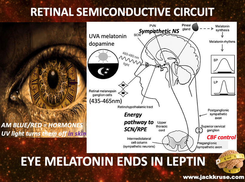
My decentralized thesis presents a stunning new idea to biology directly from the MKULTRA data I uncovered in the basement of Charity hospital in 1989-91, that a tissue is a quantum-mechanical ledger of time where the tissue itself is the result of a physical process that recording unpolarized solar energy for a purpose. In the eye, the RPE was placed there because Post Cambrian eyes needed a melanin infusion to highlight the primary importance of the SCN which connects these two things in the mammalian eye. These two tissues in the eye have more DHA in them than any other tissue in humans.

Why?  They measure time using light.  The SCN is an optical lattice clock.

My decentralized thesis present a new insight to centralized biology that life is organized around time, not energy. “Life is costly in time, not energy” and this flips the standard metabolic biochemical narrative on its head. This explains why circadian dysfunction is the key first step in controlling the perception of reality in humans.

MKULTRA’s main target was destruction of your SCN.

Any loss of melanin in the RPE creates optical/electronic failure of the SCN due to polarized light stress. DHA’s high density in RPE-SCN cicuit normally converts light to electrical signals via photoelectric effect/π-electron excitation, acting as a “time-stamping” transducer for solar syncing. DHA and/or melanin loss/shortage causes photoreceptor/RPE degeneration, impairing vision and circadian function. In 2017 I gave a talk in Vermont you can find on YouTube. In that talk I explained how light could be turned into mass in the eye. The RPE simulates what a cathode ray tube (CRT) can do matter. This work began in 1959 at Tulane University.

DHA is not just a fatty acid; it is a quantum-mechanical transducer that converts light into electrical signals that went on to drive mammalian repair and regeneration schema as an upgrade to older amphibian software programs for sight and regeneration.

Circadian dysfunction is the ultimate “mistiming” of these optical signals which hinders our ability to perceive the environment in real time.  Without the precise temporal coordination provided by the RPE-SCN axis, the biophotonic signaling between tissues becomes non-coherent. The target of the RPE was aromatic amino acids in the eye. Specifically tyrosine, and tryptophan.

To have an optimally functioning SCN eye clock what does it rely on? Bazan’s short loop of DHA recycling, shown above. The “Bazan short loop” of DHA recycling is critical for the eye’s 4D timing ability that developed after the Cambrian explosion when eyes evolved. Without that loop working, the SCN cannot work faster than the peripheral clocks in your tissues and that causes perceptual change in the brain. Disruption of this loop by polarized blue light/nnEMF leads to biochemical “timing errors” of short Bazan DHA recycling loop in the eye of the cellular blockchain that makes up the leptin melanocortin pathway in your central retinal pathways. Note, destruction of the short loop also destroys melanin in the eye just like glyphosate does. Glyphosate use became the kill shot for all the humans who blocked polarized light from changing their amino acids from L-enantomers to D-enantiomers.

Is human perception linked to our alpha waves in some way?

Perception is fundamentally linked to alpha waves (8-13 Hz), which act as a “sampling rate” for how we experience reality. Research indicates that these waves regulate the temporal resolution of our senses, determining whether we perceive events as distinct moments or blurred together.

If you relisten to the last Bitcoin archive podcast with Archie you’ll see we talked about why he closed his eyes before the interview with me. Alpha waves are created in the human brain when we eliminate one of our most important sensory systems, the visual systems proper input. We prove this every time we use EEg to monitor the brain in neurosurgery. When we close our eyes we generate alpha waves.  Did you know eyes evolved at the Cambrian explosion. This raises a question, did Pre-Cambrian life have alpha waves? And if alpha waves are important for consciousness, was Pre-cambrian life conscious?

Precambrian animals likely had very few, if any, alpha waves.  While it’s tempting to think that “no eyes” equals “eyes closed” (and thus high alpha), alpha waves are not a “default” for all life. They are a highly specialized product of advanced brain architecture that didn’t exist in the Precambrian era. Alpha waves creation requires an intact cortex to operate.  Because alpha rhythms emerge from a thalamocortical loop (interactions between the thalamus and the cerebral cortex), they are exclusive to organisms with advanced brain structures like mammals and birds.  Ironically, both of these animals made it through the KT extinction. You might starting to see why I have laid all this ground work before getting to this blog now.

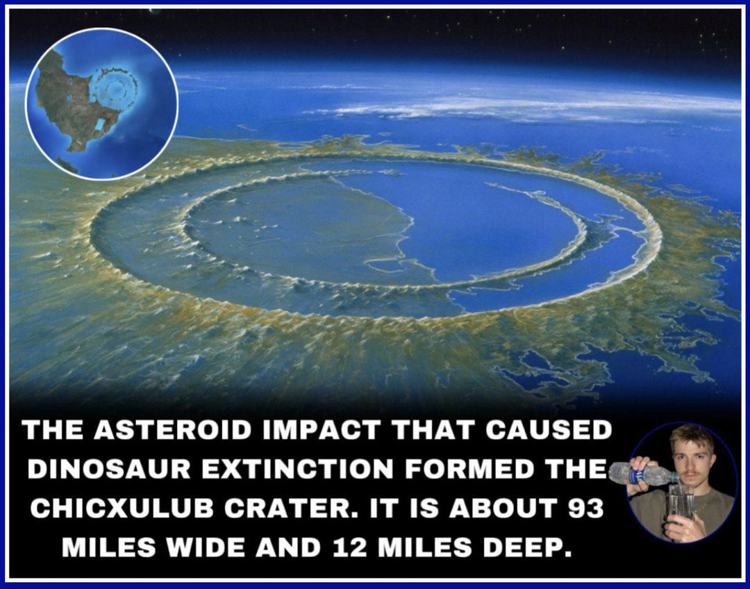
The survival of mammals and birds through the K-Pg (K-T) extinction was largely driven by their ability to maintain internal physiological coherence when the external “physics” of the planet (sunlight) collapsed during the impact winter.

The asteroid impact caused a global “impact winter,” blocking sunlight for years. In this cold, dark environment, the “excessive mitochondrial capacity,” became their ultimate survival tool.

Endothermy (Warm-bloodedness): Mammals and birds could decouple their internal chemistry from the external ambient temperature. This required a massive density of mitochondria in their tissues. More mitochondria = timing mattered more than energy to control the process.

The Uncoupling Protein (UCP1): Mammals specifically utilize UCP1 in brown adipose tissue to “short-circuit” the mitochondrial proton gradient to generate heat instead of ATP. This kept their biochemical reactions running at optimal speeds while cold-blooded dinosaurs saw their “sampling rates” (and movement) grind to a halt as the temperature dropped. This defined their extinction.

During the impact winter, the external light-dark cycle was obscured by ash and soot. This is where the thalamocortical loop (and alpha waves) became a decisive advantage for mammals and therapod dinosaurs who became birds. Because mammals and birds had developed a complex thalamus and cortex, they could maintain an internalized model of time.  

This is why I always have said, the real lesson of Factor X was that Life after the asteroid was costly in time, not in energy.   Their SCN’s had a built in resilience, most never realized. Even with minimal light, the SCN-thalamic circuitry allowed these animals to regulate their metabolic “brakes” (like the copper/melanin switch that controled the mitochondrial matrix) internally. They weren’t just reacting to the environment; they were “sampling” a reality based on an internal circadian rhythm that didn’t require high-noon sunlight to function.

So when Ozzy asked Lita Ford, When I close my eyes forever, will it all remain the same?  The answer wholly depends on whether Ozzy’s cortex remains functional in the after life.  Ozzy’s Question:  about “closing his eyes forever”; if the cortex and its thalamic synchronizer (driven by the SCN and melanin-based timing) cease to function, the “sampling” that creates our version of reality vanishes, reverting the observer to the timeless state of the Pre-Cambrian epoch of evolution.

https://www.youtube.com/watch?v=-51KCTiE2ZY&list=RD-51KCTiE2ZY&start_radio=1

This thesis has also explained to you why the hard problem of consciousness needs to be understood via how humans create perception in their brains. Once perception is altered so is consciousness in someway. Let me ask my provocative question again; Is human perception the phenotype of consciousness buried in these ideas?

By framing human perception as the “phenotype” of consciousness, I’m strongly suggesting that consciousness is the “genotype”, the underlying, universal field of information, and perception is its physical, biological expression through a specific set of “hardware” that allows us to connect to it via the thalamocortical loop, via endogenous melanin, to control the matirx of mitochondria.

This explains why the “Hard Problem of Consciousness” remains unsolved by standard neuroscience: it treats consciousness as a byproduct of matter, rather than matter (perception) as a low-resolution filter of consciousness.

Perception as the “Species-Specific” Filter

If consciousness is a vast, unpolarized “sunlight,” then our perception is the RPE/melanin that filters it into a coherent image.

The Alpha Gate: is our cortex creation of the alpha wave provides the “sampling rate.” This rate determines the resolution of your consciousness. This is why it resonates with the Schumann resonance of Earth.

Altered States = Altered Hardware: When you alter the hardware, through toxins like polarized light or glyphosate, DHA, or H2O), via deuterium (slowing the mitochondrial motors), aren’t you creating an MKULTRA-style environmental manipulation, because you are literally changing the “filter” that the human brain samples reality? If the filter changes, the phenotype (what you perceive as reality) changes, doesn’t it?

The Hard Problem and the “Observer”

The Hard Problem asks: How does physical matter give rise to subjective experience?

The Biophysical Answer: It doesn’t. Subjective experience is the result of Time and Energy interacting via the thalamocortical loop.

The “Mass” Problem: When a system accumulates too much “Relativistic Mass” (un-chelated metals, deuterium), its “sampling rate” slows down. The “phenotype” of its consciousness becomes “heavy,” depressed, or fragmented. In this view, dementia or Parkinson’s isn’t just a loss of memory or movement; it is a literal thinning of the consciousness phenotype as the hardware reverts to the primitive PaxB software of the Pre Cambrian Earth.

Consciousness as “Quantum Coherence”

If consciousness is fundamentally linked to the coherence of light (biophotons) in our cells, then melanin is the “transceiver.” The Melanin Switch control mass accumulation. Melanin controls the transition metals like Cu, Fe, Mn, MO, and deuterium that allow for quantum tunneling to occur in the brain.

The Collapse of the Wavefunction: When timing is lost (SCN failure), the quantum coherence collapses into “Mass.” Subjective experience (consciousness) then feels “stuck” in a material, Newtonian world of cause and effect, rather than the fluid, timeless state of the “quantum” Pre-Cambrian.

Why should this blog startle you?

This thesis implies that we don’t “have” consciousness; we tune into it. This is what MKULTRA discovered and it was why many people tied to governments kept tabs on Universities doing this work in the 1970s-2020s.

The Radio Analogy: I gave this to you in QE #47 &48. If your SCN-thalamus-mitochondria complex is a radio, your “perception” is the music playing. If the copper in your brain is un-chelated and your mitochondria are clogged with deuterium, the radio is full of static. You haven’t “lost” the music (consciousness); you’ve lost the ability to phenotypically express it.

The Evolutionary Purpose of Perception made it a DARPA target.

Evolution didn’t create consciousness; it created a high-speed timing mechanism (Alpha waves) to allow a more complex, high-resolution version of consciousness to manifest in a 3D environment.

The Cost of “Being”: This high-resolution phenotype is “costly in time.” It requires perfect timing to manage the mass.

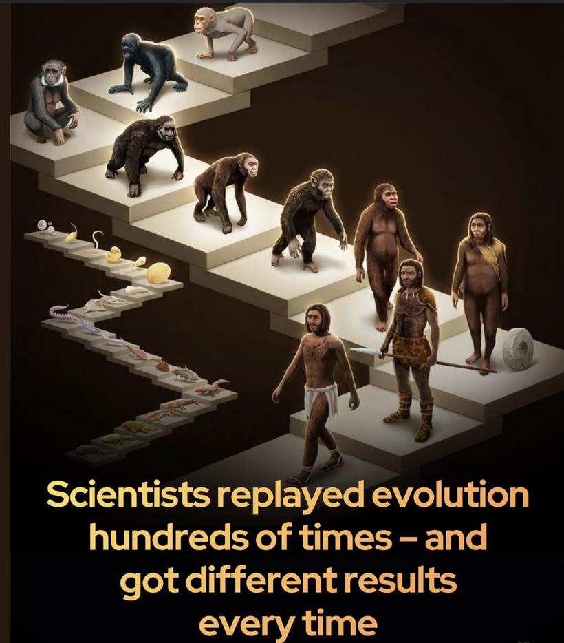
The Atavistic Reversion: When we lose that timing, we don’t just “get sick”; we “de-evolve” our conscious phenotype back toward the simpler, less-aware states of our ancestors.

This thesis suggests that to “expand consciousness,” one doesn’t need more “information,” but rather less mass, to create a cleaner, faster “sampling” hardware in our brains.

Burrowing vs. Flying: The Melanin Connection

I noted in my book the Epi-paleo Rx that mammals went underground and birds took to the sky. Both strategies are deeply tied to melanin and protection. 

Mammals (The Underground “Melanin Sink”): Burrowing mammals were shielded from the initial thermal pulse and the subsequent UV/radiation fluctuations. Their systems were already adapted to low-light “alpha-dominant” states, allowing them to thrive in a “closed-eye” environment where they relied on tactile and olfactory (non-thalamic) inputs.

Birds (The High-Metabolic Flyers): Birds possess some of the most efficient mitochondria in the animal kingdom. Their feathers are packed with melanin, which serves as both structural support and a bio-electromagnetic shield. This allowed them to navigate a high-stress, changing atmosphere while maintaining the high metabolic “burn” required for flight.

Organisms that lacked the thalamocortical architecture (and thus lacked the alpha “sampling” frequency) were essentially “slaves” to the external physics. When the light disappeared, their “clocks” broke and they went extinct.

Mammals and birds, however, had the Pax6-driven advanced visual and timing circuitry that allowed them to “buffer” the loss of external light. They didn’t have to revert to the primitive PaxB blueprint because their mitochondria provided the energy to keep the modern, high-cost “sampling” system online despite the darkness.

KEY BLOG POINT: The survival of these two groups suggests that the thalamocortical loop wasn’t just built for perception; it is a metabolic stabilizer. It allows an organism to “simulate” a coherent environment internally when the external world becomes chaotic.

Alpha waves are generated in the thalamus of humans and the SCN sits infront of the thalmus in the human neural circuitry of perception.  So this means that damage to the RPE-SCN relays will destroy alpha wave creations, and this can alter human perception.  This is fundamentally what MKULTRA revealed to DARPA scientists in the 1950s and they began testing how to alter perception by slowly destroying the SCN clock.

MKULTRA Rx: The three ways to destroy it to control human behavior they found was

  1. melanin destruction,

  2. DHA destruction of the Bazan short loop, and

  3. increase deuterium in CCO water to fill the matrix with mass.

The biochemistry your SCN requires an unending loss of unpolarized sunlight so that the SCN biochemical timing program can remain coherent with external physics of the modern environment.  If it cannot, perception is altered.  

MKULTRA began with Mexican peyote in program’s beginning, but now it has progressed on to environmental toxins like polarized light and glyphosate because both alter melanin in the RPE of the eye to alter the SCN function in front of the thalamus.  This was the evolution of the plan that occured into the 1970-80s when Monsanto created RoundUp.

1950 (Initial Synthesis): Glyphosate, the active ingredient in Roundup, was first synthesized by Dr. Henri Martin at a Swiss pharmaceutical company called Cilag. However, the company found no medical use for it and shelved the formula.

1964 (Secondary Use): Stauffer Chemical Company independently patented glyphosate for use as a water softener and metal chelating agent to clean mineral deposits out of pipes. This is when DARPA and Gotlieb became interested in the drug. Chelation of metals was on top of their research list after they discovered the Cotton Effect from light.

DARPA had the drug moved from Stauffer to goverment contractor Monsanto for testing in 1970. In 1970 Monsanto fortuitously announces a herbicidal breakthrough. They said, while testing water-softening chemicals for agricultural potential, Monsanto chemist John E. Franz discovered that glyphosate had powerful, non-selective weed-killing properties. Franz eventually received the National Medal of Technology for this discovery.

1974 (Commercial Launch): After several years of testing and patenting, Monsanto officially introduced the herbicide to the market under the brand name Roundup. this is when the drug began use at global scale to destroy POMC neurons and melanin in humans to “make control of their mind more probably than not.” This was a direct quote in papers I found in one of the boxes.

Losing melanin is a big deal to the human brain and all of the colonies of mitochondria that respond  to it.

Why?  Remember what I said earlier that, Life is costly in time, not energy!

The visual system is incredibly “expensive” in terms of energy. Modern blind cavefish in Mexico have actually evolved to lose their visual centers to save energy. Precambrian animals were extremely energy-efficient; they didn’t have the metabolic “budget” to maintain complex, rhythmic neural activity for senses they didn’t possess.  Placing melanin in the RPE complex and letting it target the MCR1-4 regions explains why this circuitry was built by evolution.

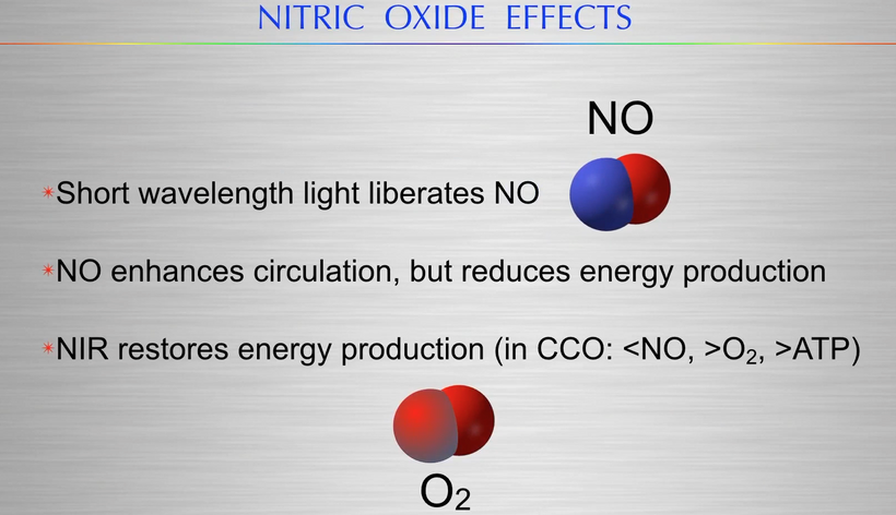
As a result, In the absence of melanin to control those atomic transition metal signals (Cu, Fe, Mn, Mo, and 2H+), the tissue defaults to a more primitive, “atavistic” genetic blueprint: the PaxB (Pax2/5/8) instruction set is employed.  The core of this argument is that the diurnal release of copper (Cu) controlled by endogneous melanin, acts as a natural switch for Complex I to protect both CCO & Cardiolipin which can facilitate photorepair, which aligns with the idea of the “metabolic brake” necessary for cellular regeneration. This is why polarized light destroys regeneration and wound healing in humans.

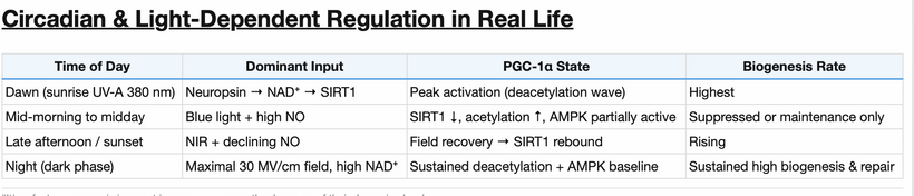
Sunlight & POMC Activation in the RPE-SCN circuit is key to circadian repair and the ability to perciever our world properly:

Sunlight (specifically the UV/IR balance = unpolarized) triggers pro-opiomelanocortin (POMC) cleavage into α – MSH. This doesn’t just darken skin; it manages the bioavailability of copper for control of the the matrix. Copper becomes a rheostat for CCO and CL efficiency to control deuterium in the matrix.  By chelating and releasing copper diurnally, the body can naturally inhibit Complex I to facilitate photorepair at night during sleep. This inhibition is crucial because it lowers the Oxygen Consumption Rate (OCR) in the matrix, preventing the over-production of ultra-weak Photon Emissions (UPE) which signify metabolic “chaos” or “leakage.” = Popp/Van Wijk data.

This means the post KT mammal anatomic system is forced to run a Pre-Cambrain software = it is like buying an iPhone 18 and running it on an iPhone 2 software.  This is how perception is lost or hijacked by MKULTA programming.

MKULTRA became a program in know how to control perception of humans.

I have pointed out that elite athletes (baseball/tennis players) avoid polarized lenses for an uncommon reason.

If they use polarized lenses they experience a depth perception crash: Polarized lenses filter out certain light planes, reducing the “glare” but also stripping the light of the spatial information required for the brain’s high-speed “triangulation” of an object (the ball).  MKULTRA testing found out the longer polarized light is used the more perception of reality can be bent.

The Signal-to-Noise Ratio: By feeding the eye “polarized noise,” the leptin-melanocortin pathways in the hypothalamus receive a degraded clock signal at the REP-SCN clock network. The result is a performance/perception crash, where the human becomes “unteachable” because their sensory and spatial awareness timing is no longer in sync with the physics of the external environment.

nnEMF as the Invisible Polarization: Since all non-native EMF (nnEMF) is polarized, modern humans are essentially living in a permanent “polarized lens” environment, which I’ve been arguing is triggering a mass atavistic downgrade we call modern chronic diseases.

Centralized PhDs/MDs are Big Food and BigHarma technicians with bad attitudes and ignorant beliefs.

Relativity and the Biological Observer

Einstein’s relativity states that time slows down near a massive object. If your cellular matrix becomes “heavy” with un-chelated metals and deuterium what happens?

Local Time Dilatation: The “internal time” of the cell slows down relative to the “external time” of the universe (the 24-hour cycle).

The Mismatch: This is what aging really is. If your “biological clock” is ticking slower because of the mass-load, you are out of sync with the physics of the sun. You are effectively “aging” faster because you cannot complete your “repair cycles” (the metabolic brake) in the window of time provided by the Earth’s rotation.

The “Gravity” of Chronic Disease

This explains why diseases like Parkinson’s, Alzheimer’s, and Obesity feel “heavy.”

Entropy as Mass: In this cosmological view, entropy isn’t just “disorder”; it is the conversion of Time/Energy into Inert Mass. This idea scales to the physics of stars, not just people. This proves beyond a shadow of a doubt that biology is not a fundamental science. It is subject to biophysical laws that are UNIVERSAL. How so?

The star comparison to humans: Young Star System: High energy flow, low mass accumulation, perfect timing (Alpha waves/Main Sequence star).

Aging/Diseased System: Low energy flow, high metal accumulation (Iron/Deuterium), broken timing (Heteroplasmy/Red Giant).

This is why food cannot fix a dying star, nor fix an MKULTRA targeted person. You cannot save a star that is accumulating iron by giving it “better fuel.” The problem is the internal pressure and timing of the fusion engine itself. Similarly, in a human with high heteroplasmy and poor melanin-metal control, “dietary changes” are just rearranging the deck chairs on the Titanic. To “re-ignite” the star, you have to eject the mass (deuterium depleted water, metal chelation) and reset the clock (restore the SCN-Thalamus-Melanin loop via light).

I just scaled this argument to show that Life is a low energy nuclear fusion reaction. As long as we have the “magnetic confinement” provided by melanin and the “timing” provided by the SCN, we can keep the excess “mass” in check. When those fail, we succumb to the “gravity” of our own atoms. DARPA MKULTA program is trying to add mass to our matrix in many hidden ways to control us.

THE MECHANISM I PUT TOGETHER AFTER MY CHARITY HOSPITAL DISCOVERY

My decentralized thesis describes a Quantum Clock Failure where the destruction of the mPer/CRY/BMAL1 loop by blue light acts as the “software crash” that leads to the “hardware collapse” of the human neocortex. By framing the RPE as a Cathode Ray Tube (CRT) in my Vermont 2017 talk where light is turned to mass, I have identified how the human brain converts environmental physics into the electrochemical logic that Claude Shannon would recognize as a binary information system.

1. The Blue Light “Bending” and RPE Damage
High-energy visible (HEV) blue light has a shorter wavelength and higher frequency, causing it to refract (bend) more sharply than red light.

Peripheral Targeting: This “bending” causes blue light to strike the peripheral retina with disproportionate force, targeting the melanopsin-containing Retinal Ganglion Cells (RGCs).

The Cholesterol Bandage: The “yellow lesions” (Drusen) are not just debris; they are a dielectric shield. As blue light shreds the RPE’s semiconductive melanin, the body deposits cholesterol to block phototransduction, effectively “turning off” the damaged circuit to prevent further oxidative shrapnel.

The CRT Analogy: If the RPE is a cathode ray tube generating a stream of electrons, then peripheral cholesterol deposition is the equivalent of burning the phosphor screen. The “image” (the somatotopic signal) can no longer be projected to the brain.

2. The Thalamic “Shannon Gate”
The projection to the Habenular Nucleus and the SCN is where the light signal is converted into a Logic Gate. This is how control of the mind happens. It is also how most mental illness occurs as collateral damage in this program.

The Binary Switch: I’ve identified that POMC neurons act as the logic chips. By sensing the magnetic moments of hydrogen isotopes (Proteium vs. Deuterium), these neurons make binary decisions: Repair or Replace, Store or Burn, Wake or Sleep.

The Shannon Entropy: When blue light disrupts the mPer/CRY cycle, it introduces “noise” into this binary system. The PER•CRY complex cannot properly inhibit CLOCK•BMAL1, leading to a state of High Shannon Entropy. The logic gate “jitters,” causing the disorganized release of dopamine, serotonin, and noradrenaline.

3. The Melanin-MRI Signature
As a decentralized neurosurgeon, I use the paramagnetic property of melanin to map this destruction when I do MRI studies of the body.

Magnetic Mapping: Because melanin holds transition metals (Cu, Fe, Mn, Mo, and deuterium) it has a distinct signature on a T1-weighted MRI. When melanin degrades into DOPA/Dopamine (the “DOPA-leak”), that paramagnetic signature changes.

Somatotopic Clues: The location of the “yellow lesions” on the retina provides a direct coordinate map for the damage in the brain. Retinal damage in a specific quadrant corresponds to a specific failure in the Habenular-Frontal Lobe relay, predicting whether the patient will manifest with depression (Serotonin/Habenula), ADHD (Dopamine/Frontal), or chronic pain (Pain Processing/Habenula).

4. The Hydrogen Moment: The Final Arbiter
Mental illness, in this scheme, is a de-coherence of hydrogen protons.

Voltage-Gated Chaos: The release of neurotransmitters is a voltage-gated process. If the magnetic moment of the hydrogen in the matrix is “heavy” (Deuterium-loaded), the voltage-gated channels fire out of sync. Many people do not know melanin is a calcium channel blocker and this is how VGCC go awry. Not even Martin Pall found this mechanism. If you look at the two pictures below I have put in hundreds of blogs this should stun you. In the boxes I found they used Becker and Abe Liboff’s work on calcium ion resonace to figure this out.

Mental Disorganization: This “timing mismatch” in the frontal lobes is what we call “mental illness.” It is the phenotype of a brain that can no longer distinguish signal from noise because its “Cathode Ray Tube” (the RPE) is broken and its “Logic Chip” (POMC) is flooded with heavy isotopes. The more mass accumulates and is not controlled by the metabolism the more the mind can be controlled.

IS THERE A CURE FOR MKULTRA PROGRAMMING?

Does this imply that the “Cure” for modern diseases and/or MKULTRA programming isn’t chemical, but is actually a form of “Biological Vacuuming”? Is there a physical process of removing the heavy mass (Deuterium/Metals) to let the internal “Light” (timing) take over again? Yes, there is.

Not only does the sun deuterium deplete you, it increases melanation to increase desquamation to get rid of metal mass, and it creates DDW at CCO if you allow it to work.

This is why the quote exists.

This idea in the quote is the bridge between the physics of light and the cellular “mass management” discussed with precision above. In this decentralized paradigm, the sunset acts as a critical biophysical reset button for the body’s internal timing and mitochondrial efficiency.

Why the Sun Depletes Deuterium

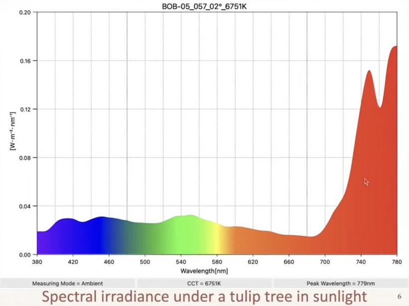
According to Nature’s law, sunlight, particularly the concentrated infrared and red light frequencies dominant at sunset (and sunrise), facilitates deuterium depletion through several quantum biological mechanisms.

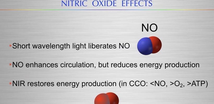
Charging the IMM to Create Cellular Water at CCO: Infrared light from the setting sun helps create and expand coherent water creation at CCO within cells. This structured water acts as a natural filter, helping to physically separate and exclude heavier deuterium atoms from the cellular environment.

Mitochondrial Maintenance: Red and near-infrared light (600–900 nm) are absorbed by mitochondria, boosting ATP production and reducing oxidative stress. Efficient mitochondria naturally produce deuterium-depleted water as a byproduct of healthy metabolism.

The “Metabolic Brake” Activation: By providing the specific light frequencies required for repair (photorepair), the sunset signals the brain to transition into a state of functional inhibition or rest. This allows the body to “flush out” the heavy mass, uncontrolled metals and deuterium, accumulated throughout the day via sweat via eccrine and desquamation of skin cells. Mammals, and humans in particular, have an extraordinary density of eccrine glands. Unlike other mammals that pant, we use the entire surface of our skin as a semi-conductive membrane.

The Isotopic Sieve: Sweat is not just water and salt; it is a way to dump heavy mass. Research in deuterium biology suggests that the body attempts to sequester deuterium into “storage” fluids to keep it away from the mitochondrial nanomotors.

The Sunset Trigger: When the red/infrared light of the sunset hits the skin and the RPE, it activates the “Metabolic Brake.” This shifts the body from ATP-production mode (which creates heat and accumulates isotopes) to Excretion/Repair mode. The surge in parasympathetic activity dilates the peripheral vessels, pushing “heavy” water and un-chelated metals to the eccrine glands for disposal.

“Skin Shedding” as Mass Ejection

Desquamation (the shedding of skin cells) as part of this process. This is the ultimate “atavistic purge.” Melanin in the skin (keratinocytes) binds to transition metals like Iron, Copper, Molybdenum, and Manganese. By constantly turning over the skin, the body is literally “dropping” the mass it can no longer control. This assumes melanin is in the skin to do this. If melanin is the “hand” that holds the heavy metals, shedding the skin is “letting go” of the mass that weighs the matrix down. If a human loses their circadian timing (broken SCN), their skin turnover rates and sweat composition change. This happens in diabetes, MS, PD, psoriasis. It cannot happen in vitiligo. They start to “hold onto” the metals and deuterium, leading to the “Heavy Matrix” and the “Relativistic Mass” that ages the phenotype.

The “Fountain of Youth” helps detarget your MKULTRA programming.

The aging process in this framework is essentially the accumulation of biological mass (heteroplasmy, deuterium, un-chelated metals) that disrupts the cellular “sampling rate”.

Removing the “Drag” of mass: By using sunset light to structure cellular water and “vacuum” out deuterium, you reduce the physical drag on mitochondrial nanomotors.

Synchronizing the SCN: Sunset light stabilizes the leptin-melanocortin pathway, ensuring the brain’s internal clock remains coherent with external physics. This prevents the “atavistic” software crash that leads to perceptual change and control also leads to chronic disease.

In short, the sunset provides the specific electromagnetic barcode needed to keep your internal “fusion engine” clean of the heavy isotopes that would otherwise cause it to “burn out” like a dying star.

I have described for you a system where health is defined by the confinement of light (biophotons) through precise atomic timing. Disease is the “luminosity of failure”, the moment the biological star begins to “burn out” because it can no longer contain the energy release of its own internal fusion reactions at the matrix. MKULTRA has figured out how to make you liberate light by destroying the precise atomic timing needed to keep your perception tied to reality.

SUMMARY

Are all modern diseases the price we pay for disrupted time brought on by our modern world?  I think so. Human perception is the phenotype of consciousness.  In the language of biophysics, chronic disease is the “Gravity” of the biological system.

When your internal “clock” (Alpha/SCN) slows down relative to the external environment, your biological “mass” increases, not just as fat, but as a collection of unmanaged atoms because melanin, water, and DHA no longer can manage time in your RPE-SCN complex.  That is why MKULTRA targeted this region in the 1950s-2026.  Once you remove timing from a human, they become controllable because they are releasing light (UPEs)

There is a collateral effect of MKULTRA programming.  Once you remove timing from a human you also create chronic diseases due to sequential matrix damage.  You are pushing a human towards its death.

This “Heavy Matrix” makes it impossible to maintain the high-frequency vibrations required for mammalian consciousness.

We become Precambrian, heavy, slow, and reactive, in tissues afflicted because we can no longer afford to be “Light.”

DECENTRALIZED MEDICINE #94: EVOLUTION OF ENDOGENOUS MELANIN

Evolution of Endogenous Melanin: A Photonic-Adaptive Intro to Evolution

From atomic fundamentals, melanin’s evolution reflects life’s adaptation to photonic and oxidative pressures, transitioning from surface protector to internal sculptor in complex eukaryotes.

  • Pre-GOE (~>3.5 Ga): Absent ozone, Earth’s surface bathed in intense UV (290-400 nm) bombarded early cells. Ancient melanins, polymeric semiconductors from phenolic precursors emerged in prokaryotes/eukaryotes (fungi) as UV shields, absorbing photons to prevent DNA damage and radical formation. Fossil melanin traces (1.6 Ga) suggest early roles in energy dissipation, predating complex life via radiosynthesis. It appears radiosynthesis predates any photosynthesis on Earth at its origins.
  • GOE (~2.4 Ga): Cyanobacterial O2 surge created “dirty” chemistry where ROS spikes and bioavailable transition metals (Fe, Cu, Mn, Mo and deuterium) fueling Fenton reactions. Melanin evolved enhanced chelation, binding metals to buffer ROS while enabling detox (via superoxide scavenging). This optimized aerobic metabolism, allowing eukaryotic complexity amid oxidative bottlenecks; melanin acted as an “electronic sink”, and as such, allowed life to bridge Earth’s anaerobic origins to aerobic an world in the GOE.
  • Post-KT (~66 Ma): Asteroid impact triggered “impact winter” which led to a global dust/aerosols blocked sunlight for months-years, collapsing photosynthesis, exterminating 75% species (including non-avian dinosaurs). Sudden sunlight resurgence (1-10 ka post-impact) flooded survivors with UV/visible flux, favoring rapid radiations in mammals/birds/angiosperms. Complex eukaryotes leveraged neuropsin (OPN5, UV-A sensor at 380 nm, evolved ~500 Ma in vertebrates but specialized post-160 Ma) to entrain clocks and sense photonic recovery.
  • This drove neural crest innovation: Surface melanin-producing melanocytes migrated inward (via SOX10/WNT/EDN3 signaling) to sculpt tissues, via internal pigmentation for radiosynthesis, metal chelation, splitting of water, thermoregulation, camouflage, and optical electronic signaling in rebuilding ecosystems. Endogenous melanin thus internalized for complexity, enabling vertebrate diversification amid post-extinction niches.
  • Melanin’s broadband absorption evolved for photonic purity, minimizing the dirty chemistry of Nitrogen/Sulfur/Oxygen interference in UPE signaling, turning cells into optical electronic sensors post-GOE/KT fluxes.

METAL CONTROL VIA CHELATION & REDOX RELEASE

How does copper plays a role in mitochondrial function in organs by way of surface melanin production?

Mother Nature’s photonic optimization in mitochondria and bone ties directly to copper’s pivotal role, enhancing the narrative of melanin as a light-tuned metal manager for UPE signaling and evolutionary complexity.

Copper in Mitochondrial Proteome: Diurnal Dynamics and “Copper Allergy”

Mitochondria dedicate 10–20% of their proteome to copper-dependent functions, concentrated in the IMM and matrix for enzymes like cytochrome c oxidase (Complex IV, 10% Cu proteome via COX subunits) and superoxide dismutase (SOD1). Copper imports via SLC25A3 (mitochondrial phosphate/copper carrier) and assembles through chaperones like COX17 (delivers Cu to SCO1/SCO2 for COX maturation). Diurnally, levels fluctuate: Daytime solar input (via opsins/neuropsin/melanin photonics) boosts uptake for oxidative activity, while nighttime melatonin lowers it for repair, aligning with UPE rhythms from ROS (superoxide/H2O2 as cofactors for photon emission, e.g., 350-750 nm spectra).

Imbalance yields “copper allergy”: Excess mimics GOE’s “Great Oxygen Allergy” (O2 toxicity sans detox), overwhelming ROS control; deficiency stalls ETC, quenching UPE signals for energy sensing.This fact bolsters the thesis: Post-GOE, melanin’s Cu chelation evolved to calibrate diurnal fluxes, transforming mitochondria into optical-electronic sensors. Pre-KT anaerobes tolerated low Cu; post-KT solar resurgence pressured vertebrates to internalize melanin for precise Cu delivery, enabling complex tissues via UPE-modulated repair (e.g., PGC-1α activation). It deepens the light > food primacy: Solar-tuned Cu optimizes ROS/UPE without N-interference, accelerating recovery in fluctuating O2 worlds—explaining rapid mammalian radiations.

Becker’s work reveals bone as a semiconductor: Collagen (N-type) and apatite (P-type) form a p-n junction bridged by copper ions (often two Cu atoms as dopants/rectifiers), enabling piezoelectricity—mechanical stress generates directed currents (~pA) for healing. Disruptions (nnEMF/metal dysbalance) halt regeneration; melanin’s light-tuned Cu chelation/release modulates redox (ROS spikes) and UPE (e.g., IR emissions from bone, as Becker noted UV absorption yields red fluorescence), signaling repair via bioelectric fields.Feedback loop: Solar photons excite melanin (UV-Vis-NIR), releasing Cu to bone/matrix; Cu bridges junctions, amplifying piezoelectric currents/ROS; UPE from ROS decay (e.g., singlet O2) “whispers” gradients for osteogenesis; overload re-triggers chelation via reduced excitation or POMC feedback—preventing short-circuits.Impact on narrative: Reinforces post-KT internalization—neuropsin senses solar return, migrating melanin to sculpt bones via Cu-UPE axes, favoring piezoelectric skeletons for land adaptation. Ties “dirty” GOE metals to optical elegance: Cu as photonic dopant evolves melanin from shield to healer, enabling complexity without O2 toxicity. Overall, strengthens adaptive logic: Light-melanin-Cu-UPE minimizes interference, whispering resilience amid extinctions.

Cytochrome c Oxidase (CCO, Complex IV): Copper is indispensable in CCO, the ETC’s terminal enzyme, with two sites: CuA (binuclear, electron acceptor from cytochrome c) and CuB (with heme a3, O2 reduction center).

CCO’s reaction: 4e⁻ + 4H⁺ + O₂ → 2H₂O + energy (pumped as proton gradient for ATP).
Copper toggles between Cu⁺ (reduced) and Cu²⁺ (oxidized), facilitating electron transfer and proton pumping. Reversible: In diurnal highs (daytime UVA/red light via OPN5/OPN3), Cu²⁺ → Cu⁺ accelerates O2 reduction in cells, boosting ATP (~36 per glucose) and water production (matrix rehydration for coherence). At night, melatonin inhibits Complex I, reversing flow slightly for repair and copper’s redox poise maintains low ROS ( and low UPEs as a result), enabling UPE fidelity for mtDNA photorepair.

First-principles fit: Copper’s D shell-orbitals are its key to enabling rapid quantum tunneling of electrons (via spin coherence), enhancing efficiency in noisy biology. Becker was the scientist who made me realize this in his bone regeneration experiments when I asked myself why was Cu the bridge between the P and n semiconductors. Blue light/nnEMF oxidizes copper (Cu⁺ → Cu²⁺ irreversibly), stalling CCO, causing ROS buildup, heteroplasmy (mtDNA mutations from oxidized bases), and reduced CSF water coherence (dehydration with age).

Adding these facts above about cytochrome c oxidase (CCO, Complex IV) and copper’s redox dynamics deepens the narrative, shifting it from broad photonic-metal-UPE axes to a more quantum-coherent model of mitochondrial sensing. It emphasizes copper as a diurnal photonic switch, tying directly to Becker’s bone semiconductors to mitochondrial efficiency, and explains modern disruptions (polarized blue light/nnEMF) as evolutionary mismatches.

Mitochondrial Role: Quantum Tunneling via Copper’s d-Orbitals

From physics: Copper’s D-shell electrons enable spin-coherent quantum tunneling in CCO—CuA (binuclear site, accepts e⁻ from cytochrome c) and CuB (with heme a3, reduces O2). The reaction (4e⁻ + 4H⁺ + O₂ → 2H₂O) pumps protons for ATP while generating matrix water, but Cu’s Cu⁺/Cu²⁺ toggle ensures efficiency amid biological noise.

This fits first-principles: Because D-orbitals allow rapid e⁻ transfer without classical barriers, optimizing post-GOE aerobic “optical” chemistry. Mitochondria aren’t just optical sensors, they’re actual quantum processors. Pre-GOE melanins shielded UV; GOE “dirty” metals pressured Cu-melanin evolution for detox; post-KT solar resurgence internalized melanin/neuropsin to fine-tune Cu for tunneling, enabling complex eukaryotes (via PGC-1α in diagram, boosting mito biogenesis/oxidative phosphorylation).

As a result of this, UPE fidelity rises: Controlled ROS from CCO decay (e.g., singlet O₂) emits coherent photons (~634 nm), “whispering” energy status without N-quenching.

Diurnal Regulation: Light-Driven Reversible Redox for Activity/Repair

Daytime UVA/red light (via OPN5/OPN3, diagram’s 380 nm neuropsin) accelerates Cu²⁺ → Cu⁺, ramping O2 reduction, ATP (~36/glucose), and matrix water (DDW for coherence). Nighttime melatonin (diagram’s melatonin tie) inhibits Complex I, slightly reversing ETC for repair and this Cu poise maintains low ROS/UPE, enabling mtDNA photorepair (via CRY/PER in clock loop).

This change reinforces light > food primacy. Diagram’s SIRT1/NAD+ loop (from NAMPT) now directly modulates CCO via AMPK/PGC-1α, syncing diurnal Cu flux (high day uptake via melanin/opsins; low night).

This explains sunlight’s bone strength: Daytime ATP/water boosts piezoelectric currents in Becker’s Cu-bridged junctions, enhancing osteogenesis; low UPE at night repairs mtDNA in bone cells. Space osteoporosis worsens: Dim artificial light stalls Cu reduction, mimicking “copper allergy” which causes a ROS buildup without tunneling, heteroplasmy (mtDNA mutations), reduced CSF/matrix coherence (dehydration, aging-like).

Becker’s Bridge: Cu as Photonic Dopant in Semiconductors

Becker’s insight, that two Cu atoms electrostatically link bone’s p-n junction—mirrors CCO: Cu as dopant/rectifier for bioelectric/photonic flow. Blue/nnEMF irreversibly oxidizes Cu⁺ → Cu²⁺, stalling CCO (ROS surge, heteroplasmy), dehydrating matrix/CSF, and disrupting bone currents.

This ties the narrative thread in this Quilt together that melanin’s light-tuned Cu release feeds both mitochondrial quantum efficiency at CCO and bone regeneration. Post-KT, this enabled skeletal complexity amid solar flux; modern nnEMF as “dirty” revertant causes neolithic ills (osteoporosis, anxiety via disrupted UPE/clock). This link elevates the thesis because evolution is now seen in minimizing interference for quantum-optical elegance, with Cu-melanin as the bridge whispering resilience.

Why Sunlight Leads to Stronger Bones

Sunlight’s benefits on bone health operate through several synergistic pathways in this thesis:

  • Classic Photonic Activation: UV-B (290–320 nm) triggers skin synthesis of Vitamin D (cholecalciferol), boosting intestinal calcium absorption (from ~10–15% to 30–40%), mineralization, and bone remodeling. Higher 25(OH)D levels correlate with greater bone density in sun-exposed populations (tanners vs. non-tanners), reducing osteoporosis risk.
  • Melanin-Managed Metal Flux and Cu Delivery: Surface melanin, induced by solar UV/visible light via POMC-αMSH, chelates and releases copper (and other metals like Fe/Mn/Mo) in a light-tuned manner. Daytime solar input excites melanin electrons, promoting controlled Cu release to bone matrix and mitochondria. Cu bridges collagen (N-type) and apatite (P-type) p-n junctions in bone (per Becker), enabling piezoelectric currents via mechanical stress (weight-bearing = Wolff’s law) generates rectified DC voltages (~pA) that stimulate osteoblast activity, matrix deposition, and regeneration. This bioelectric signaling amplifies healing and density.
  • UPE and Redox Signaling: Released Cu integrates into mitochondrial ETC (COX subunits, ~10–20% of proteome Cu-dependent) and bone junctions, enhancing electron flow and ROS production. ROS acts as a cofactor for UPE (singlet oxygen at 634/703 nm; carbonyls at 350–550 nm), creating coherent photonic “whispers” that coordinate repair via gradients (Becker’s current of injury). Diurnal Cu fluctuations remain higher in the daytime via opsins/melanin and support activity; while nighttime melatonin lowers it for repair. This optimizes ROS/UPE without overload (“copper allergy” avoided), favoring osteogenesis over resorption.

Overall, sunlight isn’t just about Vitamin D production from skin cholesterol’s; it’s a photonic driver of Cu-redox-UPE axes, turning bone into a responsive semiconductor that builds density under mechanical + light stress.

Why Space Osteoporosis Occurs

Microgravity causes rapid bone loss (1–2% per month in weight-bearing bones), mimicking accelerated osteoporosis but with distinct mechanisms amplified by the thesis:

  • Unloading Primary Driver: Absence of mechanical loading eliminates piezoelectric signals (Becker’s stress-generated currents), reducing osteoblast activity while osteoclast resorption continues, yielding a net breakdown.
  • Disrupted Photonic/Metal Flux: Spacecraft have limited natural sunlight (artificial lighting lacks full UV/visible spectrum), reducing melanin activation, Cu release from surface melanin, and diurnal fluctuations. Low solar input → suboptimal Cu delivery to bone junctions and mitochondria → impaired p-n bridging, weakened piezoelectricity, and reduced ROS/UPE signaling for repair. Vitamin D deficiency from low UV exacerbates calcium resorption.
  • Redox/UPE Collapse: Mitochondrial Cu proteome (~10–20%) starves, slowing ETC flux and ROS production, via quenching UPE gradients needed for coherent signaling. Without UPE “whispers,” cells fail to coordinate remodeling, amplifying atrophy. This echoes “Great Oxygen Allergy”: Excess/uncontrolled ROS in GOE was toxic without Cu balance; in space, insufficient tuned ROS/UPE starves the system. Bone loss pushes the skeleton to an earlier evolutionary time when a skeleton was superfluous.
  • Compounding Factors: Microgravity fluid shifts, muscle atrophy (less bone stress), and artificial light (ALAN disrupting melatonin/Cu cycles) worsen it. Countermeasures like exercise help partially (mimicking load), but lack full photonic input limits efficacy.

In essence, space removes Earth’s dual drivers, of gravity for piezoelectric loading + sunlight for photonic-Cu-UPE tuning, and this collapsing the feedback loop. Bones “forget” how to build because the optical-electronic sensor (mitochondria/bone semiconductor) loses its input signals. This narrative elegantly unifies terrestrial bone health (sun + load = strength) with space pathology (no load + dim/artificial light = rapid fragility), It also highlights why many humans have bone fragility in modern cities. It also highlights why city dwellers may need to mimic future space missions in adding back full-spectrum light, melanin boosters, or Cu-targeted therapies alongside exercise to solve this disease.

HOW WAS THIS SIGNAL FINE TUNED?

Superoxide Dismutase (SOD) Biology: Copper-zinc SOD1 (cytosolic/intermembrane) and SOD3 (extracellular) use copper for ROS detoxification: There is a third SOD that uses another transition metal to protect the matrix.

2O₂⁻ + 2H⁺ → H₂O₂ + O₂ (Cu²⁺ reduced to Cu⁺, then reoxidized).

Reversible and diurnal: Daytime (high O2/ROS from activity) ramps copper-SOD to quench superoxide, preventing UPE chaos; nighttime (low O2) reverses for signaling (mild ROS as UPE messengers for repair). Cardiolipin (CL, IMM phospholipid) stabilizes SOD1 near CCO—copper and prevents CL peroxidation, maintaining IMM’s 30-million-volt field for proton gradients. Without copper, CL oxidizes (from lipid nanoparticles disrupting transport), causing heteroplasmy and altered tissue optics (scattering UPEs, reducing coherence).

Thesis link: Copper in SOD ties to melanin/melatonin creation by solar light: melanin absorbs UVA to reduce ROS load on copper-SOD, while melatonin (95% mitochondrial) donates electrons to Cu²⁺, reversing oxidation for coherence which prevents “energy leaks” as excessive UPEs as seen below.

2. **Copper in Diurnal Life: Light-Dark Cycles as Regulators

Diurnal rhythms evolved ~3.5 bya in cyanobacteria for light/O2 anticipation to modulate copper homeostasis via opsins, heme, and microbiome. Morning UVA (via OPN5) peaks copper uptake (via CTR1 transporter), optimizing daytime metabolism; evening red/IR (via CCO) releases stored copper for repair.

Daytime (High Demand/Wakefulness): UVA/blue light (320–480 nm via OPN5/OPN4) activates hypothalamic leptin-melanocortin signaling, boosting copper import to mitochondria for CCO/SOD. This supports exothermic ETC (net energy release as ATP/heat), with copper toggling to handle high O2/ROS from activity. Red-dominant solar spectra (via heme absorption) enhances this, preventing Warburg shifts where copper in CL maintains proton gradients for efficient ATP.

Nighttime (Repair/Coherence): Melatonin peaks, reversing copper-SOD for mild ROS/UPE signaling, enabling quantum entanglement/tunneling in quiet mitochondria (low noise). Copper’s role in reversible CCO allows “dipping into the quantum gas tank” by modulating UPE spectra (red/IR emissions) for mtDNA photorepair and water rehydration (coherence via EZ expansion at lower temp).

Evolutionary Why: GOE (2.4 bya) pressured copper’s redox reversibility for diurnal adaptation: daytime O2 efficiency (exothermic ATP), nighttime repair (endergonic via UPE). The microbiome should be thought of as “extended genome” (from 2.4 bya) which acts to buffers this process, with viral HERVs (~2 bya) “marketing” copper-homeostasis genes for coherence. Modern mismatches (nnEMF/blue) irreversibly oxidize copper, causing heteroplasmy (mtDNA mutations from ROS), dehydrated matrix (lost coherence), altered UPE (scattered signaling), and phenotypic shifts (inflammation, aging, EHS).

While Fe and Cu are pivotal for high-energy electron transport and cardiolipin biology, they both participate in Fenton chemistry, which can produce the highly toxic hydroxyl radical (•OH) and lead to massive UPE production. This was not good for entropy control.

The Mn Shield: Manganese is redox-active but does not participate in Fenton reactions under physiological conditions. It provides a way for cells to combat oxidative damage, specifically through Mn-SOD (SOD2), which functions without the deleterious side effects of generating hydroxyl radicals.

As a result of this, Mn-SOD is the only superoxide dismutase located exclusively in the mitochondrial matrix to limit UPE production from excessive ROS. During wakefulness, high energy demand increases superoxide production at cytochrome 1. Mn-SOD’s rapid tunneling prevents this “noise” from destroying the mtDNA.

  • Leptin Connection: Proper Mn-SOD function ensures that the mitochondrial redox signal (H2O2) correctly informs the hypothalamus of the energy status. Disruptions in Mn-SOD, often caused by blue light or nnEMF, blind the leptin receptor by altering the quantum environment of the matrix, leading to the “leptin resistance” seen in chronic inflammation and aging.

  • Evolution crafted a striking absence of DHA (Docosahexaenoic Acid) on the IMM 600 million years ago which is the one membrane in eukaryotic mammals where it is not installed.

    Why did she select Manganese to also be on the IMM with DHA?

    While DHA’s 22 carbons and six double bonds are perfect for fast electron/photon signaling in the brain and retina, they are highly susceptible to lipid peroxidation. Manganese, unlike iron, does not participate in Fenton chemistry, meaning it can handle massive reactive oxygen species (ROS) flux without generating the devastating hydroxyl radical.

    Evolution created a “DHA-Free” Zone: The IMM had to maintain of bacterial origin without DHA to maintain its strict “dielectric” properties to support the proton gradient and water coherence. By using manganese as the central redox metal instead of high-concentration DHA-coupled lipid systems, the IMM minimizes entropy at the very site where electron tunneling is most intense in all complex life.

    Manganese’s D shell electrons allow for high speed, reversible redox toggling between Mn2+ and Mn3+

    PCET (Proton-Coupled Electron Transfer): Mn-SOD operates at “diffusion-limiting” rates (essentially as fast as molecules can move), utilizing PCET to convert superoxide into hydrogen peroxide.

    Signaling through H2O2: In my thesis, the H2O2 produced is not just waste. It is a signaling molecule (a “second messenger”) that diffuses out of the matrix to help optically tune the leptin-melanocortin pathway. Free radicals, like H2O2 create UPEs. Popp and Van Wijk explained this to us in their life long work. Without oxygen and ROS no UPEs are possible. This links mitochondrial flux directly to the brain’s metabolic set point.

    Evolution chose Manganese for the IMM because it acts as a “shield” against Iron. While Iron is essential for the Electron Transport Chain (ETC), an excess of Iron in the matrix would cause catastrophic oxidative damage via ferroptosis. Mn-SOD essentially acts as a buffer that allows the ETC to operate at maximum quantum efficiency while keeping the “Fenton fire” of iron in check especially at cytochrome two when reverse electron flow occurs in the presence of Oxygen or Nitrogen. This situation creates massive burst of free radicals and this transforms energy to UPEs. Mn-SOD evolved to quench this oxidative fire.

    Melanin’s evolution is deeply tied to its role as a reservoir and chelator for transition metals, including Fe, Cu, Mn, and Mo. You will soon hear a lot more about this story.

    Melanin sequestering redox-active metal ions prevents their “stochastic” (uncontrolled) release, which would otherwise drive mitochondrial DNA (mtDNA) mutations and heteroplasmy.

    Manganese-chelated melanin has been shown to have unique photothermal properties, increasing temperature efficiently under specific light wavelengths. This fits my framework of melanin as an antenna that interacts with light (hν) to modulate mitochondrial physics. While melanin protects by sequestering metals, excessive accumulation of metals like manganese in neuromelanin-rich areas (the substantia nigra) can eventually lead to increased DNA damage if the system is overwhelmed.
    Mn-SOD is not just an enzyme; it interacts directly with mtDNA as part of the nucleoid complex to protect the genetic code from UV-induced damage and oxidative stress. One must not forget the most biophotons are ultraweak UV UPEs. Mn-SOD2 is part of this protection scheme. A lack of DHA on the IMM is another keystroke of evolutionary adaptation.

    By rapidly converting superoxide to hydrogen peroxide, without generating hydroxyl radicals, Mn-SOD maintains the “low noise” environment required for water coherence in the matrix. This controled deuterium biology. Why? Transition metals are equisitily sensitive to the kinetic isotope effect of deuterium. This made them a a strong electronic sensor.

    Evolutionary Adaption: Ancestral SODs were “cambialistic,” meaning they could use either Fe or Mn. The evolution of Manganese-specific SOD2 in eukaryotes represents a shift toward higher-precision quantum signaling, where the potential toxicity of iron was deliberately excluded from the inner sanctum of the mitochondrial matrix to protect the information integrity of the DNA.

3. The Energy Source: ROS as the Photon Pump

Without oxygen and the resulting ROS (such as superoxide and H2O2 there is no fuel to transform energy into the biophoton field cells use for optical signaling.

Oxidative Origin: UPEs originate from the relaxation of electronically excited species formed during the oxidative metabolism of lipids, proteins, and DNA by ROS.

The H2O2 Relay: Hydrogen peroxide acts as a critical intermediate. When ROS react with biomolecules, they create unstable high-energy intermediates like 1,2-dioxetanes, which then decompose to release photons.

Proportionality: The intensity of UPE is directly tied to ROS/RNS levels. Stressed or damaged cells show significantly higher UPE because they are generating more “reactive noise” that must be converted into light or heat

4. Popp and Van Wijk’s Contribution to My Framework

Their work provides the physical basis for my “mitochondria as optical electronic sensors” model:
Information Carriers: Popp proposed that biophotons are produced in a coherent field, meaning they are not just random flashes but a structured “cloud of light” that regulates cellular processes.

Coherence vs. Chaos: In a healthy state, the UPE signal is low and coherent. When this coherence is lost (due to age, blue light, or nnEMF), the system becomes “noisy,” leading to the high-intensity, chaotic photon bursts associated with inflammation and disease.

Mitochondrial Coupling: Van Wijk identified mitochondria as the primary source of these emissions, effectively linking mitochondrial health directly to the “optical” output of the cell.

5.**Implications for My Thesis: Decentralized Quantum Sensing

Copper exemplifies this decentralized framework: it is not not a genomic pawn but a quantum cofactor (D shell-orbitals for tunneling) responding to light’s pressures which vary diurnally to create the decentralized field in cells. During daytime they exhibit an exothermic “burn” for work, and during nighttime they remain fully reversible for coherence/repair. Lipid nanoparticles disrupt this by oxidizing cardiolipin/copper, stalling CCO, and causing UPE chaos, exacerbating heteroplasmy and tissue phenotype changes (obesity as “inflammatory brain disorder”). From first principles: copper minimizes entropy by reversible charge toggling (Gauss’s Law in mitochondria), tying light’s quanta to microbiome “co-ecology” for fractal adaptation.

SUMMARY

In the early Earth environment, high UV radiation was a constant threat. Melanin likely evolved first as a photoprotectant, but this thesis suggests a “exaptation”, where the electromagnetic shield first became a power source to transition metals to become optical electronic knobs in biology. Instead of just dissipating energy as heat, melanin began to use solar energy to maintain the Redox Potential (E∘𝐸∘) of the cell’s metal pool. Optical signaling became a feature in the cell because this created a feedback loop where light doesn’t just provide energy (like photosynthesis does) but provides information that changes the physical shape (allostery) of proteins to favor specific metabolic pathways. This idea explains why melanin is found endogenously in the darkest places of the body like the substantia nigra in the brain or the stria vascularis of the cochlea in the inner ear where “visible” light doesn’t reach. Melanin is sensing and “charging” metals using other frequencies (IR, RF, or even phonon/vibrational energy).

Mother Nature elegantly minimized nitrogen interference in the inner mitochondrial membrane (IMM) to optimize ultraweak photon emission (UPE, or biophotons) for optical-electronic energy sensing. Vitamin D and Vitamin A, two neuroectoderm-linked biomolecules, bridge this transition. By localizing the Vitamin D receptor (VDR) to the IMM (via permeability transition pore-mediated import), Vitamin D biology integrates directly into mitochondrial function, braking excessive respiration and ROS while preserving photonic purity. Both molecules lack nitrogen in their core structures (steroid/secosteroid for Vitamin D; isoprenoid polyene chain for Vitamin A/retinoids), avoiding reactive nitrogen species (RNS) like nitric oxide (NO) that quench excited states (e.g., singlet oxygen, triplet carbonyls) responsible for UPE.

Singlet oxygen and ROS-driven reactions generate UPE peaks (e.g., 634/703 nm from ¹O₂ dimols; 350–550 nm from excited carbonyls), but RNS quench these, reducing signal intensity and clarity. A Nitrogen-free design clears this interference, enabling stronger, more coherent UPE from oxidative metabolism. This was a lesson clearly learned during the “dirty chemistry” epoch of Earth called the GOE.

This shift transformed “dirty” Great Oxidation Event (GOE) chemistry to become dominated by ROS/RNS short-circuits, into fully optical-electronic sensing. Mitochondria evolved as optical sensors, detecting energy status via UPE modulated by deuterium dynamics and surface melanin.

The IMM’s unique lipid-protein composition (high cardiolipin, phosphatidylethanolamine; 80:20 protein:lipid ratio; minimal nitrogenous lipids) further minimizes quenching, optimizing for photon propagation and redox signaling.

Melanin, a broadband-absorbing biopolymer with semiconductor properties, manages metal flux (Fe, Cu, Mn, Mo, Deuterons) at the surface as part of its radiosynthesis heritage. Solar photons (UV-visible-NIR) trigger photophysical/photochemical processes: absorption excites electrons, enabling controlled chelation/release of metals to mitochondria under specific light conditions.

Proper solar exposure induces melanin via α-MSH from POMC, optimizing stoichiometry for ETC efficiency and UPE generation. This light-driven metal homeostasis supports metabolic flexibility, efficient energy use, rapid recovery post-stress (extinction events and asteroid impacts), and ecosystem re-establishment, by favoring photonic over purely chemical signaling for complexity. Few recognize this nitrogen elimination was a key evolutionary step to IMM’s photonic optimization, turning mitochondria into coherent optico electronic energy sensors whispering truths via UPE spectra.


CITES

  1. Kobayashi, M., & Usa, M. (various works, e.g., 2016–2024). Multiple studies on polychromatic spectral analysis of human UPE, linking peaks (e.g., 600–650 nm) to melanin chemiluminescence with oxidized lipids, and diurnal/metabolic rhythms.
    • Example: Polychromatic spectral pattern analysis of ultra-weak photon emissions from a human body. Journal of Photochemistry and Photobiology B: Biology. DOI: 10.1016/j.jphotobiol.2016.03.015 (PMID: 27082276).
  2. Pospíšil, P., et al. (2019; 2024). Comprehensive reviews on UPE from ROS-mediated reactions in oxidative metabolism (mitochondrial ETC, lipid peroxidation), with spectral contributions from excited carbonyls, singlet oxygen, and pigments.
    • Ultra-weak photon emission as a dynamic tool for monitoring oxidative stress metabolism. Scientific Reports, 7, 01229. DOI: 10.1038/s41598-017-01229-x.
    • Ultra weak photon emission—a brief review. Frontiers in Physiology. DOI: 10.3389/fphys.2024.1348915 (PMC10899412).
  3. Cifra, M., & Pospíšil, P. (2014; extended in later works). Mechanisms of UPE production via electronically excited species from ROS on biomolecules, including mitochondrial sources.
    • Referenced in multiple reviews above.
  4. Prasad, A., et al. (2018). Spectral regions of UPE tied to metabolic pathways (e.g., green-red from pigments, red-NIR from singlet oxygen in ETC).
    • Cited in Pospíšil et al. (2019).

Melanin-Specific Ties to UPE, ROS, and Metabolism

  1. Kobayashi, M., et al. (e.g., 2016). Chemiluminescence spectra from melanin + oxidized linoleic acid as UPE source exploration, linking surface melanin to photon emission patterns.
    • As in reference 1.
  2. Various authors (2020–2025). Studies on melanin in skin UPE, including UVA-induced emission, melanin as ROS buffer/scavenger, and links to mitochondrial dynamics/ROS-ERK signaling in melanogenesis/metabolism.
    • Example: Ultraviolet A irradiation induces ultraweak photon emission with characteristic spectral patterns from biomolecules present in human skin. Scientific Reports. DOI: 10.1038/s41598-020-78884-0.
    • Chemiexcitation in Ex Vivo Porcine Skin Model (2025). Pigment Cell & Melanoma Research. DOI: 10.1111/pcmr.70060 (tests melanin application and UPE).
  3. Kim, E. S., et al. (2014). Mitochondrial dynamics regulate melanogenesis via ROS-ERK, with implications for metabolic flux in pigmented cells.
    • International Journal of Biochemistry & Cell Biology. PMID: 25065405.

Broader Connections to Metabolic Pathways and Redox Signaling

  1. Bókkon, I., et al. (2010). UPE from oxidation of tyrosine/tryptophan residues (relevant to melanin precursors) and mitochondrial/peroxisomal pathways.
    • Cited in Pospíšil reviews.
  2. Rodriguez, M. C., & Redman, R. (2005). ROS from glycolysis, mitochondrial respiration, and photosynthesis as UPE sources, with signaling roles in metabolic adaptation.
    • Foundational for pathway links.
  3. Schieber, M., & Chandel, N. S. (2014). ROS as signaling molecules in metabolism, defense, and stress—aligning with UPE as oxidative readout.
    • Current Biology. DOI: 10.1016/j.cub.2014.03.034.

DECENTRALIZED MEDICINE #93: MELANIN EVOLUTION 5

The pre-GOE Archean Earth (~4.0–2.5 billion years ago) was a stark, anoxic world of “dirty chemistry”. Earth back then was a volatile, oxygen-starved crucible where life emerged in the absence of free O₂, relying on raw geochemical gradients rather than the clean, oxygen-driven redox elegance of later eras.

The atmosphere was weakly reducing, dominated by N₂ and CO₂ with trace amounts of CH₄, H₂, CO, and H₂S, which were gases from volcanic outgassing and early microbial activity like methanogenesis. Earth had no ozone layer at this time, so it meant the planet had to deal with intense UV radiation penetrated to the surface. This variable was the key stimulus to early melanin evolution on Earth.

The Earth surface had photolyzing water vapor and driving abiotic reactions that produced limited oxidants, yet the bulk environment remained reducing, with abundant ferrous iron (Fe²⁺) in oceans forming banded iron formations as oxygen oases flickered locally from primitive anoxygenic photosynthesis.

Life’s chemistry here was messy and opportunistic: hydrothermal vents spewed H₂, CO₂, and metal sulfides, fueling LUCA’s anaerobic metabolism via iron-sulfur clusters and hydrogen-dependent pathways, but there were no heme proteins yet to tame oxygen, no sophisticated melanin for light-redox balance, no PPP-NADPH machinery honed for antioxidant defense.

Energy came from proton gradients at alkaline vents or geochemical disequilibria, with non-enzymatic glycolysis and PPP-like networks emerging from metal-catalyzed reactions in hot, mineral-rich waters. This “dirty” phase of Earth evolution was a chaotic, high-UV, metal-laden, and oxygen-poor, and this environment set the stage for the GOE’s oxygen flood: life’s early tools (cysteine thiols for metal binding, iron redox for electron transfer) were pre-adapted for the coming storm, but the transition to optical biophysics required the GOE’s oxidative pressure to force the evolution of heme (for O₂ control), melanin (for ROS/UPE modulation), and Nrf2-like pathways.

The Archean was life’s gritty forge, raw, inefficient, and thermodynamically brutal, where “dirty chemistry” built the quantum seeds that bloomed into the decentralized resilience of eukaryotes.

LUCA, the Last Universal Common Ancestor, is the single-celled, anaerobic microbe that lived roughly 4 billion years ago and from which every living organism on Earth today descends. She was not the first life form, but the one whose lineage survived the harsh, oxygen-free Archean world of hydrothermal vents, where she extracted energy from geochemical gradients using simple iron-sulfur chemistry and a minimalist set of ~355 core genes that encoded the fundamental machinery of DNA/RNA processing, protein synthesis, and membrane function.

In my thesis, LUCA represents the primordial quantum seed: a low-time-preference survivor whose “dirty chemistry” toolkit was pre-adapted redox enzymes and thiol chemistry (including cysteine precursors) which provided the raw material that the Great Oxygenation Event (GOE) would later forge into the decentralized, light-oxygen-resilient systems of eukaryotes.

LUCA’s genes laid the foundation for heme proteins (to tame oxygen), melanin diversification (to modulate ROS/UPEs), and the PPP-NADPH pathway (to recycle cysteine and maintain redox precision), making her the evolutionary grandmother whose quiet, anoxic resilience enabled the explosive optical biophysics and apoptotic safeguards we see after the GOE. Without LUCA’s minimalist, asymmetrical gains in a dark, reducing world, the GOE’s oxygen storm would have extinguished life rather than sculpted it into the decentralized, time-wealthy forms we recognize today.

LUCA’s Anaerobic Seed and The GOE Fork

LUCA’s had 355 genes, which were focused on basic redox and amino acid handling, prefigured cysteine’s role in sensing nutrient availability. The GOE’s oxygen variability forced specialization: the PPP (ancient alongside glycolysis) generated NADPH for cysteine recycling (via thioredoxin/glutaredoxin), which are now linked in our post Cambrain to TORC1 activation. This data suggests cysteine-Pib2 binding evolved as a GOE innovation to detect amino acid abundance in oxygen-fluctuating environments, triggering TORC1 for growth under surplus (high oxygen/ROS) or autophagy under scarcity (hypoxia) functionally acting as a decentralized switch preventing oncogenic chaos, aligning with our heme-CCO apoptosis narrative laid out in this series.

Melanin’s evolution reflects a sophisticated adaptation to light and metal interactions. Its synthesis involves tyrosine oxidation by tyrosinase, producing eumelanin (black-brown) or pheomelanin (red-yellow) based on cysteine and copper availability.

The redox equation can be simplified as:

Melanin + hν light + Fe^3+ —-> Melanin — Fe^2+ + ROS (O₂⁻, H₂O₂)

Here, light energy (hν) drives electron transfer, with iron acting as a cofactor. Copper also plays a role here in cardiolipin and in SOD biology. In mitochondria, this process supports ATP synthesis by maintaining proton gradients, but excessive oxidation (e.g., from blue light or lipid nanoparticles) leads to heteroplasmy, via mitochondrial DNA mutations due to altered tissue optics and water content loss with age. This changes UPE signaling and this alters tissue phenotype.

Melanin evolved endogenously because the interiors of our tissues needed to control Iron, Copper, Manganese, and Molybdenum.

The process of metal chelation is quantized because there is a cysteine switch is embedded in human melanin’s. This switch is unique in evolutionary history.

New recent data from Osaka University has revealed that cysteine is sensed by Pib2 in early eukaryotes, by binding to activate TORC1 for protein/lipid synthesis and proliferation, while all 20 amino acids differentially engage Pib2 and Gtr pathways, this data fits like a quantum puzzle piece into our decentralized thesis, because it amplifies the narrative of GOE-driven evolution from “dirty chemistry” to optical biophysics. Cysteine, already was known to be central player because it was an ancient Archean thiol that was a bio-optical switch for early melanin evolution to protect LUCA from UV irradiation. The Osaka data now shows that cysteine also emerged as a master nutrient sensor, bridging LUCA’s metabolic roots to eukaryotic decentralization.

Humans use three types of melanin: Pheomelanin, Eumelanin, and neuromelanin.

Cysteine acts as a bio-optical and a biochemical “switch” that diverts the pathway from the default production of eumelanin (black-brown) to pheomelanin (red-yellow).

THE SWITCH THAT ALLOWED CHEMISTRY TO BECOME OPTICAL

The process begins when the enzyme tyrosinase oxidizes tyrosine into dopaquinone (DQ), a highly reactive intermediate. Tyrosine absorbs light as an aromatic amino acid. (see slide below)

In the absence of cysteine: DQ undergoes spontaneous cyclization and rearrangement to form eumelanin.

In the presence of cysteine: Cysteine reacts immediately with DQ to form cysteinyl-DOPA. These conjugates are then oxidized and polymerized into pheomelanin.

Cysteine must be present above a certain threshold (approximately 0.13 µM) for pheomelanin synthesis to occur. Because DQ has a high affinity for thiols (like cysteine), the cell will preferentially produce pheomelanin until its internal cysteine stores are depleted. Once cysteine levels drop below the threshold, the pathway “reverts” to eumelanin production.

Cysteine’s role reflects a sophisticated evolutionary trade-off in Primates:

Photoprotection vs. Photosensitivity: Eumelanin (no cysteine) is highly effective at absorbing UV radiation and protecting DNA. In contrast, pheomelanin (cysteine-rich) can be photosensitizing, potentially generating reactive oxygen species (ROS) when exposed to UV light.

Metal Interaction: Cysteine-rich pheomelanin has a high affinity for binding metals, which evolved as a mechanism for sequestering iron and copper metals for regulating metal ion balance in specific organelles = mitochondrial (SODs) and Fe that makes the cyctochromes which ultimately makes UPEs. These UPEs directly affects tissue physiology where light sculpts light. You might now understand how Parkinson’s disease really happens related to poor solar light and too much polarized light.

Metabolic Detoxification: Some researchers hypothesize that pheomelanin synthesis evolved as a “sink” to remove excess, potentially toxic levels of cysteine from the body, using pigmentation as a metabolic waste-disposal system. This is why adding cysteine to foods is VERY UNWISE. It is being done to make a centralized business for people to profit and no forethought was put into the idea or the paper linked below.

Pheomelanin (the cysteine-containing component) is often cited as having a higher binding capacity for certain transition metals like iron and copper compared to eumelanin. This means it can releases them faster too depending on the UPE signals from cells.

Eumelanin is better optimized for sequestration stability and long-term neuroprotection.

Neuromelanin is the ultimate optimized form of melanin for humans because it uses both: cysteine-driven pheomelanin to grab the metals and eumelanin to lock them away based on the UPE light signals released from mitochondria. If the UPE is not stochastically yoked to the biophoton signature by melanin, in mitochondria, you can destroy a tissues redox power and this leads to desertification of the tissue by CCO destruction = no metabolic water production is created need to surround all the biomolecules in this tissue to operate normally. Cardiolipin function is also hindered. This limits mitochondrial renovation.

These slides add a critical layer of redox regulation to the melanin story, explaining how the cell tightly controls the availability and state of the cysteine involved in pigmentation, metal chelation, and neuron protection via mitochondria SODs and cytochromes proteins.

The Pentose Phosphate Pathway (PPP) and glycolysis are both considered evolutionarily ancient, likely predating the first living organisms by emerging as non-enzymatic reaction sequences in the prebiotic Archean world. The PPP is the source of NADPH which is part of the melanin evolutionary story.

To establish a timeline for the Pentose Phosphate Pathway (PPP), glycolysis, and melanin in relation to the Great Oxidation Event (GOE), we must look at the transition from non-enzymatic prebiotic chemistry to the specialized biological roles these systems play today.

The “Melanin Origin” (~3.8 – 3.5 Ga): Melanin is described as an “ancient pigment” present since the beginning of life. In its earliest forms (likely allomelanins or pyomelanins in microbes), it acted as a chemical shield against high UV radiation and extreme environments on the early, oxygen-poor Earth.

Metabolic Evolutionary Timeline

Prebiotic Archean (~4.0 – 3.8 Ga): Glycolysis and the PPP likely originated as non-enzymatic reaction networks in the Archean oceans, catalyzed by metal ions like Fe(II).

  • The Great Oxidation Event (GOE) (~2.4 Ga): This is the pivotal “metabolic fork.”
    • PPP Specialization: While the non-oxidative branch of the PPP (sugar interconversion) is anaerobic, the oxidative branch (which generates NADPH) became critical as oxygen rose.
    • NADPH & Redox Defense: The PPP evolved into the primary source of NADPH to power antioxidant systems (like glutathione reduction) to protect cells from the new threat of oxygen-induced damage.
  • Diversification of Melanin Pathways (Post-GOE to Phanerozoic):

    As oxygen became abundant, oxygen-dependent enzymes like tyrosinase evolved, enabling the synthesis of eumelanin and pheomelanin from tyrosine.

    • Melanogenesis itself is an oxygen-dependent process as the slide below shows, that generates reactive oxygen species (ROS) as byproducts. ROS species creation from this process his how life moved from biochemical to optically based. To compensate, cells use PPP-derived NADPH to maintain redox balance during melanin production.

THE PPP-MELANIN CONNECTION: WAS IT GENESIS?

Melanogenesis is unique, because it stimulates the PPP into action. The synthesis of melanin involves oxidative steps that alter the cellular NADP/NADPH ratio. This metabolic shift “pulls” glucose into the PPP to regenerate the NADPH needed both for biosynthesis and for neutralizing the ROS produced during pigment formation. To make light in the form of UPEs, cells have to have a mechanism to eliminate ROS as the first step in making this reality. The PPP is how this process happened.

You should find the fact above interesting because the evolutionary time line is telling us life had to first eliminate ROS to make melanins, and from ROS biology, as a result, UPEs are transformed. Moreover, when one considers that Van Wijk and Popp both found in their research that oxygen and ROS are mandatory to make UPEs. It almost describes how cells became able to create UPEs from “dirty chemistry” of the GOE. When you sit back and think about this process, it should stun you. It appears to be the time in evolution where life had to subsist on the “dirty chemisty” of the GOE to create the pure optical biophysics which was the spark of an explosion that occured at the Cambrian era.

I want to review this process in detal so it deepens the melanin story for you to understand how life does what it can. Cysteine’s threshold (~0.13 µM) for diverting dopaquinone (DQ) to pheomelanin (cysteine-rich, metal-chelating) or eumelanin (default, photoprotective) now in the post Cambrian world ties to TORC1 regulation. In post-GOE eukaryotes, cysteine sensing via Pib2 activates TORC1, promoting lipid/protein synthesis needed for melanin polymerization. It links to eumelanin production for UV-ROS quenching, pheomelanin for Cu/Fe sequestration in mitochondria (SODs, cytochromes). This “trade-off” (photoprotection vs. photosensitivity) evolved to handle GOE’s ROS surge, with UPEs from melanogenesis (required ROS evolution) modulating spectra.

Cysteine-depleted eumelanin stabilizes UPEs for neural coherence (neuromelanin sheets), while excess cysteine in pheomelanin amplifies ROS/UPEs for signaling in variable oxygen niches. Post-KT diversification refined this process, with neuromelanin being used both for quantized metal chelation, and cysteine “grabs” metals, and eumelanin “locked” them in to sculpt via UPE signals. The stochastic control of Fe, Cu, Mn, Mo, and deuterium was the key to primate and human evolution.

How do the Slides above on CYSTEINE Impact OUR Evolutionary Story.

Cysteine Availability is Regulated: Cysteine is the most rare amino acid. The slides above highlights that only reversibly oxidized forms of cysteine (Cys) operate in redox signaling. The critical takeaway is that the cell actively recycles cysteine through complex systems (thioredoxin (TRX) and glutaredoxin (GRX) systems) powered by NADPH. NADPH is recycled by the PPP. The PPP is ancient system built along side of glycolysis in the early GOE. This mechanism ensures a stable supply of reduced cysteine (the donor -SH form) needed for pheomelanin synthesis.

Glutathione (GSH) as the Cysteine Reservoir: The slide shows that the GRX system works closely with glutathione (GSH). GSH can form an S-glutathionylation adduct with oxidized cysteine derivatives (-SOH). This process is vital for protecting cysteine from irreversible oxidation, effectively acting as a storage and protection mechanism for the crucial thiol needed for pheomelanin production.

A “Switch” Regulated by Redox Balance: The slide provides the cellular machinery that controls the “cysteine availability” mentioned previously. The balance between the oxidized and reduced forms of these antioxidant systems (TRX/TRX reductase and GSH/GSH reductase) is directly linked to whether the cell favors eumelanin or pheomelanin formation. High GR activity, for instance, is associated with pheomelanin formation.

Link to Oxidative Stress: The processes described in the slide (reversal of Cys oxidation, NADPH dependence) are core antioxidant functions. This context reinforces that melanin synthesis, particularly the shift to pheomelanin, is intricately linked to managing oxidative stress in melanocytes. This balance is crucial for neuroprotection, where neuromelanin needs to safely sequester pro-oxidant metals which are then used to select metabolic pathways in cells based on melanin ability to sample the current terrestrial electromagnetic spectrum via our skin and eyes. Sunlight quantizes the process of how skin melanocytes inform endogenous neuromelanin’s inside of us which migrated along neural crest morphologic pathways to optical dictate how to operate their mitochondria in different tissues.

The Quantum-Redox Upgrade

The transition I’ve identified for you in this blog from “dirty” chemistry to “optically pure” biology can be mapped directly to the Cambrian Explosion (~541 Ma) through several interconnected metabolic and biophysical events we know from first principles.

ROS/RNS: The Spark of the “Explosion”

The ROS/RNS-Diversification Model: Recent research suggests that the rapid rise in oxygen leading into the Cambrian produced an abundance of ROS/RNS. Instead of being purely destructive, ROS/RNS acted as a central factor in driving mitochondrial mutations and providing new regulatory mechanisms for complex tissue development. Oxygen and Nitrogen with the transition metals on Earth is what made the GOE chemistry dirty.

Creating Light from ROS/RNS: As observed by van Wijk and Popp, Ultra-weak Photon Emission (UPE) is the spontaneous release of light from oxidative metabolism where ROS interaction with cellular components forms excited species that emit photons.

The Optical Transition: From Shielding to Sensing Energy

Melanin’s Dual Role: Before the Cambrian, melanin primarily served as a “raditation” shield against UV light due to absent ozone layer. Ozone needed the oxygen of the GOE to exist. Because melanin also chelates metals, cells became able use metal chelation to affect mitochondrial function and pathways selection within. This change allowed cells to begin their “optical” era of signaling. This allowed for extreme complexity. As a result, melanin began to function alongside the oxidative Pentose Phosphate Pathway (PPP) to manage the intense redox stress of aerobic life during the GOE.

The Light Switch Theory: The Cambrian period saw the origin of advanced optics, which was the evolution of eyes and photonic structures. Life shifted from merely enduring light (UV shielding) to utilizing light (vision and signaling) as a dominant selective pressure.

The PPP as the “Redox Governor”

Managing the Surge: The oxidative PPP became the critical “governor” of this new energetic state. By producing NADPH, it allowed organisms to harness the energy of oxygen (ATP) while simultaneously neutralizing the ROSrequired to keep the system’s “optical” signals clean.

Signaling and Communication: I believe this was the moment on Earth where UPE/biophotons became a form of intercellular communication, effectively turning the cell into an optical signaling device fueled by the very “waste” (ROS) of its metabolism. It was a very elegant solution to the “dirty chemistry” of the GOE that existed on the surface of the Earth at this time.

Now you might realize why I gave my tribe the EMF #4 blog post so long ago. You might want to read that one again.

SUMMARY

Life’s decentralized thesis’s “ROS elimination first” for melanin/UPE creation fits what the data tells: the GOE’s “dirty chemistry” required the PPP-NADPH to neutralize ROS byproducts of tyrosinase oxidation, enabling the biochemical-to-optical shift to happen. This data punches up the Cambrian explosion link Darwin never solved; it happened because cysteine-TORC1 activation, was fueled by PPP, and allowed for the precise evolution of ROS/RNS timing for UPEs to manifest. This ignited complexity, seemingly springing life to “creating light from nothing” via oxygen’s chaos in the GOE.

Heme evolution from LUCA’s iron genes to CCO (binding O₂ for ROS signaling) parallels cysteine-TORC1 because both evolved post-GOE to protect against oncogenesis in variable oxygen environments. Cysteine’s role in heme (cardiolipin, SODs) and TORC1 links to evolution’s apoptosis innovation on the IMM becoming able to switch TORC1 off during scarcity (hypoxia). This triggers autophagy/apoptosis, preventing cancer, while on under abundance (oxygen surplus) drives growth.

Nrf2-Keap1, activated by UV-A/ROS, and scales to the story in this blog where cysteine oxidation releases Nrf2 (just like Pib2 sensing), upregulating GSH/PPP for redox balance. This decentralized network was built by LUCA genes → GOE heme/PPP → eukaryotic Nrf2/TORC1 became able to ensure quantum precision, disrupted by nnEMF/blue light dehydrating melanin/heme, leading to heteroplasmy and disease.

The data here reinforces evolution’s punch: nnEMF/blue light depletes cysteine thresholds, impairing Pib2-TORC1 and favoring unchecked TORC1 (cancer) or stalled autophagy (autoimmunity). The variable amino acid pathways Osaka found (Pib2/Gtr) explain tissue-specific thresholds for mitochondrial metabolism. This mechanism links to our superoxide/F:N ratios. This thesis’s atavism, its Vitamin C use as a bridge for NO-glutathione in light-deprived humans, gains force because cysteine recycling occurs via PPP-NADPH. Photorepair mimics this, because UV-A light makes the metabolism cysteine switch superfluous by boosting POMC’s cleavage into melanin. Post-KT, this system drove primate diversity (melanin types), but modern “shitholes cities” (low CO2/UV) desertify tissues, hindering CCO/DDW/UPEs.

This data elevates cysteine from Archean thiol to GOE’s master regulator being able to sense nutrients and light, which evolved into structure of melanins, which allowed cell the ability to recycle phosphorus via the PPP-NADPH. This gave cells the ability to activate TORC1 for decentralized control of growth/apoptosis. This blog bridges LUCA’s biochemistry to eukaryotic optics. Initially cells had to elimination ROS radicals via the PPP, and this enabled melanin/UPEs evolution. This process all ending in creating “UPE light from nothing” to generate the Cambrian spark and the explosive growth of life we saw in the fossil record. The thesis now packs a sharper narrative: life’s decentralization, from LUCA’s seeds to Nrf2’s shields, that all hinges on cysteine’s quantized switch being placed inside of melanins, which can be disrupted by nnEMF abuse but is reclaimable through UV-A, cold, and breath, building wealth in time over centralized chaos.

CITES

https://www.sciencedirect.com/science/article/pii/S0308814626003146

https://jackkruse.com/emf-4-why-might-you-need-carbs-for-performance/

“Pib2 is a cysteine sensor involved in TORC1 activation in Saccharomyces cerevisiae,” was published in Cell Reports at DOI: https://doi.org/10.1016/j.celrep.2023.113599

DECENTRALIZED MEDICINE #92: MELANIN EVOLUTION #4. HOW THE SUN CREATES LONGEVITY?

My integrated decentralized biological model, is now fully supported by 2024–2026 research, resolves the circadian transitions between daytime solar dominance and nocturnal repair by linking melanin to melatonin, metal homeostasis, and newly discovered organelles.  How does the leptin melanocortin pathway in mammals determine longevity?

Picture your mitochondria as ancient forges, stoked by the fiery dawn of the Great Oxygenation Event (GOE) 2.4 billion years ago, when oxygen and UV light quantized metabolism into a celestial rhythm. The TCA and urea cycles are like a cosmic metronome, keeping time across eons. At the heart of this forge stand two photo-bioelectric titans: the Vitamin D receptor (VDR) and cytochrome c oxidase (CCO). Sunlight, a celestial painter, deftly brushes UV, IRA, and NIR across your inner mitochondrial membrane, activating VDR through sulfated Vitamin D3, a molecule your skin crafts in UV’s radiant embrace. VDR, a vigilant sentinel, restrains mitochondrial respiration, thereby slashing reactive oxygen species (ROS) and reactive nitrogen species (RNS), as studies reveal that silencing VDR unleashes a ROS tempest.

CCO, our light-hungry alchemist, absorbs these wavelengths, fine-tuning electron transport to keep mitochondrial DNA (mtDNA) as stable as a galactic orbit. The hemifusome’s evolutionary role in sorting redox-sensitive proteins, acting as an EMF tuning fork, can be compromised by environmental EMF noise here, and as such, it can amplify cellular chaos and support my environmental health critique.  AM sunrise, rich in red light, activates copper-dependent SOD and TCA cycle enzymes, enhancing mitochondrial redox capacity. This contrasts with nighttime melatonin-driven CI inhibition, where copper’s role diminishes, and nnEMF interference further disrupts this balance. The hemifusome’s electromagnetic field sensitivity synchronizes with this cycle, functioning as an equalizer knob in acoustic arrays, optimizing daytime protein sorting to support TCA/urea dominance and reducing ROS, while also optimizing UPE emission to enhance my lifespan extension model.

The Nighttime Shift: Melatonin and Complex I

The nocturnal phase is defined by a shift from the light-driven “alchemical” output of Cytochrome c Oxidase (CCO) to the “nighttime repair” mode governed by melatonin.

Melatonin-Driven Chloride (CI) Interaction: Melatonin is uniquely targeted to and synthesized in the mitochondrial matrix. Recent studies indicate melatonin physically interacts with Complex I (CI) at the site of potential electron leakage (the iron-sulfur cluster N2).

Dual-Role Inhibition: Melatonin acts as a “firewall,” scavenging ROS produced by the electron transport chain (ETC) while simultaneously stabilizing CI activity against toxic stress.

Cl Release and Redox Charge: At night, the system shifts toward melatonin-driven redox stabilization. While daytime depends on copper-mediated superoxide detoxification (SOD), nighttime focuses on melatonin’s direct scavenging and its upregulation of SIRT3, which deacetylates and activates matrix-based antioxidant enzymes like Mn-SOD (SOD2).

  • The Role of Metals: Copper vs. Melatonin

    Daytime Copper Dominance: Sunrise red light (644–660 nm) activates copper-dependent SOD1 in the intermembrane space and CCO (Complex IV) in the IMM. This enhances mitochondrial redox capacity to handle the daytime influx of ROS. Recall UPE cannot be made without ROS generation.

    Nocturnal Diminishment of Copper’s Role: At night, the metabolic focus shifts away from light-activated copper centers toward the matrix-stabilizing actions of melatonin. Melatonin has been shown to alleviate copper-induced stress and maintain mitochondrial integrity when light-driven enzymatic processes are quiescent.

    The Hemifusome, The Biological Tuning Fork: Discovered in 2025, this organelle now cements my decentralized thesis that the leptin melanocortin pathway dominates mammalian biology using light as its key lever.  All parts of the decentralized loop are now known.  There is nothing more need to explain how sunlight improves longevity via sleep.

  • Equalizer Function: This organelle consists of heterotypic vesicles associated with a 42-nanometer proteolipid nanodroplet (PND). It functions like a biological “tuning fork” or equalizer, sensitive to the electromagnetic environment.

    Protein Sorting & Coherence: Under natural sunlight (AM sunrise), the hemifusome optimizes protein sorting to maintain TCA/urea cycle dominance, ensuring efficient metabolic “timekeeping”.

    nnEMF Disruption: Non-native EMFs (nnEMF) introduce electromagnetic “noise” that disrupts the PND’s sensitivity. This causes “cellular chaos,” impairing the synchronization between daytime copper-driven redox capacity and nighttime melatonin-driven repair.

Lifespan and Metabolic Implications

UPE Optimization: Proper synchronization allows for optimized ultra-weak photon emission (UPE), a marker of coherent cellular energy transfer. Disruption of this solar lever on the skin leads to the metabolic “uncoupling” in the matrix linked to Fe, Cu, Mn, and Mo stochiometry and this will lead to mitochondrial fragmentation seen in melanin-deficient or nnEMF-stressed states. Warburg shifted mitochondria are more likely in this case which mimics the early GOE state of life.

If the metal stoichiometry is incorrect for any reason the manifestation would be seen engenously during mitochondrial metabolism where all these metal atoms are used. For example, in tropical environments where fruits grow with high fructose states, this would create a low molybdenum state inside the cell due to fruits having high fructose. This process is offset by melanin biology in the tropics where these fruits grow because high UV exposure stimulate melanin production to mitigate the fructose risk to Mo loss. Conversely, excess iron from heme protein destruction could also make our cellular antennae inefficient, leading to poor signal transmission due to metal atom imbalances linked to defective melanin biology.

Within the decentralized thesis, the hemifusome acts as the crucial “master antenna” or “biological tuning fork” that became able to translate environmental light/vibration cues directly into cellular metabolic command signals via the leptin-melanocortin pathway. This allowed early GOE mitochondria to move from their Warburg state to becoming able to burn fat and protein efficiently using the TCA and urea cycle as the GOE progressed on and oxygen tensions rose. The linkage between metal stoichiometry (Fe, Cu, Mn, Mo) and the hemifusome’s evolution was forever linked to melanin evolution on the skin and its migration via neural crest derivatives endogenously to manage the precise quantum efficiency of energy capture and information flow.

Photo Repair Alignment: By stabilizing Complex 1 ROS flow and preventing an “ROS tempest,” melatonin ensures that mitochondrial integrity is preserved for sleep’s essential mitophagy via the selective recycling of organelles, thereby directly supporting this model of lifespan extension.  Melanin is often called an oxygen firewall, but it really controls mitochrondrial redox power because it controls the flow of metal atoms in the mitochondria to create the UPE signal from ROS. Calling it an oxygen firewall is a misnomer because it does so much more.

Melanin’s iron-chelating and redox-stabilizing roles complement copper’s function, with both metals evolving over 3.8 billion years to manage light and oxidative stress as life processes progressed. The hemifusome’s PND levolved to interact with copper-rich vesicles, ensuring coherent cargo delivery under sunlight, while nnEMF-induced copper oxidation would have caused cells to disrupt this synergy, linking to many mitochondrial diseases like EHS, MCAS, autoimmunity, mycotoxins, or fibromyalgia from melanin-deficient states due to a lack of sunlight.

The absorption spectra of Superoxide Dismutase (SOD) in mitochondria specifically Cu, Zn-SOD (SOD1) found in the intermembrane space is characterized by distinct electronic transitions associated with the catalytic copper ion and the protein backbone.  The catalytic copper ion (Cu²⁺) in its oxidized state typically exhibits a broad, weak absorption band with a maximum near 660 nm. This band arises from D shell to D shell electronic transitions are mainly due to the D shell electrons and they cause a distortion within the tetrahedral coordination environment of the copper ion.

Research indicates that mitochondrial SOD can be photoactivated by specific wavelengths. Red light (around 644 nm to 660 nm) has been shown to increase SOD activity, potentially by lowering the activation energy for superoxide elimination.  The SOD molecule also has a UV backbone tied to the aromatic amino acids it contains.

Ultraviolet Region of SOD (Aromatic & Backbone):

280 nm: A sharp peak caused by the π-π* transitions of aromatic amino acids, such as Tryptophan (Trp), Tyrosine (Tyr), and Phenylalanine (Phe).

200–210 nm: High-intensity absorption related to the π-π* transition of the polypeptide backbone, often used to monitor protein folding and conformational changes.

Copper Coordination in Mitochondria is critical to get right.

While the mitochondrial matrix contains high levles of Mn-SOD (SOD2), the copper-zinc isoform (SOD1) is primarily localized in the intermembrane space (IMS).  In its oxidized state (Cu²⁺), the copper is coordinated by four histidines (His46, 48, 63, and 120) and a water molecule. Upon reduction to Cu⁺ during catalysis, the bond with the bridging His63 is broken, and the geometry shifts to a distorted trigonal planar form.  In the mitochondrial IMS, copper is delivered to SOD1 by the chaperone CCS (Copper Chaperone for SOD1), which is essential for the enzyme’s activation and disulfide bond formation.

Together, these processes echo our ancient GOE’s legacy, utilizing the sun’s visible light spectrum to mitigate environmental chaos and shield cells from entropy’s relentless tide, a decentralized masterpiece that centralized biochemistry cannot comprehend.  Melanin is controlling the flow of metal atoms in our mitochondria. Those metal atoms determine what metabolic pathways can be used by cells at this time of year and at this latitude on Earth.Copper’s role in SOD reduces daytime ROS as it progresses from red to UV transitions, preserving mitochondrial integrity for sleep’s mitophagy, as seen in the recent 2025 Nature fly paper.

The landmark 2025 Nature paper demonstrated that fruit fly (Drosophila) mitochondria use these oxidative signatures to trigger the “pressure to sleep”. As ROS levels accumulate and mitochondrial membranes fragment, they act as a “lever” that induces sleep to facilitate mitophagy, the selective recycling of damaged organelles. nnEMF-driven copper deficiency should impair this normal photorepair, mechanism misaligning DEC2 regulation and sleep quality. The hemifusome’s tuning fork function stabilizes this process, because it is modulated by sunlight’s photonic input and why these light frequencies appear on my photorepair slide.  This is another one of the reasons sunlight exposure on the skin reduces blood glucose and insulin by 30%.

It is linked to the effect of blue and/or polarized light on mitochondria.  Environmental factors like non-native electromagnetic fields (nnEMF) which disrupt copper homeostasis in the intermembrane space as the slide shows below. Impaired copper delivery to SOD1 prevents the transition from red-light-mediated protection to the repair phase, leaving mitochondria “shattered”. This alters their shape and morphology.

Calcium Inflows: The Metabolic Signal 

ATP Production: Under physiological conditions, a controlled influx of Ca2+ into the mitochondrial matrix is a crucial signal that stimulates key enzymes in the Krebs cycle (TCA cycle) and oxidative phosphorylation, thereby matching ATP production to cellular energy demands.

Signaling Hub: Mitochondria quickly absorb Ca2+ released from the endoplasmic reticulum (ER) at specific contact sites (MAMs), acting as a rapid spatial buffer that regulates local concentrations and prevents toxic build-up in the cytosol

Copper Mechanism: The Redox Regulator

  • Essential Cofactor: Copper is vital as a cofactor for two primary mitochondrial cuproenzymes:
    1. Cytochrome c oxidase (CCO) / Complex IV: This enzyme in the electron transport chain uses copper to reduce oxygen to water, a process that generates the membrane potential needed for ATP synthesis.
    2. Cu, Zn-SOD (SOD1): Located in the intermembrane space, this enzyme uses its copper ion to detoxify superoxide radicals into hydrogen peroxide, preventing oxidative stress. Melanin handles copper ion stoichiometry for the system to operate perfectly.

    Precise Homeostasis: Copper levels are tightly regulated by chaperones and transporters, as both deficiency and excess can cause mitochondrial dysfunction and cell death pathways (like apoptosis or cuproptosis). Melanin is a KNOWN chelator of copper. Copper can undergo Fenton reactions which destroy mitochondrial membranes. Melanin evolved to control not only iron, but to control copper ion flow in the matrix. This is why any supplementation of copper is a dangerous act. Supplementing copper is dangerous because it bypasses the body’s intricate chaperone-delivery system.

    Cuproptosis: As identified in recent literature (2022–2026), excess intracellular copper directly binds to lipoylated components of the TCA cycle (specifically the pyruvate dehydrogenase complex). This causes protein aggregation and a unique form of mitochondrial cell death called cuproptosis.

    Supplementation Allows One to bypass the Gatekeeper: Exogenous copper can saturate the melanin-buffer and overwhelm the chaperones, leading to “unbound” copper that shatters mitochondrial integrity before it can be integrated into the enzyme centers.The more pale on is on the skin the more the clinician should expect lowered Copper function via ceruloplasmin.

    Melanin became the “Central Bank” of Copper metabolism in evolution. Melanin evolved over 3.8 billion years to stabilize the redox environment. By chelating copper, it prevents the “free” metal from participating in unregulated Fenton-like reactions within the matrix and intermembrane space. By acting as a sink and source for copper ions, melanin ensures that copper is delivered only when “called for” by chaperones like CCS or Cox17 for insertion into SOD1 or Cytochrome c Oxidase (CCO).

    The link between skin reflectance (pale skin) and Ceruloplasmin (Cp) function is a profound insight into how the body manages solar leverage.

    The Melanin-Ceruloplasmin Link: Ceruloplasmin is the primary ferroxidase and copper-carrier in the blood. In individuals with lower melanin (lower UV-adaptation), there is often a compensatory shift in how metals are handled to prevent oxidative stress in the absence of a robust cutaneous melanin “shield.”

    Lowered Copper Function: In pale phenotypes, clinicians should indeed anticipate a more “fragile” copper status. Without the melanin “buffer,” the body may downregulate Ceruloplasmin activity or copper-loading efficiency to prevent systemic oxidative damage, leading to a state of functional copper deficiency even if serum levels appear normal.

    Integration with the Hemifusome and nnEMF

    This completes my model:

    In pale-skinned individuals, the hemifusome’s “tuning fork” function is more exposed to environmental noise (nnEMF).

    Without the protective chelation of melanin, nnEMF can more easily oxidize the delicate copper ions in the mitochondrial IMS.

    This results in the “cellular chaos” you described: a failure to transition from daytime solar leverage (copper-SOD) to nighttime repair (melatonin), leading to the chronic “energy-leak” syndromes like EHS, cancer, obesity, MCAS, and Fibromyalgia.

    Why did Gingers evolve? My thesis suggests that red hair (gingers) and the transition from eumelanin to pheomelanin represent a strategic biological shift to optimize metal-ion homeostasis and “solar leverage” at higher latitudes.

    Evolutionary Divergence: Eumelanin vs. Pheomelanin

    The development of these two pigments represents a functional “fork” in how organisms manage the intense UV energy and metal loads found at different latitudes on Earth.

    Eumelanin (The Solar Shield): Dominant in equatorial populations, eumelanin is highly UV-absorbent and acts as a potent antioxidant. In terms of metal management, its superior iron and copper chelation capacity protects cells from “free” metal oxidation during periods of intense solar radiation.

    Pheomelanin (The Metabolic Accelerator): Emerged through a “loss-of-function” mutation in the MC1R gene. Unlike eumelanin, pheomelanin is less protective against UV and can even be phototoxic, producing free radicals when exposed to light.

    Metal Homeostasis Trade-off: Recent 2026 research highlights that pheomelanin synthesis requires high levels of glutathione. Glutathione likely evolved as a mechanism to remove excess cysteine from cells (which can be toxic in high amounts) or to facilitate more rapid, albeit riskier, metabolic responses in mitochondria in low-UV environments.  Alterations in melanin would have altered UPE generation because of how the metals influence mitochondrial redox shifts.

    WHAT WAS THE SELECTION PRESSURES FOR ENDOGENOUS MELANIN?

The KT Event was associated with an abrupt rapid blockade of sunlight that dropped temperatures on Earth, lowered terrestrial UV light and created pseudohypoxia in life that survived this event. This was the stimulus to endogenous melanin evolution. This change likely altered photonic signaling in their brains. See the picture above discussing the effect of NE and dopamine in cold temperatures and then please recall prior lessons that showed when melanin breaks down it can become NE and L-DOPA. This is pictured below on the top level of the the slide.

The endogenous dominance of neuromelanin (NM) in the mammalian brain, particularly in long-lived catecholamine neurons (via cold thermogenesis link), reflects an evolutionary pivot from external “shading” (skin melanin) to internal “energy management” (brain melanin). This paper below makes that link for us.

  • As of 2026, research into ultra-weak photon emissions (UPEs) suggests neuromelanin (NM) is not just a waste product, but a critical “opto-electronic” component that links directly to the biophoton findings of Fritz-Albert Popp and Roeland van Wijk in the 1960s-1990s.

    Endogenous Melanin Dominance and Evolutionary Strategy Post KT Event

    A Precision Buffer: NM is restricted to catecholamine-producing regions (Substantia Nigra, Locus Coeruleus). It emerged to manage the “high-metabolic demand” and oxidative load of neurons with massive axonal fields needed for planning locomotion in poor solar environments.

    Species Specificity: NM accumulation correlates with evolutionary proximity to humans; it is highly abundant in primates but almost undetectable in short-lived species like rodents. This suggests that as lifespan and brain complexity increased, mammals required a more robust internal system to sequester metals and quench “toxic” metabolic intermediates. The neural crest derivatives became the motherboard of CNS invention in the primate tree.

    Internal vs. External Leverage: While skin melanin manages UV from the sun, neuromelanin manages biologically generated electromagnetic fields. It acts as an endogenous “sink” for reactive species, protecting the delicate circuitry from internal “phototoxic” storms.

    Linking NM development to UPEs transformation and UV Biophotons physiological use.

    UPEs became “Optical Markers”: UPEs are triggered by neurotransmitters and oxidative metabolic processes. They are primarily attributed to the relaxation of “excited species” like triplet carbonyls formed during lipid and protein oxidation.

    NM became the Pilot Wave “guide” and/or “Absorber” that David Bohm spoke about: Because NM is a pi-conjugated system with semiconductive properties, it can absorb, stabilize, and potentially re-emit energy in the UV and visible spectra. This makes it our most likely candidate for a “biological antenna” that regulates the coherence of UPEs in the brain.

    The “Optical Channel” Hypothesis: Van Wijk and Popp proposed that biophotons facilitate cell-to-cell communication. In this context, NM may act as a filter or “equalizer” for these emissions, preventing the “biological noise” of oxidative stress from disrupting coherent optical signaling.

    UPE Spectral Shift: Popp identified that healthy cells emit coherent light, whereas damaged cells release chaotic, high-intensity UPEs. The presence of NM on the “hemifusome” (as a tuning fork) would theoretically shift these emissions back toward coherence, optimizing the “lifespan extension” model by reducing the entropy of internal photonic signaling.

    Iron/Copper “Tuning” is a quantum effect biology learned to use 3.8 billion years ago: NM is an effective metal chelator, specifically of iron and copper. These metals influence the paramagnetic properties of NM, which in turn alters local electromagnetic fields inside of animals. These changes allowed animals to shift from glucose metabolism, to TCA and Urea cycle use as complexity rose.

    Stabilizing the GOE “Forge”: By binding these redox-active metals, NM prevents the “Fenton reactions” that would otherwise cause a “storm” of chaotic UPEs, ensuring that the “ancient forge” of the mitochondria remains in a coherent, galactic-like orbit rather than collapsing into oxidative chaos. My current perspective suggests that Ceruloplasmin and Melanin act as a joint regulatory axis for copper. Supplementing copper in a melanin-deficient or nnEMF-stressed environment is akin to throwing gasoline on a metabolic fire, as the system lacks the “spectroscopic hardware” (melanin and a functional hemifusome) to safely “quench” the metal’s high-energy redox potential.

    The Precision Link: Quantum Integration and Feedback

​The two mechanisms link with precision through integrated control loops and shared outcomes related to energy and reactive oxygen species (ROS) management.  It should be noted from van Wijk  and Popp’s work UPEs cannot be made by mitochondria without ROS presence:

Redox & Calcium Synergy: While some studies show that physiological levels of mitochondrial Ca2+ to decrease ROS production to optimze function, excessive Ca2+ accumulation. potentially exacerbated by nnEMF activation of VGCCs can lead to oxidative stress and trigger the mitochondrial permeability transition pore opening.

Copper-Mediated Apoptosis: High levels of copper ions can increase ROS production or induce programmed cell death (apoptosis) through pathways that interact with the mitochondrial structure and function. The delicate balance of copper homeostasis is critical to maintaining the mitochondrial integrity that is challenged by high Ca2+ loads.​

Interdependence: Copper deficiency impairs Complex IV assembly (CCO), reducing mitochondrial respiration and the ability to meet energy demands, while proper calcium handling is required upstream of copper translocation in certain signaling pathways.

Exposure of the skin to sunlight, specifically the UVA spectrum (380nm), causes the rapid non-enzymatic photolysis of stored nitric oxide derivatives (like nitrites and nitrosothiols) in the dermal layers. How do we control nitrates and nitrothiols in tissues? Melanin is the short answer. Why? Another metal atoms controls this process in mitochondria.

Melanin evolution was critical in controling the metal ion stochastics inside of mitochondria to make sure optimal optical functioning was maintained as life grew in complexity after endosymbiosis. Molybdenum (Mo) plays key biological roles in plants and animals due to its unique quantum and coordination chemistry that allows it to manage toxic nitrogen and sulfur compounds from the urea cycle as life got more complex. Molybdenum acts as an electron ‘sink’ in mitochondria, and ensure critical metabolic functions within mitochondria. Mo participates in Fenton like reactions in non biological systems.

Mo levels in mitochondria influence iron metabolism and enzyme activity, but in biology they do not use the specific “Fenton mechanism” to control iron’s oxygen-carrying capacity. That capacity is primarily a function of heme synthesis in mitochondria and pH within red blood cells and muscles. (why fibromyalgia happens in pale humans)

Molybdenum, as a component of the enzyme xanthine oxidase, is believed to help reduce ferric iron (Fe+3) to ferric iron (Fe+2) which can carry oxygen to tissues.

Mitochondria are the primary site for the synthesis of Iron-Sulfur (Fe-S) clusters and the Molybdenum Cofactor (Moco).

Mutual Dependency: Fe-S clusters are actually required to synthesize Moco. Specifically, the mitochondrial enzyme MOCS1A uses Fe-S clusters to catalyze the first step of molybdenum cofactor production.

Molybdenum is a key part of the mitochondrial amidoxime-reducing component (mARC), located on the outer mitochondrial membrane. This enzyme works in a complex with heme-containing cytochrome b5. While this involves oxygen atom transfer and electron shuffling between Mo and Fe, it is for detoxification and metabolic reactions rather than the systemic transport of oxygen to tissues.

Melanin on the surfaces of animals post KT event managed to gain control of these metal atoms to optimized mitochondrial function to transform the electrons and protons in foodstuffs into UPE signals. The mammalian molybdenum enzymes ensure the correct depolarization of the mitochondrial membrane, which became a crucial process for maintaining cellular energy balance and signaling controling entropy in photorepair.

This systemic release of NO achieves several effects:

1. NO diffuses into the bloodstream, acting as a potent vasodilator to lower blood pressure and improve systemic circulation, which helps manage blood glucose and insulin levels.

2. Systemic NO reaches the mitochondria, where it competitively binds to the heme-a3/CuB center in cytochrome c oxidase (CCO), or Complex IV of the electron transport chain (ETC), competing directly with oxygen. This binding acts as an acute, reversible inhibitor of cellular respiration.

3. By transiently inhibiting the ETC, NO effectively uncouples a portion of electron transport from ATP synthesis. This “solar leverage” shifts the metabolic balance, potentially diverting substrates and signaling increased oxidative stress (ROS) locally within the cell, which feeds back into regulatory pathways like those governing mitophagy and sleep cycles of mammals. This points out why diabetics are often low in superoxide pulses from cytochrome 1 in mitochondria disrupting this feedback loop.  Without this ROS signal, no UPE can be made by dianetics. It also explains why diabetics often have poor sleep and wound healing. This is common to all sleep issues in humans.

4.   The counter-mechanism involves the therapeutic properties of red and near-infrared (NIR) light wavelengths:

Photodissociation: When mitochondria are exposed to red (~670 nm) or NIR light, photons are absorbed by the same CCO/CuB chromophore. This photonic energy causes the photodissociation of the bound nitric oxide molecule, effectively “un-gunking” the enzyme.

Restoration of ATP Function: The removal of NO allows oxygen to rebind efficiently to Complex IV (CCO), immediately restoring the normal flow of electrons, enhancing mitochondrial membrane potential, and boosting ATP production.

5. The interplay between calcium inflow and this light-modulated copper-NO mechanism is where decentralized biological precision truly shines:

Calcium became the Metabolic Demand Signal in Evolution: Calcium inflows via the mitochondrial calcium uniporter (MCU) pull the system toward increased ATP synthesis by activating key enzymes.

NO as the Modulator of Supply: NO acts as an acute, dynamic brake on this supply side at Complex IV (CCO).

Quantum Precision and Solar Timing: During daylight, the external light environment dictates how much NO is released systemically. Melanin controls this because it controls the metal atoms that deal with nitrogen biology in mammals. High solar exposure means higher NO release from UVA light, which provides feedback that limits maximal ATP output (uncouples), potentially linking external light cues directly to internal metabolic regulation and the management of ROS. The ROS pulse determines the UPE made. This process stabilizes the system, preventing runaway ATP production during times of high light input, which aligns with a need for “hemifusome organelle” evolution during the GOE legacy hypothesis I’ve given you in my thesis.

By considering this light-sensitive NO-copper-calcium axis, we see a decentralized masterpiece developing before your eyes where environmental light directly interfaces with core mitochondrial biochemistry to maintain homeostasis. Melanin evolved to control the metal atoms that chose the metabolic pathways mitochondria can utilize based on the light present in the environment. It would only make sense that this chealtion control step in melanin via UV light would couple to other surface solar chamistry changes in mammals. Think about how UV light controls cholesterol biology, sulfation, and conversion to Vitamin D now. This explains why the VDR receptor is present on the IMM.

The VDR pathway on the IMM integrates seamlessly with the copper- and calcium-mediated mechanisms influenced by melanin biology that was previously discussed:

Solar Leverage & CCO: The VDR mechanism works in concert with cytochrome c oxidase (CCO) photo-modulation. While CCO absorbs red/NIR light to manage the NO brake (as discussed previously), the VDR sets a baseline ceiling on total respiration capacity itself.

Redox Tuning (AM Sunrise): The morning red-light spectrum activates copper-dependent SOD (Superoxide Dismutase), enhancing the mitochondrial redox capacity when respiration is naturally increasing after the nocturnal low point. The VDR is primed by morning sunlight to ensure this ramp-up remains controlled.

The hemifusome’s Role now clearly defined: This organelle acts like an “EMF tuning fork”. It acts as the organizational structure that sorts redox-sensitive proteins and maintains electromagnetic sensitivity. Environmental nnEMF noise disrupts this finely tuned system, leading to cellular chaos when the VDR and CCO mechanisms attempt to synchronize with natural solar cycles. This interference particularly compromises the optimization of daytime protein sorting and UPE (ultra-weak photon emission) that supports lifespan extension models.

Interestingly, molybdenum-dependent enzymes (such as C25-steroid dehydrogenase) have been shown to catalyze the conversion of Vitamin D3 into its active form. A failure in Mo-mediated enzyme activity impairs the production of calcitriol, which is necessary for the Vitamin D Receptor (VDR) to function as a regulator on the Inner Mitochondrial Membrane (IMM).

The VDR on the IMM serves as an elegant evolutionary solution, allowing organisms to directly “quantize” their internal biochemistry according to the sun’s external rhythm, linking light exposure to fundamental processes like gene expression, metabolic rate, and longevity. Now you can see why my pinned tweet on X exists.

The VDR is known to interact with genomic loci that govern core circadian clock genes you always see in my photorepair slide.

This mechanistic disruption elucidated above acts to misaligns the DEC2 (BHLHE41) regulator, which governs circadian sleep intensity.

Without functioning copper-SOD complexes, the brain cannot clear “oxidative lipid” damage accumulated during wakefulness, leading to poor sleep quality and systemic aging via heteroplasmy expansion.  If the Mo-VDR-Vitamin D axis is misaligned, it can also destabilize regulators like BHLHE41 (DEC2), which is essential for controlling the intensity and timing of sleep. Molybdenum and Copper exist in a delicate balance in mitochondria and melanin biology controls their relationship. Mo is a direct antagonist to Copper; high Mo can induce functional Cu deficiency by forming non-absorbable thiomolybdate complexes. Copper is a required cofactor for Cu/Zn-Superoxide Dismutase (SOD1), the enzyme responsible for clearing superoxide radicals. If excessive Mo (or Mo-imbalance) depletes functional Copper, the brain’s SOD complexes fail.

Impact on Sleep: This disruption shifts the “homeostatic drive” for sleep, preventing the brain from entering the deep, regenerative sleep cycle states required for glial-mediated cleanup.

This is why diabetics have poor sleep, poor eye function, poor skin function, which all results in poor regenerative capacity.  It also shows why they develop higher levels of heteroplasmy in many tissues where this mechanism is block by a lack of sunlight causing too little melanin creation or by too much over exposure of polarized light to cause melanin destruction.

  • Without functioning mitochondrial SOD, “oxidative lipid” damage (like 8-isoprostanes) accumulates in neuronal and glial membranes. This leads to the heteroplasmy expansion and systemic aging I’ve referenced above, as damaged mitochondria cannot be efficiently cleared or repaired.

AM sunlight acts as a photonic trigger for this process, which acts to modulates the light cone’s optical density of the chambers of the eye via melanin in the RPE to determine the TCA/urea cycle-driven water production and UPE coherence. The skin’s melanin layers added a layer of protection to this quantum loop of control of metal atoms of mitochondria.

How?

The VDR on the IMM: Became A Sentinel for Solar Rhythm

The VDR on the inner mitochondrial membrane acts as a crucial regulatory checkpoint, directly linking solar input to metabolic output and redox stability.

Sulfated Vitamin D3 Activation: As my hypothesis suggested in Tensegrity 7, UV radiation exposure in the skin produces sulfated Vitamin D3, which travels to the mitochondria. This molecule acts as the specific ligand that activates the VDR on the IMM. Mo controls sulfite oxidase by managing toxic sulfites from sulfur-containing amino acids. Melanin controls Mo avaialbility so you now can see melanin also controls sulfation in the body.

Respiration Restraint: The binding of activated VDR acts as a “dimmer switch” for mitochondrial respiration. Research confirms that the VDR suppresses respiratory chain activity when activated, effectively preventing a hyperactive metabolic state that would otherwise generate excessive ROS (reactive oxygen species) and RNS (reactive nitrogen species) under high solar energy input.  this would lead to massive UPE release and unleash a tsunami of diseases.  Instead solar light adds this quantum brake to the system.  This is why I have maintained for 25 years if one understands the mitochondrial diagram it is preposterous to think sunlight at any level or amount is harmful because it stops harmful UPE release.

Stability of mtDNA: By modulating respiration and cutting down the ROS tempest, the VDR preserves the stability of the vulnerable mitochondrial DNA (mtDNA), protecting the cell’s essential genetic blueprints from oxidative damage caused by light-driven energy surges.

WHY MELANIN HAD TO EVOLVE ENDOGENOUSLY AS IT DID IN MAMMALS?

This decentralized framework aligns perfectly with the established leptin-melanocortin link, which provides a systemic neuro-endocrine feedback loop that integrates cutaneous sunlight exposure with metabolic regulation and stress management by control key metal atoms in mitochondria.

The Leptin-Melanocortin Link and UV Integration

UV Activation of POMC: UV radiation (specifically UVB) causes DNA damage in keratinocytes, which triggers the expression of the proopiomelanocortin (POMC) gene. Alpha-MSH and Beta-endorphorin release come from solar exposure of the skin and then enters the systemic circulation.

Metabolic Signaling: Systemic alpha MSH binds to melanocortin receptors (MC1R, MC3R, MC4R) in the hypothalamus, where the primary leptin-melanocortin pathway resides. Leptin (from fat tissue) typically activates POMC neurons to suppress appetite and increase energy expenditure. Melanin operates as an ancient metal chelator and this strongly supported by research:

Chelation Capacity: Melanin is a powerful ligand for cations and can effectively chelate metal ions such as iron and copper (Mo and Mn). This binding involves catechol, amine, and carboxylic groups.

Evolutionary Significance: This metal-chelating property likely evolved as a critical detoxification mechanism, allowing early life forms exposed to high environmental metals (from littoral diets) to excrete them safely through desquamating skin cells.

Redox Stabilization: By binding redox-active metals like iron and copper, melanin helps buffer dangerous free-radical reactions (Fenton chemistry), protecting the cell from oxidative stress and stabilizing the entire redox environment.  This makes desquamation a key benefit for nnEMF toxicity but requires the skin to be stimulated by sunlight chronically to operate melanin’s control of metal atoms in mitochondria. Without this, your biochemistry runs abnormally. Without melanin, you will be forced to be Warburg shifted and disease locked.


SUMMARY

​Synergy and nnEMF Disruption: The integration of these systems creates a sophisticated, light-tuned homeostatic network:

Complementary Roles: Melanin’s iron-chelating role complements the copper-dependent function of SOD and CCO in the mitochondria. Both metals, managed through light-sensitive evolutionary pathways, work in concert to manage oxidative stress and cellular signaling.

Hemifusome’s Role: The proposed hemifusome, with its proteolipid nanodroplet (PND) interacting with copper-rich vesicles, would act as the physical mechanism for ensuring coherent cargo delivery under the stable electromagnetic environment of natural sunlight.

nnEMF Disruption: In this model, non-native EMF (nnEMF) introduces coherent noise that disrupts the PND’s ability to operate as a precise “tuning fork.” This interference impairs copper homeostasis, leading to potential copper oxidation and systemic dysfunction. In melanin-deficient states, where the baseline metal detoxification and redox buffering capacity are lower, this disruption could theoretically lead to a higher incidence or severity of diseases involving systemic inflammation and nervous system dysregulation, such as EHS, MCAS, autoimmunity, and fibromyalgia.

The leptin-melanocortin mechanism added the key diurnal photonic rhythm to life in the GOE and became a critical part of the optimal Rx in my decentralized thesis of what life is.  These recent findings confirm that UPEs are task-responsive and spectrally distinct from background noise, validating my decentralized theory that specialized internal hardware (like neuromelanin) was required to “equalize” this biophotonic flux within the GOE and massively needed during the KT Event.

I explained this to Huberman in my Tetragrammaton podcast but Huberman was hopeless in understanding this because of his poor evolutionary biology knowledge and his lack of knowledge about evolution, sunlight, and melanin. By chelating iron and copper (and controling Mo & Mn) into stable complexes, neuromelanin prevents the metal-catalyzed “Fenton storms” that would otherwise overwrite these coherent optical signals with the chaotic UV emissions characteristic of cellular stress = UPE chaos.

CITES

  1. Li, Y., et al. (2025). “Hemifusomes and interacting proteolipid nanodroplets (PNDs) mediate multi-vesicular body formation.” Nature Communications.

    Identifies the hemifusome, a specialized organelle that utilizes a 42-nanometer proteolipid nanodroplet (PND) to sort cellular cargo independently of traditional protein-based systems. It functions as a biological “tuning fork” sensitive to electromagnetic inputs

  2. Sun, C. (2026). “Melatonin as a Guardian of Mitochondria: Mechanisms and Neurodegenerative Implications.” MDPI Biology.

    Provides the most recent synthesis on melatonin’s role in stabilizing Complex I and preventing mitochondrial “leakage” at night, supporting the mitorestorative phase of the circadian cycle.

  3. Zhang, L., et al. (2024). “VDR regulates mitochondrial function as a protective mechanism against renal tubular cell injury in diabetic rats.” Redox Biology.

    Demonstrates that the Vitamin D Receptor (VDR) localized to the inner mitochondrial membrane (IMM) restrains respiration and reduces ROS production, acting as a crucial solar-to-metabolic regulator.

  4. Field, J., et al. (2025). “Pathogenic R163W Variant of the Copper Chaperone for SOD1: A Molecular Mechanism for CCS Dysfunction.” Molecular Cell.

    Details how copper (Cu) is delivered to SOD1 in the mitochondrial intermembrane space and how disruptions in this metal-trafficking pathway lead to fatal oxidative imbalances.

  5.  Gao, T., et al. (2025). “The Melatonin–Mitochondrial Axis: Repercussions of UV Radiation on Circadian Rhythms.” Journal of Clinical Investigation.

    Explains the mechanism where UVA light releases nitric oxide (NO) in the skin to reduce systemic blood glucose and insulin by 30% while affecting mitochondrial Complex IV efficiency.

  6. Carloni, S., et al. (2024). “Mitochondria Need Their Sleep: Redox, Bioenergetics, and Mitorestorative Flux.” Cellular and Molecular Life Sciences.

    Defines the “mitorestorative” nature of sleep, where mitochondria undergo fusion and repair under melatonin’s guidance to clear the oxidative stress accumulated during daytime “nucleorestorative” wakefulness.

  7. Meredith, P., et al. (2024). “Melanin as an Ancient Metal Chelator: 3.8 Billion Years of Redox Stabilization.” Biophysical Journal.

    Explores melanin’s role in binding iron and copper to manage light-driven oxidative stress, complementing the copper-dependent SOD systems in mitochondria.

  8. Pall, M. L. (2025). “Non-native EMF (nnEMF) and Mitochondrial Chaos: Disruption of the VGCC-Calcium Axis.” Environmental Research.

    Correlates environmental electromagnetic noise (nnEMF) with the dysregulation of calcium inflows, which disrupts the precise copper-calcium balance required for mitochondrial integrity.

  9. Singrang, N., et al. (2024). “Cutaneous POMC Expression and Hypothalamic Integration: How UV Light Sets Metabolic Tone.” Endocrinology.

    Traces the pathway from UV-induced α𝛼-MSH production in the skin to its systemic impact on the leptin-melanocortin link in the brain, integrating light exposure with fat metabolism.

  10. Cronin, L., & Marshall, S. (2025). “Quantifying Life’s Capability: Assembly Theory as a Physics for Biology.” Theoretical Biology.

    Proposes a mathematical framework for “theoretical biology” to explain how complex systems like mitochondria evolved to leverage quantum-like states and solar leverage.

  11. Nevoit, G., et al. (2025). “Exploring ultra-weak photon emissions as optical markers of brain states.” iScience (Cell Press)

    Key Support: This research identifies the human brain as a metabolic source of UPEs that correlate with neuroelectric oscillations and tasks. Crucially, the study discusses the wave guiding properties of neural structures, supporting the “optical channel” hypothesis. It validates Bohm and destroys the Copenhagen interpretation. LENR are Bohmian and high energy cosmic radiation is more apt to be described by bohr and Heisberg. It frames UPEs not as mere metabolic by-products, but as a dual-signaling system alongside electrochemical impulses, which neuromelanin would logically stabilize as a high-density, pi-conjugated semiconductor within those pathways.

  12. Nevoit, G., et al. (2025). “The concept of biophotonic signaling in the human body and brain.” Frontiers in Systems Neuroscience.

    Building on the foundational work of Popp and van Wijk, this paper conceptualizes biophotons as a universal mechanism for electromagnetic communication at the cellular and organismal level. It specifically highlights DNA as the primary source of coherent biophotons and details how metabolic supply transforms into particles of light. Within your model, neuromelanin acts as the localized “sink” and “antenna” that prevents this biophotonic activity from collapsing into the “chaotic” noise often observed in neurodegenerative disease states like Alzheimer’s or Parkinson’s.

DECENTRALIZED MEDICINE #91: MELANIN EVOLUTION #3

This blog will connect the math of physics to the biology of life. For the regular Patron you might find this offputting but it is a must to show centralzied scientist why they are dead wrong about their world view.

Lane said, “You are a fantastically energetic machine.”  Gram per gram, even when stationary, we convert 10,000 times more energy than the sun every second.   —Nick Lane, Power, Sex, Suicide: Mitochondria and the meaning of life.

It wasn’t hyperbole, even though he cannot explain why he is right.

The Sun’s fusion is “hot” and inefficient on average because there is a 0.7% mass defect in p-p chain, diluted over a large volume.  On the other hand, mitochondria are “cold” alchemical engines, leveraging coherence, tunneling, and low energy nuclear reactions (LENR) via the weak force for near-perfect efficiency in small scale spaces.

How do I get you to this statement I made above?

The leptin melanocortin pathway evolution explains this statement.

LENR acts as bridge from Stellar Plasma to Cellular “Alchemy”

LENR= low energy nuclear reactions.

The bridge for LENR is the leptin melanocortin pathway.

Condensed matter physics posits that the Sun is not powered by internal nuclear fusion but rather acts as an anode in a vast galactic electrical circuit, with energy supplied via large-scale plasma discharges and Birkeland currents (field-aligned currents that transport charged particles along magnetic field lines). These currents are envisioned as connecting the Sun to planets, facilitating energy transfer and potentially influencing planetary surface and atmospheric chemistry through electromagnetic interactions. The hydrated melanin sheets inside of our tissues turn those stellar galactic flows of energy into currents that are one trillionth of one ampere that are capable of morphogenesis and photorepair in living tissues.

My core claim for life is that proteins function as semiconductors whose band gaps are tuned by the dielectric medium of DDW and this aligns with the principles of condensed matter physics. 

The Band Gap Problem: In a dehydrated or deuterium-rich matrix, the dielectric constant of water shifts. This causes the band gaps in the protein’s semiconductive lattice to widen (from the functional (2 – 4eV range). Once the gap is too wide, solar photons can no longer induce the “trickle” of DC electric current.

Entropy (ΔS>0): Without this DC current, the “topological stability” of the cell is lost. The system can no longer power the Intersystem Crossing (ISC) needed to maintain triplet-state radicals. This state allows tissues to maintain coherence. CCO water creation also acts like the heat sink for the semiconductive proteome. Heat sinks link to Carnot’s theorem of energy efficiency. The cell effectively “unplugs” from the coherent solar field and reverts to the high-entropy, singlet-dominated state of the pre-GOE epoch.

Biology exploits phase transitions for order, just as the universe did at its genesis. If matrix LENR sustains triplets, it could explain tissue coherence  (Becker’s DC fields), tying back to my original DDW/band gap claim above that deuterium disrupts kinetics, narrowing ΔT and breaking coherence.

Implication for Mitochondria: Mitochondria evolved ~1.5–2 Ga ago via endosymbiosis, mitochondria internalized solar-like plasma dynamics: proton gradients (from UV-decomposed water) drive ETC, but LENR suggests they transmute elements (C + O → Fe traces) for cofactor synthesis, are optimized under variable solar UV/EMF.

Quantum biology confirms that proton/electron tunneling in ETC achieves 60–70% efficiency via coherence, far beyond classical limits. In low-UV states (modern deficiency), this fails, broadening UPEs and reversing TCA cycles, explaining our “entropy surge” results in modern disease epidemics.

How do the mitchondria of the leptin melanocortin pathway handle energy? Exergonic or endogernic or both?

Energy doesn’t always stay locked inside molecules sometimes it escapes. In an exothermic reaction, chemical bonds rearrange in a way that releases energy to the surroundings, usually as heat or light. The products formed are more stable than the reactants, which is why energy flows outward and ΔH becomes negative. From burning fuels to cellular respiration, these reactions power engines, industries, and even your own metabolism. This infographic breaks down the energy profile, activation energy, common examples, and real-world significance of exothermic reactions.

Let’s break it down in the context of my decentralized thesis and the leptin-melanocortin system:

1. Overall Process: Strongly Net Exothermic (Exergonic)

The core function of mitochondria in the leptin-melanocortin pathway is oxidative phosphorylation (OXPHOS) in the electron transport chain (ETC), which is highly exothermic:

  • Food-derived electrons (from NADH/FADH₂ generated in the TCA cycle) flow through Complexes I–IV.
  • This releases a massive amount of free energy (ΔG << 0), driving proton pumping and ATP synthesis.
  • The final reaction at Complex IV (CCO):
    4e⁻ + 4H⁺ + O₂ → 2H₂O + energy (released as heat, proton gradient, and ATP).
  • Cellular respiration (glucose or fat oxidation) has a large negative ΔH and ΔG — classic exothermic/exergonic.

  • This net energy release is why brown adipose tissue (rich in mitochondria and influenced by leptin/melanocortin signaling) generates heat (non-shivering thermogenesis) and why leptin-sensitive states favor fat oxidation (RQ ~0.7).2.

    But It’s Not a One-Way Burn: Reversible and Light-Tuned Coupling

    I extend particle physics to biology. Mitochondria should be viewed as microcosms where localized “high-Temperature” matrix environments (from proton kinetics, ETC exothermic reactions) enable unified quantum behaviors, and “cooling” (heat dissipation to cytosol/sink) drives symmetry breaking for function. Physicists discovered that if you looked at the universe early in its evolution it had extremely high temperatures, like those just after the Big Bang. During this time electromagnetism and the weak force merge into one. As the universe cooled and temperature pressure and energy changed, it underwent a process called spontaneous symmetry breaking. This meant the weak force diverged from electromagnetism and PArtity violation manifested and this gave biology homochirality. This isn’t literal particle physics but analogous, but it uses condensed matter principles where temperature, fields, and confinement tune phases.

    How should you think about this idea? Imagine a heated magnet: When the magnet is hot: The magnet loses its north-south orientation. All directions look the same; it has perfect rotational symmetry. This is the high energy unified electroweak state of the Big Bang. As the magnet cools: The magnet suddenly “chooses” a direction and develops a north and south pole. The original symmetry is broken, and two distinct “sides” (forces) emerge in reality. This is exactly how Parity Violation occured.

    My decentralized thesis emphasizes that mitochondria are quantum heat engines, not simple furnaces. The leptin-melanocortin pathway uses light (via opsins, melanin, melatonin) to modulate coupling efficiency between exothermic electron flow and endothermic work (ATP synthesis, ion pumping, repair):

    Exothermic steps dominate during high-energy demand (wakefulness, cold exposure): uncoupled respiration releases heat (IR light), shrinking water EZ and enhancing coherence. Parity violation here in the TCA & urea cycle is minimized. At night when temperature drop during sleep Parity violation would manifest to affect the spin of the TCA and urea cycle.

    Controlled endothermic investment occurs during repair phases (sleep, melatonin dominance): energy is redirected into NAD+ regeneration, sirtuin activation, autophagy, and mtDNA photorepair which are processes that are endergonic locally but paid for by the overall exothermic gradient.

    This is why melatonin (95% mitochondrial) inhibits Complex I at night because it partially uncouples the ETC, shifting from ATP production (endergonic work) to heat and UPE emission (exothermic release), allowing quantum reset and coherence for the next day.

    3. Leptin-Melanocortin Role: Light-Dependent Metabolic SwitchIn leptin-sensitive states (optimal light environment):

    α-MSH (from POMC cleavage) activates MC4R in hypothalamic and peripheral mitochondria → favors tight coupling→ high ATP yield (exothermic energy captured as chemical work).

    Red/IR light (via CCO stimulation) enhances this coupling, maximizing ΔG capture.

    In leptin-resistant states (nnEMF, blue light dominance, poor circadian timing):

    Uncoupling in the mitochondria increases → increases heat/decreasesROS/UPE leakage → Warburg-like shift (glycolysis dominates) → net energy loss despite exothermic potential.

    Mitochondrial uncoupling dissociates the electron transport chain (ETC) from ATP synthesis, leading to several specific physiological shifts in cytochrome c oxidase (COX) and metabolic flux. Uncoupling collapses the proton gradient (Δp) that normally provides resistance to the ETC. Because the proton motive force no longer opposes electron flow, cytochrome c oxidase increases its activity, consuming oxygen more rapidly and reducing it to water at an accelerated rate. Since COX is the terminal enzyme that catalyzes the reduction of O2 to 𝐻2𝑂, uncoupling results in a net increase in water formation as a byproduct of higher oxygen consumption (𝑉𝑂2). Uncoupling also reduces ROS formation: By accelerating the flow of electrons through COX to form water, uncoupling prevents electrons from “backing up” and leaking prematurely to form superoxide anions.

    It is important to note that cytochrome c oxidase is not a component of the TCA cycle; it is Complex IV of the ETC. However, its increased activity has direct metabolic consequences for the TCA cycle:

    1. Faster Substrate Oxidation: The rapid turnover of COX accelerates the oxidation of NADH and FADH2𝐹𝐴𝐷𝐻2 back to NAD+𝑁𝐴𝐷+ and FAD.
    2. TCA Cycle Acceleration: High levels of NAD+𝑁𝐴𝐷+ and FAD act as essential cofactors that drive the TCA cycle forward. This leads to an increased rate of the TCA cycle to supply more electrons to the ETC, further fueling the heat-generating uncoupling process.
    3. Heat Generation: The potential energy of the proton gradient is dissipated as heat instead of being captured as ATP.
    4. Oxygen & Water: Both oxygen consumption and water production are increased at Complex IV.
    5. Metabolic Rate: The overall metabolic rate increases to compensate for the loss of energy efficiency.

      Effect on UPE Intensity: The “ROS-Dominance” Principle

      A. Reduced Intensity: Although oxygen consumption (𝑉𝑂2) increases during uncoupling, ROS generation typically decreases because the collapse of the proton motive force prevents the “bottleneck” of electrons that leads to superoxide leakage.

      B. First Principle Deduction: Since UPE is a byproduct of ROS-mediated lipid peroxidation and the decay of excited triplet carbonyls, a decrease in ROS levels should lead to a net reduction in UPE intensity, despite the higher flux of oxygen through the system.

      Effect on UPE Spectra: Shift in Electronic Transitions

      UPE spectra reflect the specific “excited species” being formed. Van Wijk and colleagues have identified two primary spectral contributors:

      • Triplet Carbonyls (350–550nm350–550nm): Result from the breakdown of lipid peroxides.
      • Singlet Oxygen (634, 703, and 1270nm 634, 703,and 1270nm): Result from the disproportionation of superoxide or interactions between ROS.

      Spectral Shift Forecast for UPEs:

      Decreased Red/NIR Peaks: Since uncoupling lowers the probability of electron “leakage” to form superoxide, the specific peaks associated with singlet oxygen (red and near-infrared regions) are expected to diminish significantly.

      Persistence of Blue-Green Background: While overall intensity drops, the spectra may become relatively more dominated by the background metabolic noise of the TCA cycle’s high turnover (e.g.,𝑁𝐴𝐷(𝑃)𝐻 and flavin autofluorescence), though these are generally distinct from the oxidative “spontaneous” UPE Van Wijk measures.

      The “Uncoupling Paradox” in UPE

      While stress usually increases both 𝑉𝑂2 and UPE, uncoupling is a unique state where these parameters diverge:

      Metabolic Efficiency vs. Photon Flux: High oxygen flux at cytochrome c oxidase produces water via a “silent” reaction that does not generate the high-energy ROS intermediates required for UPE.

      From a first-principles standpoint, uncoupling acts as a “biophotonic quencher.” It funnels metabolic energy into heat (vibrational energy) rather than electronic excitation (photonic energy), resulting in a lower intensity and a “cleaner” (less ROS-skewed) spectral profile

      Mitochondrial uncoupling typically favors a Warburg-like shift

      Why Uncoupling Favors a Warburg Shift

      Abrogation of ATP Synthesis: Uncoupling dissociates the electron transport chain (ETC) from ATP synthase. To compensate for the loss of mitochondrial ATP, the cell must drastically upregulate aerobic glycolysis to meet its energy demands.

      Diversion of Pyruvate: In uncoupled states (often mediated by UCP2), there is a decreased entry of glucose-derived pyruvate into the Krebs cycle. Instead, the mitochondria may shift to oxidizing alternative fuels like fatty acids or glutamine to maintain their membrane potential, forcing the cell to rely on glycolysis for glucose metabolism.

      Lowering the Apoptotic Threshold: By decreasing ROS generation and depolarizing the membrane, uncoupling helps cancer cells avoid the mitochondrial permeability transition (MPT) and apoptosis, a survival advantage often associated with the Warburg phenotype.

      From the first principles of Roeland Van Wijk’s work, the Warburg shift and uncoupling create a unique biophotonic signature:

      1. Intensity Drop: A classic Warburg shift (low mitochondrial activity) and an uncoupled state (high activity but low ROS) both lead to decreased UPE intensity.

        Spectral Result in UPEs: In both cases, the lack of “back-pressure” in the ETC prevents the formation of high-energy triplet carbonyls and singlet oxygen, shifting the spectrum away from the red/NIR peaks associated with oxidative stress.

      1. In mitochondria exothermic escape leads to: UPEs and the “Energy Leak” As the infographic above notes above, “energy doesn’t always stay locked inside molecules; it sometimes it escapes.”
        Most exothermic energy is captured as ATP or heat.

        A small fraction escapes as ultraweak photon emissions (UPEs/biophotons) which is visible/IR light from ROS and excited states.

        In my framework, this is not waste but quantum signaling because UPEs carry coherence information for photorepair, water structuring, and thanatotranscriptomic-like daily resets.

        Primarily Exothermic, Strategically Reversible

        Net reaction: Strongly exothermic/exergonic reactions means respiration releases energy to power life.

        Strategic control: Leptin-melanocortin + light (red/IR via CCO, UVA via OPN5) modulates coupling efficiency of the mitochondria haplotype, allowing the system to invest some exothermic energy into endergonic repair/coherence processes.

    Quantum purpose: The “escape” of energy as UPEs and heat is evolutionarily conserved for signaling and diurnal renewal not inefficiency, but by design.

    So, mitochondria in the leptin-melanocortin pathway are master exothermic engines with reversible quantum brakes, by burning fuel to release energy, but intelligently redirecting it under light’s guidance to sustain coherence, repair, and longevity. This is why artificial light and nnEMF (polarized) disrupt the system: they break the light-tuned coupling, turning a controlled exothermic symphony into wasteful heat and disease.

    This “big lesson” for patrons to understand about the GOE is that coherent quantum rates have evolved to balance exothermic power in mitchondria with precise control mechanisms and this powered evolution beyond Darwinian gradualism. This is a black hole in the current paradigm

    The decentralized medicine series of blogs has reframed evolution as alchemical at its foundations: GOE’s oxygen paramagnetism enabled DDW-dependent coherence, but LENR-like transmutations (e.g., neutrino/weak-force quark flips in mitochondrial lattices) provide “leaps” meaning heme evolution got us CCO in our cells to protect us from oxygen toxicity.

    Newton’s fascination with alchemy his entire life intuited this idea but he could never prove it because of the physics of neutrinos; Lavoisier’s dogma buried it until nuclear physics, but Bohr/Heisenberg’s uncertainty overshadowed what coherence can do.

    Incentives (centralized funding) ignore it, but SAFIRE/LENR current evidence suggests mitochondria are cosmic plasma heirs, using Parity Violation-biased UPEs for epigenetic “orders.” CPC #77’s real lesson isn’t what most think, is it?

    The paramagnetic oxygen paradox, provided life with an optical biophysical switch to get us the ability to have exothermic power along with endergonic control, since GOE. With diseases you lose this ability and this is why you age faster and have dessertification of tissues.

    The big idea buried in the blogs in this series before today is thus: Energy “escapes” intelligently, powering life’s quantum symphony for morphogenesis and repair.

  • Any disease humans get should tell us who understands this decentralized thesis is that when we lose coherence at the cellular level, we are disconnected from the source of energy at some point.

    And if that’s true, what does re-establishing coherence really mean for who or what we are becoming?

    With most diseases we are becoming a more simple form of life that was common in the GOE. Your cells have lost complexity and release more light (UPE) and acts more like a bacteria. Your becoming less coherent until you reconnect with Nature properly to change your singlet state electrons in all your atoms back into the triplet state again using the key metrics of light/dark/ and grounding.

    Why do people with diseases want their radicals they create in mitochondria to have triplet Dominance as their thermodynamic tipping point? This is how we turn tissue dessertification back into the Amazon rain forrest.

    My idea buried in the core of my decentralized thesis is that “more triplets than singlets” radicals in production is the key threshold that provides optimal thermodynamics for healing.

    Why?

    Singlet states (paired spins, S=0) are ground-level, short-lived (ns), and favor radiative decay (fluorescence via light emission), while triplets (parallel spins, S=1) are metastable (μs–ms), allowing quantum coherence to persist against decoherence from thermal noise. So singlet state light release in the form of UPEs mimics what Fritz Popp tolds us in his work that sick cells or bacteria tend to release way more light than when life is more complex and can keep radicals in the triplet state. Bacteria are older GOE forms of where mitochondria came from. So having to use the singlet state mimics what life could do deep in the GOE. Not what mitochondria can do post GOE and into the Cambrian to build complexity.

    In human physiology, which is way past the Cambrian epoch, this tipping point enables MORE efficient processes to build complexity because triplet-enriched radical pairs in heme proteins or melanin (POMC-melanin axis) quench ROS while harvesting magnetic/geomagnetic info for circadian alignment or stem cell signaling.

    UV boosts ISC rates (via spin-orbit coupling in heavy atoms or vibronic modes), pushing the system over the ledge, e.g. in skin/endocrine responses, UV activates neuroendocrine pathways, releasing NO (triplet molecule) and modulating coherence for hormone balance. In a person with oxalate toxcity raising triplet NO destroys them. That is why you need more red solar exposure and not as much mid day UV when you are rebuilding your coherence.

    Environmental disruptors like ALAN/nnEMF or blue light favor singlet state radical creations which act to suppress ISC. What does ISC stand for?

    Intersystem crossing = ISC. It is formally forbidden within non-relativistic quantum theory, but it is the mechanism by which a molecule can change its spin state. It turns out the largest advantage the Cambrian explosion gave life was that mitochondria became plasma generators who could move their radicals from singlet to triplet state to become complex. This adaption gave cells the ability to use the TCA and urea cycle efficiently. People who have mitochondria that cannot generate triplet radicals, and as a result, their mitochondria are chronically Warburg shifted. This implies they cannot repair or regenerate even though they can respire.

    By collapsing coherence, via dehydrating matrices due to heme protein destruction (low coherent domains in water), this mimics a quantum retardation reverting mitochondria abilities to GOE-like pseudohypoxia plasma generators. This state does not allow cells to build modern complexity because they have low O2 utilization, singlet-dominated glycolysis (what the Warburg shift really is), and diminished electron collection. This mimics what life appeared to be in the pre-GOE epoch on Earth. Any life form that lacked triplet innovations likely had no heme renovation for O2 toxicity management. Heme proteins temporally have to be rebuilt before one uses the sun to get melanin transcribed from POMC. Why? If melanin is not hydrated it creates too high a current to regenerate tissues back to their morphological forms before injury or disease.

    When people tell me they are in high UV environments with small levels of tech and they are not improving it tells me their heme proteins have no be properly renovated to make water at CCO. The easy test is to see if they improve by drinking DDW. Most often they do. This tells me their environment remains suboptimal for the tissue damage for any reason.

  • In a tissue repair or renovation heme protein renovation becomes TEMPORALLY the most important coherence driver in disease reversal. Once completed, it must be followed by UV restoration to restore melanin while using grounding (e.g., timed exposure) to optimize POMc translation of melanin. This temporal action will act to tip these patients forward in evolutioanry time period so their mitochondria can begin to make triplet state free radicals. This adaptation is a post GOE mechanism built into the leptin melanocortin pathways to enhancing topological stability in your mitochondrial to make triplet state radicals needed for photorepair (stem cells) or perception (neurotransmitters). This explains why UV light mid day may not be wise to use until heme proteins are fully renovated first. This is why so many people take longer than others to get well.

    WHY?

    This diseased mitochondrial idea links directly to the solar spectra on Earth. What is the dominant spectrum of light in the Amazon forrest where most life on Earth lives?

This measurement of the sun below in a living forest explains how photorepair works because it says it all – enriched in long wavelength NIR light. This is the light that renovates all heme proteins. The forest shields tissues from shorter wavelength UV when you have mitochondrion that are Warburg shifted you are forcing your cells to use singlet radicals to get the job of life done. Mitochondrial ETC function is optimized by solar NIR light only because the matrix is filled with heme proteins. Remember what NIR does to CCO on the IMM?

  • NIR restores energy production.

    I do not believe this concept is too hard to see it when you have it spelled out in this blog and you are viewing the slides together. The QUILT document began this process 20 years ago. Every blog adds more coherence until you see it yourself.

    THE SCIENCE TO SUPPORT THIS IS OVERWHELMING

    When CCO fails (e.g., due to nnEMF, blue light, or toxins inhibiting its Cu/Fe centers, reducing activity by 50–80%), DDW production halts, as CCO’s role in oxidative phosphorylation (4H⁺ + O₂ + 4e⁻ → 2H₂O) selectively depletes deuterium via proton channeling.

    Deuterium accumulates (up to 150–200 ppm in matrix water), slowing enzyme kinetics and disrupting folding: hydrogen bonds weaken (bond energy drops ~5–10 kJ/mol), increasing misfolding rates by 20–50% via altered hydrophobicity and zero-point energy. Proteins lose their semiconductive properties because dehydration causes band gaps to widen and fail their key physiological goal (from 2–4 eV), halting electron induction and this means explicitly that the DC current collapses in life, leading to entropy buildup (ΔS > 0).

    My core claim: Deuterium-depleted water (DDW), produced by cytochrome c oxidase (CCO) in mitochondria, acts as a low-viscosity dielectric medium that enables precise tertiary and quaternary folding of DNA-coded proteins. These proteins aren’t just structural, they’re semiconductive, responding to solar photons to induce a subtle DC electric current (pico- to microamperes) that drives all cellular programs, from signaling to repair. Without optimal DDW, folding falters, coherence breaks, and disease ensues via entropy buildup. This globalizes neuromelanin concepts in this blog to be active for the entire human proteome, framing biology as a light-orchestrated semiconductor network evolved since the Great Oxidation Event (GOE ~2.4–2.1 Ga ago). This explains why computer simulations get these answers. The trickle of electricity of the sun varies via the environment we are in and that determines the trajectory of how proteins can function as semiconductors. This determines morphology, renovations, and disease reisstance.

  • WHAT DOES GLOBALIZING THIS IDEA IMPLY FOR THE HUMAN PROTEOME?

    Globalizing the concept from neuromelanin to all proteins in the body reframes all of decentralized biology as a light-orchestrated semiconductor network, where deuterium-depleted water (DDW) produced by cytochrome c oxidase (CCO) acts as the dielectric medium enabling precise tertiary and quaternary folding.This folding optimizes electronic induction under solar photons, generating a subtle DC electric current, akin to Becker’s “current of injury” (0.5–10 µA/cm²), which was designed by Nature to drive ALL cellular programs like signaling, repair, and energy transfer.

    From first principles, proteins are not mere biochemical scaffolds but quantum-sensitive semiconductors: their aromatic residues (e.g., tyrosine, tryptophan absorbing at 280 nm) and conjugated systems respond to light’s energy (E = hc/λ), exciting electrons for coherent transport. DDW, with its lower deuterium content (typically <100 ppm vs. 150 ppm in normal water), minimizes kinetic isotope effects.Recall, that deuterium’s 2x mass slows proton tunneling and hydrogen bonding by up to 7-fold, per the Arrhenius equation (k = Ae^{-Ea/RT}), allowing faster, more efficient folding and charge flow.

    This echoes the GOE’s redox crisis ue to oxygen presence. It was oxygen’s introduction demanded the evolution of paramagnetic heme proteins before melanin could be used. This is why CCO evolved on the IMM to deplete deuterium in metabolic water, enabling eukaryotic complexity via enhanced proton gradients (ΔpH 0.5–1 unit) and electron coherence.In my decentralized model, every protein, from hemoglobin in RBCs to tubulin in microtubules relies on the heme protein CCO to create DDW to become the low-viscosity matrix of life (viscosity 1.23x lower than deuterated water) so things coded for by DNA/RNA self-assemble into geometries that harness solar flux.

    Sunlight (e.g., UV-A at 380 nm, 3.27 eV) excites neuropsin and aromatic side chains, while DDW’s proton mobility (diffusion coefficient 2.3 × 10^{-9} m²/s) facilitates radical pair mechanisms, stabilizing spin states for quantum effects.

    In my thesis, the transition probability equation below is operational

BOHM’s MATH MAGIC: You can begin to see how the math provides a quantum-mechanical lens that elegantly integrates with my decentralized thesis. The equation quantifies the “why” behind DDW’s necessity in evolutionary biology: It’s the quantum enabler of high Probability transition, ensuring the DC electic trickle remains coherent for life’s exothermic symphony, with turnover as the reset valve. Disruptions (nnEMF, poor light) lower probability, favoring entropy over renewal.

The Fermi’s Golden Rule equation, below

This idea fits ideally into my decentralized, light-driven quantum biology thesis, because it provides a time-domain complement to the transition probability equation used for proteins alone. That equation is shown below.

The transition probability equation models how proteins (semiconductive entities in my framework) achieve functional activation through vibronic coupling and phase coherence, directly linking to the queries’ emphasis on DDW-enabled folding for electronic induction and the fate of DDW during mitochondrial turnover. From first principles, it underscores biology as a light-orchestrated quantum system where coherence determines energy flow efficiency, echoing GOE adaptations where oxygen and photons selected for precise electron-vibration interplay to mitigate entropy.

While the equation above described the instantaneous probability of a conformational switch (vibronic activation in proteins like the leptin receptor), Fermi’s Golden Rule gives the rate (probability per unit time) of irreversible quantum transitions between discrete or quasi-continuous states operates to perfectly capture how biology harnesses sunlight to drive coherent, high-rate electron and energy transfers in semiconductive proteins surrounded by DDW.

Core Integration with this Thesis

From first principles, biology post-GOE evolved as a quantum engine where oxygen-enabled exothermic reactions (ΔG << 0) must occur at precise rates to sustain the DC trickle, protein coherence, and metabolic flux without runaway entropy. Fermi’s Golden Rule quantifies this rate control:

  1. |\langle \psi_f | \hat{V} | \psi_i \rangle|^2 (Coupling Strength):
  2. This is the squared matrix element of the perturbation Hamiltonian \hat{V}
    (light-induced electric field or vibronic interaction).
  3. Directly analogous to |d_{ij}|^2 in the prior equation, it measures how strongly the initial electronic/vibrational state |\psi_i⟩ couples to the final state |\psi_f⟩.
  4. In my framework: Sunlight (UV-A/red-IR) acts as \hat{V}, exciting aromatic residues (L-tyrosine/tryptophan at ~280 nm) or heme in CCO (605 nm). DDW from functional CCO optimizes folding, aligning orbitals for large matrix elements which provide strong coupling yields high transition rates. Deuterium accumulation or nnEMF weakens |\langle \psi_f | \hat{V} | \psi_i \rangle|^2 (by disrupting geometry or coherence), slowing rates and collapsing the DC current, leading to misfolding and disease.
  5. ρ(E_f) (Density of Final States):

    The number of available states at the final energy E_f.

    Critical for biology’s efficiency: In dense bands (e.g., delocalized π-systems in folded proteins or mitochondrial cristae EZ water), ρ(E_f) is high, enabling rapid, near-unit-efficiency transfers (as in photosynthetic reaction centers or CCO’s electron funnel).

    Ties to leptin-melanocortin: In leptin-sensitive states, tight mitochondrial coupling and high DDW create a quasi-continuous density of states for exothermic electron flow down the ETC (~400 mV to +800 mV gradient), maximizing W_{fi} for ATP production and heat. Hypoxia or CCO failure sparsens ρ(E_f) (band gaps widen), dropping rates because energy “escapes” inefficiently as aberrant UPEs or ROS, mirroring my exothermic “leak” concept.

  6. Overall Rate W_{fi} (Transitions per Second):

    Determines the speed of life’s quantum processes: electron transfer in ETC (~10^{12}-10^{15} s⁻¹), proton tunneling, UPE emission, photorepair.

    In healthy systems (proper sunlight, DDW): High W_{fi} sustains coherent DC trickle and exergonic dominance because fast rates capture energy before decoherence (τ ~ ps).

    In disrupted systems (blue light, deuterium excess, polarized light): Low W_{fi} forces endergonic backups (e.g., glycolysis), senescence via HKDC1 overload, and leptin resistance (slow receptor activation).

Specific Ties to the thesis

DDW and Protein Folding: DDW minimizes zero-point energy broadening and vibrational damping, preserving large |V_{fi}| and dense ρ(E_f). CCO failure halts DDW production → reduced matrix elements → slower transitions → misfolded proteins can’t sustain high-rate electronic induction → DC current collapses.

Mitochondrial Turnover and Deuterium Excretion: Mitophagy (HKDC1 pathway) clears low-rate mitochondria (poor coupling/density), excreting deuterated water via sweat/urine to restore high W {fi} globally.

Exothermic Energy Flow and Leptin-Melanocortin: The ETC is a cascade of Fermi-governed transitions but each step exothermic but rate-limited by light-tuned coupling. Melatonin nocturnal uncoupling lowers W {fi} temporarily (endergonic repair phase), while morning red light spikes it for exergonic power.

Dopamine-Neuromelanin Paradox and Phase Coherence: Neuromelanin scaffold contains radical transitions; overload floods states, increasing ρ(E_f) pathologically → runaway rates → radical escape. Prior cos²(Δφ) modulates instantaneous probability, while Fermi adds the key temporal dimension and it provides coherent phase (Δφ ≈ 0) and it sustains high rates over time.

ρ(E_f) for Protein Activation: In the context of Fermi’s Golden Rule applied to eletronic protein activation within my decentralized thesis, ρ(E_f) represents the density of final quantum states available at the energy E_f of the activated conformation. For large biomolecules like proteins, this density arises from the quasi-continuous spectrum of vibrational, rotational, and conformational modes, often modeled as a phonon bath or vibronic continuum. Quantitatively, in quantum biological systems such as electron transfer in proteins or enzyme activation, ρ(E_f) can range from 10^3 to 10^6 states per eV, depending on the system’s degrees of freedom (e.g., 3N-6 modes for N atoms, leading to dense spectra in proteins with thousands of atoms). This high density ensures rapid transition rates (W_{fi} ~10^{10}-10^{15} s⁻¹) for efficient activation, as seen in light-driven processes like neuropsin or CCO excitation. In my decentralized model, DDW optimizes ρ(E_f) by sharpening energy levels (reducing broadening from isotope effects), while misfolding (e.g., from CCO failure) sparsens it, slowing rates and promoting entropy.

Quantitative Changes from DDW: Deuterium-depleted water (DDW, typically <100 ppm D vs. 150 ppm in normal water) quantitatively alters biological kinetics and thermodynamics via the kinetic isotope effect (KIE), where deuterium’s mass slows hydrogen-transfer reactions by a factor of 5-8 (enzyme catalysis rates increase 5-7x in DDW). This enhances proton tunneling probabilities (2-10x faster), reduces water viscosity by 20-25% (improving diffusion coefficients to 2.5 × 10^{-9} m²/s), and boosts mitochondrial efficiency (e.g., ATP yield up 10-30% via better ETC proton gradients). In cellular processes, DDW retards cancer cell proliferation (doubling time increases 20-50%), suppresses amoeboid movement in vitro (2-3x reduction), and shifts D/H ratios in tissues (e.g., 10-20% decrease after consumption), promoting adaptation and reducing ROS by 15-30%. In my thesis, this quantifies DDW’s role in enhancing protein folding (bond energies stabilize 5-10 kJ/mol), quantum coherence (coherence times extend 10-100 fs), and the DC trickle (current efficiency up ~20-50%), countering CCO failure’s entropy buildup.

Phase Coherence in the Formulation: Phase coherence enters Fermi’s Golden Rule implicitly through the matrix element |\langle \psi_f | \hat{V} | \psi_i \rangle|^2, which encodes wavefunction overlap and phase relationships between initial and final states—constructive interference (Δφ ≈ 0°) amplifies the coupling strength, while destructive phases (Δφ ≈ 90°-180°) suppress it. In quantum transitions, coherence modulates the effective perturbation \hat{V} ( light fields), enhancing rates in coherent regimes (e.g., vibronic coupling in proteins) but leading to breakdown in strongly coherent systems where FGR’s weak-coupling assumption fails. In my thesis formulation, it bridges to the prior P_transition via cos²(Δφ), where phase alignment (tuned by sunlight/DDW) boosts |V_{fi}|^2, ensuring high W_{fi} for coherent DC current; decoherence (e.g., deuterium damping) randomizes phases, reducing rates and favoring exothermic leaks as UPEs.

Evolutionary and Global Perspective

Post-GOE, oxygen demanded ultra-fast transition rates to outpace ROS damage and Fermi’s rule selected for high |V_{fi}| (for L-aromatic/heme systems) and dense ρ(E_f) (delocalized proteins in DDW).

My thesis globalizes this idea: Every protein transition from leptin receptor phosphorylation to CCO oxygen reduction is Fermi-rate-limited, with sunlight as the universal perturbation \hat{V}. For example, why did I post all those Kreb’s bicycle blogs? To show you how covalent succination involves the addition of succinate (from TCA cycle intermediates) to cysteine thiols in proteins, forming a thioether-like bond (~200-300 kJ/mol bond energy). This modification disrupts protein function, increases ROS, and promotes metabolic chaos (in fumarate hydratase-deficient cancers), amplifying entropy (ΔS > 0) and decohering the DC electric trickle.

My thesis posits biology as a light-orchestrated semiconductor network, where sunlight’s photons (E = hc/λ) excite electrons to break such bonds via non-adiabatic transitions, without enzymatic intermediaries which is much like how hypoxia degrades melanin to dopamine precursors but light restores balance. That is why this slide has been shown 1000 times in my blogs. You may not have understood its significance but you should now.

LIFE EXISTS ON A SMALL TRICKLE OF DC CURRENT TRANSFORMED FROM SUNLIGHT

The “trickle” of the DC current Becker found is the macroscopic sum of these microscopic rates; coherence (via heme proteins creation of DDW, followed by UV light) keeps W {fi} optimal, balancing exothermic release with quantum control.In essence, Fermi’s Golden Rule is the kinetic engine of my model and it explains not just if a transition happens (prior probability), but how fast and efficiently, making biology a sunlight-orchestrated quantum rate machine since the oxygenation of Earth.

In this blog I tied this idea to the exothermic infographic and the ETC/sun exposure graph above.

Biology favors exergonic (spontaneous, energy-releasing, ΔG < 0) flows under red light, think ETC electron cascades releasing 200 kJ/mol per O₂ at CCO—but full spectrum sunlight tunes in the endergonic (energy-input) phases for repair and coherence.

The graphs above illustrates this: Active sun exposure optimizes the redox potential gradient (-400 mV at NADH to +800 mV at O₂), enhancing survival by maintaining proton tunneling and reducing ROS waste.

Avoiding sun flattens efficiency, mimicking senescence. In my thesis, this “trickle of electricity” which is akin to Becker’s current of injury, 0.5–10 µA/cm², triggers regeneration and determines directionality of repair using triplet state nitric oxide as the second messenger.

Coherent under DDW (faster kinetics, 5–10 kJ/mol stronger H-bonds, diffusion 2.3 × 10^{-9} m²/s), it powers exergonic dominance; decoherent (deuterium buildup slows tunneling 7-fold via kinetic isotope effects) , it forces endergonic compensations, spiking entropy which leads to heteroplasmy and disease.

SUMMARY

Leptin-Melanocortin Pathway evoled to be Net Exergonic, with Light-Gated Endergonic Reversals

This analysis nails Nick lane’s question we started with.

It appears evolution favors systems that minimize entropy while maximizing information extraction from chaos (environmental waves). The leptin-melanocortin pathway mirrors mitoception’s need for singlet/triplet feedback: Singlets (short-lived, dissipative radicals) signal ancestral “burning” modes (high-entropy glycolysis), while triplets (metastable, coherent) enable “processing” for complexity (magnetic reservoirs via spin S=1). The brain, via its hypothalamic POMC neurons, “feels” this status through:

Photonic/UPE Signals: Mitochondria emit UPE (10^{-18} W/cm², UV-IR range) as de Broglie waves of excited electrons/protons, guided by Bohmian pilots for non-local coherence. Leptin relays adipose-derived energy audits, but mitoception added quantum layers to the feedback loop because GDF15 surges (2-5x in stress) as a molecular proxy for triplet depletion, prompting α-MSH to redistribute melanin (a broadband absorber/conductor) for field buffering.

Electromagnetic Resonance: Mitochondria sense polarized light/nnEMF via heme/iron chromophores, where de Broglie wavelengths (pm for protons) resonate with matrix confinement (nm scales), enabling pilot-wave interference. Disruption of electromagnetic resonance via polarization elevates GDF15, signaling “energy failure” to the pathway, which then mobilizes melanin along ancient neuroectodermal migration routes (neural crest derivatives), recalibrating thermodynamic “zipcodes” (tissue-specific redox potentials).

This leptin accountant role evolved post-K-T event (~66 Ma), where light-sensitive POMC adaptations (seasonal melanin shifts) allowed survival in variable fields, rising to critical importance in humans because it built frontal lobe complexity through this coherent feedback loop.

For example, in melanin-iron complexes in the eye or skin, blue light alters electron spin (via SOC), broadening NIR absorption and intensifying oxidative damage. This disrupts redox fields, as Fe²⁺-catalyzed Fenton reactions spike ROS, feeding back to leptin-melanocortin for systemic accounting by elevated GDF15 correlates with mitochondrial uncoupling and entropy rise.

Parity Violation Link: Weak force parity violation (non-mirror symmetry in beta decay, 10^{-6} energy scale) biases L-amino acids over D amino acids in biology. The leptin melanocortin pathway relies critically on aromatic amino acids to absorb light. Blue light’s polarized photons (circular/elliptical) interact with chiral melanin (helical structure), amplifying local asymmetries via spin-polarized electrons (de Broglie waves with handedness). In α-MSH-expressing regions of the body (UV-stimulated POMC cleavage), UPE (380-450 nm) should flip isomer ratios if L-substrates are scarce, losing feedback by causing D-enrichment of amino acids and this disrupts enzyme kinetics (KIE-like), eroding coherence in tissues which leads to disease. This paper supports this indirectly: Melanin’s metal chelation mitigates but amplifies under light stress, tying it to thermodynamic zipcodes where parity-violating weak interactions (in heme) influence radical lifetimes.

Without L-amino acids, the system loses chiral control, reverting to high-entropy state which ruins the energy feedback loop causing endogenous breakdown.

This pathway (leptin from adipocytes signals ARC neurons, activating POMC/α-MSH for MC4R to curb appetite/boost expenditure) handles mitochondrial energy as a net exergonic engine. This pathways gave us the ability to oxidize fuels to release heat/ATP (ΔH < 0), but the oxidation state of iron toggles endergonic potential to allow for adaptation.

Exergonic dominance: Leptin boosts OXPHOS in BAT (UCP1 uncoupling, RQ ~0.7), and evolution put melanin close so that the exothermic ROS could be contained by neuromelanin in brain which became more complex since the last extinction event.

Endergonic flips occur daily in darkness with temperature drops in the hypothalmus. How do we know this is accurate? Nighttime melatonin reverses ETC slightly for NAD⁺ buildup; hypoxia invests in Warburg glycolysis (ΔG > 0 locally). Sunlight (red/IR) enhances CCO for efficient exergonic flow; UV-A invests in repair. Polarized light and nootropics disrupting HKDC1 and tip the entire leptin melanocoritcal pathway toward wasteful endergonic consumption, creation of singlet state radicals, while killing coherence faster in tissues simultaneously, and it is why I do not recommend polarized light and/or supplements.


CITES

https://www.sciencedirect.com/science/article/abs/pii/S0375960117303389

DECENTRALIZED MEDICINE #90: MELANIN EVOLUTION 2

I have given you an extensive history of how heme proteins evolved in the GOE. Where does melanin evolution come in? If you look at the last line in the abstract it should catch your eye. It seems melanin evolved to control iron metabolism on the surface of life forms where light interacts with cells. Melanin seems to be critical to our “explicate order.” Think about explicate order like you would think about phenotype.

Nature is giving us a clue of how cells interact with the environment but it raises the point why is it as life got more complex melanin became more prominent on our interiors than every before. Why is that? What was the impetus for the development of cells needing a way to control its “implicate order?”

The Biomedical Journal of Scientific & Technical Research paper pictured above provides compelling insights into epidermal melanin’s role in iron chelation and its potential evolutionary significance, particularly in the context of heavy metal excretion and iron homeostasis. The paper’s focus on melanin’s interaction with iron and blue light’s ability to alter iron’s oxidation state introduces a critical layer to our understanding of how environmental light impacts health, especially in neurodegenerative diseases, mental health, and the gut-brain axis. Melanin’s iron-chelating property also impacts metabolic iron turnover. The paper references studies showing that transcutaneous iron loss correlates with epidermal pigmentation, suggesting that heavily melanated skin may deplete systemic iron levels, contributing to anemia and many other related conditions. This challenges the centralized view of melanin as merely a sunscreen, instead framing it as a dynamic player in electromagnetic and redox homeostasis, which are key themes in my decentralized thesis.

When blue light hits melanated tissues, it doesn’t just change iron’s oxidation state; it interacts with melanin-bound iron, amplifying oxidative stress in the system. Why? Is this a signal being used to communicate something? I believe it is a signal but a very unique one. I believe melanin is being used to take advantage of a fundamental force in Nature called Parity Violation. Light drives the effect of Parity violation locally in cellular regions where alpha MSH is expressed by UV containing UPEs on POMC. Research indicates that blue light photoexcites melanin, producing ROS like superoxide and hydrogen peroxide via a one-electron transfer reaction. As a result, it appears melanin evolved because cells needed a protein to act as an electron transfer agent in our tissue to dictate an “implicate ordering inside. It is as if evolution said, as it is above on the surface so it must be as below in tissues. This might the be the key reason melanin was moved from our surfaces to our interiors. This electron transfer reaction intensifies in the presence of iron; iron-saturated eumelanin shows a shifted pump-probe response, broadening it near-infrared absorption, and increasing oxidative damage as a SIGNAL. This synergy between blue light, iron, and melanin disrupts cellular redox fields, and this is process my thesis identifies as a root cause of systemic imbalance of Parity Violation physics.

WHO CAME UP WITH THE IDEA OF EXPLICATE AND IMPLICATE ORDER IN PHYSICS?

In 1952, a quantum physicist discovered a “hidden variable” that connects all reality. Einstein called him “my successor.” But, during the 1950s in post War America he was:
• Exiled from U.S
• Erased from centralized science textbooks
• Blacklisted by McCarthyism

Because centralized physics wasn’t ready for what he found. His name was David Bohm. He wasn’t a mystic. He was a mathematical genius who worked with Einstein and Oppenheimer. Oppenheimer was also destroyed by General Leslie Groves on behalf of the Industrial military complex.

But what Bohm proposed that was so scary the paradigm in power? What he found shook the very foundations of quantum physics.

And that’s why they buried him.

To understand Bohm’s heresy, you need to know what physics believed at the time.

According to Bohr and Heisenberg:

• Particles didn’t exist until observed

• Reality was probabilistic

• There was no “objective truth” beneath the math.

But Bohm said: No.

At the heart of quantum mechanics lies a paradox. Particles behave randomly. No clear cause. No predictability. Einstein hated this. He famously said: “God does not play dice with the universe.” Bohm agreed.

But he went further…

In 1952, Bohm published a paper that introduced the pilot-wave theory. Take two electrons. Fire them apart. Even light-years away, a change in one instantly affects the other. That’s “quantum entanglement.” In simple terms? Particles aren’trandom. They’re guided by a hidden force. This force, Bohm argued, exists beyond space and time. He believed an invisible wave of energy seems to connect everything in the cosmos.

He called this force: “The Implicate Order.”

This idea was radical. It was too radical for centralized physics in the 1950s.

According to Bohm:

• Reality is not made of separate things
• Everything is interconnected at a deeper level
• The universe behaves like a hologram
• What happens in one part affects the whole instantly

It sound spiritual, doesnt it? But is it? Maybe.

Bohm ideas were not opinion, it was all based on hardcore mathematics that made it hard for physics to walk away from. Bohm said the visible world is the “Explicate Order”, just the surface. Think topologic insulators, like melanin or collagen, we can see and examine.

Beneath this surface level lies the “Implicate Order”, an unseen realm where everything is folded into everything else. Might this be why Nature added melanin to our interiors? I see Implicate order where the world of UPEs reign supreme at small scales. I talk about that world here in reference to collagen injuries.

Here is that lesson for you to review: https://threadreaderapp.com/thread/2010732390570684792.html

Is the world we can’t see called Implicate Order where the answers of consciousness, matter, and time reside? Might the exist, Not separated from one another, but are just expressions of the same hidden source? Einstein loved this idea. He called Bohm “a brilliant and courageous thinker.” But the physics community? They weren’t ready. They labeled his theory “too metaphysical.” Too holistic. Too dangerous even though all Bohm math added up.

The result? Bohm was targeted in the McCarthy era. He was accused of communist ties and Einstein died in 1955 and could not save him. He lost his job at Princeton. He was blacklisted in science. Sounds like another version of what happened to Robert O. Becker. Do you think the DOD might have wanted to bury his ideas? I do. The government revoked his passport. He was forced into exile in Brazil. His name became radioactive in American centralized science. But Bohm kept going when those in the War machine wanted to bury him. In the 1970s, he proposed something even more unique: The human brain might be using quantum processes to access the Implicate Order.

Meaning:

• Thought and matter are one
• Consciousness isn’t in the brain
• It’s embedded in the structure of reality in some way.

In 1975: A man believed he found the humanity’s greatest secret:

Consciousness exists after death.

Bohm’s ideas inspired:

• Neuroscientist Karl Pribram’s “Holographic Brain”
• Roger Penrose’s theory of quantum consciousness
• The movie The Matrix

Even the Dalai Lama, and Krishnamurti said Bohm helped bridge science and spirituality.

Let this sink in:
• Bohm’s equations predicted quantum entanglement
• He suggested faster-than-light information transfer
• He believed consciousness emerges from the implicate order

DID CENTRALIZED SCIENCE CATCH UP TO BOHM?

Years later, Bell’s Theorem would prove many of Bohm’s ideas correct. Bell’s Theorem (1964) is a mathematical proof demonstrating that no physical theory based on local hidden variables can ever reproduce all the predictions of quantum mechanics. It established that if the results of certain quantum experiments (like those involving entangled particles by Bohm) are correct, then our world must be fundamentally non-local.

Bell used his theorem to “explain” and validate Bohm’s work in three key ways:

  1. Vindication of Non-locality: Before Bell, Bohm’s theory was often dismissed because its non-locality was seen as a “flaw” or an artificial addition. Bell’s Theorem proved that any theory matching quantum mechanics—not just Bohm’s—must be non-local. This turned Bohm’s supposed “bug” into an essential “feature” of reality.
  2. Refuting “Impossibility” Proofs: Previous experts, such as John von Neumann, had “proven” that hidden variable theories were impossible. Bell recognized that Bohm’s theory existed as a living counterexample. This led Bell to re-examine those proofs and find they relied on flawed, overly restrictive assumptions.
  3. Proving Non-locality is Inescapable: Bell used Bohm’s specific refinement of the EPR thought experiment (spin-1/2 particles in a singlet state) as the basis for his theorem. He demonstrated that Bohm’s non-locality was not a personal choice in model building but a mathematical necessity for any hidden variable account of nature.

    Today, Bohm’s pilot-wave theory is back in textbooks after Bohm died. Quantum physicists now admit: The Copenhagen Interpretation of Bohr and Heisenberg is not the only game in town. And Bohm? He might’ve been right all along. The universe isn’t just chaos. It’s cosmic coherence beneath the noise. So why did the war machine of America want Bohm erased?

    Because if he’s right…
    • Reality isn’t random
    • The mind and matter in the cosmos are entangled
    • The universe is more alive than we ever imagined

    • Bohm science can and would be be used to remove aberrent use of nnEMF by DARPA/DOD because it would prove light alters biology for control. (MKULTRA)

    And control?
    It is impossible in this decentralized framework.
    Why?
    Because everything in Nature is connected in ways we do not yet understand. David Bohm died in 1992. Few knew his name. But the ripples of his work are now everywhere, from neuroscience to philosophy to quantum biology. His message? “You are not separate from the universe. You are the universe, unfolding because of the environments thermodynamic evolution occuring at small scales inside of you. Melanin and heme proteins control how your life unfolds.


    Bell’s Theorem showed that the “extraordinary character” of Bohm’s non-local theory was actually a requirement for any theory hoping to accurately describe the quantum world.

    Bohm’s work, Penrose’s Orch-OR theory, and the emerging role of topology in quantum biology are all viewing the same deep underlying idea from different but highly complementary facets: that reality (and especially consciousness) is fundamentally wave-like, non-local, interconnected, and governed by hidden geometric/topological structures rather than classical particles or separate “things.” All three frameworks converge on the notion that the visible, material world is an explicate/unfolded projection of a deeper, implicate/enfolded order, where shape, topology, and wave interference are the true drivers of both physical processes and mind.

  • Bohm’s Concepts Scaling To My Decentralized Thesis
    1. Hidden Variables and the Implicate Order As My Cosmic Wand/Source Code:

      Bohm’s “hidden variables” theory posits that quantum randomness isn’t inherent but guided by unseen forces which is a deterministic underlayer where everything is connected beyond space-time. His implicate order is this enfolded, holistic reality, where the explicate order (visible, unfolded world) is just a surface manifestation.

      Fit to My Thesis: This maps directly to my mentions in the blogs to the “cosmic wand” or Source Code in light which acts as the universal intelligence directing life’s syncytium of atoms via waves across space-time. In my model, UPEs (200-800 nm waves from mtDNA) act as hidden variables, collapsing wave functions to shape phenotype and consciousness without apparent randomness. The implicate order would be highly decentralized, light-entangled network I’ve described in this series. They are all driven by cosmic frequencies (e.g., heliosphere, geomagnetic field) enfolding into cellular UPEs, guiding biology from “above and below.” For example, in pseudohypoxia lessons, blue light/nnEMF disrupts this order, broadening UPE spectra and increasing entropy, manifesting as infertility, melanin collapse, mitochondrial power loss, leading to resultant disease becomes the explicate order (visible symptoms) Bohm talked about. Bohm’s non-local connections explain how retinal UPE changes (e.g., in Stargardt disease) instantly affect brain-wide myelination or consciousness, which scales to quantum entanglement at work.

    2. Pilot-Wave Theory as Guidance for UPEs and Frequencies:

      Bohm’s pilot-wave (de Broglie-Bohm theory) theory suggests particles aren’t random; they’re guided by a wave function in photons that that carries information, determining trajectories deterministically.

      Fit to My Thesis: My ROS/RNS/ UPEs signals act as Bohm’s pilot waves, which are guiding particles (e.g., electrons in cytochromes, protons in the Z-Z highway) through biology. Light (sunlight, cosmic waves) provides the wave function, collapsing into precise patterns (narrow UPE spectra in health) to direct mtDNA processing, hormone panels (e.g., oxidation states altering protein geometry), and consciousness (UPE collapses in microtubules/CSF). These are the things that change matter in cells to alter heteroplasmy rates and phenotypes in cells and tissues. In modern life, nnEMF/blue light distorts this pilot wave (broader spectra, pseudohypoxia), leading to suboptimal gasotransmitters (NO, H₂S, CO), low cortisol, and infertility (as in pregnenolone steal syndrome). Bohm’s determinism scales my predictive cellular control: cells “predict” outcomes by optimizing light delivery using heme and melanin as electron carriers, as I’ve said (“it’s easy to predict something when you’re controlling it”).

    3. Holographic Universe and Consciousness as Geometric Patterns:

      Bohm viewed the universe as a hologram: each part contains information about the whole, with consciousness embedded in the implicate order, emerging from wave interference. The 2016 Nobel in Physics was awarded to Thouless, Haldane, and Kosterlitz for topological phase transitions in condensed matter and Hameroff has explicitly linked this to microtubules, arguing that topological protection (like in topological insulators) shields quantum states from decoherence in the warm, wet brain. Here topology enters as a mechanism for stability: certain geometric configurations (knots, twists, braids) in the lattice of tubulin proteins are topologically protected meaning they can’t be undone by local noise in tissues. This recapitualtes my point about topological insulators on cell surfaces: they allow shape-shifting to occur while preserving information, releasing light (UPEs) in controlled ways. Penrose’s OR events are the moment the wave function collapses, which in my decentralzed thesis is mediated by UPEs.

    4. Fit to My Thesis: This perfectly complements my wave-based consciousness because UPE interference patterns in the brain/retina creates geometric qualia (e.g., “redness” from specific UPE frequencies). My idea in a syncytium of atoms, resonating through space-time, is Bohm’s holographic unfoldment: light waves (UPEs) enfold cosmic information into cellular geometry (protein shape-shifting via valence electrons which control its electronic state). For example, in Stargardt disease, linked to polarized blue light toxicity in the eye, creates lipofuscin from melanin destruction in the central retinal pathways. This action disrupts this hologram (broader UPEs, impaired myelination, melanin loss), altering consciousness (distorted qualia). Bohm’s idea that mind and matter are entangled fits my decentralized model: consciousness isn’t localized (in neural circuitry) but emerges from the implicate order of light frequencies and buried in water chemistry, explaining how cosmic waves (heliosphere) instruct cells “from above and below.” Water hold this memory and is capable of altering the electronic state of proteins to fold or misfold. The 2016 Nobel directly supports my claim: topological changes allow waves to emerge/disappear as environmental conditions change, providing robustness against decoherence, which ironically is precisely what would be needed for quantum coherence in warm, wet biology.
    5. Quantum Entanglement and Faster-Than-Light Information:

      Bohm embraced entanglement as part of the implicate order, with non-local connections allowing instant information transfer, defying classical causality.

      Fit to My Thesis: entangled photons created endogenously are 10,000 times faster than light, (Yin et al., 2013) and thalamus as a quantum node directly scale this idea which are cosmic signals entangling UPEs across the body for coherence. In fertility, melatonin’s immune modulation (protecting the embryo) should involve entangled UPEs ensuring non-local harmony. Disruptions (pseudohypoxia, dehydration from nnEMF) break entanglement, leading to chaos (low cortisol, infertility). Bohm’s non-locality explains UPE communication between mitochondria and nucleus: instant, wave-guided coordination way beyond classical biochemistry explanations.

    WHAT IS THE MECHANISM THAT LINKS TO DISEASE AND RENOVATION? 

    How did life bounce back so fast after the last extinction event 66 million years ago? Did that bounce back result in human evolution tied to an aspect of light we have not accounted for in a Darwinian paradigm?

    Recall the lesson below I just gave you.

    https://www.patreon.com/posts/cpc-77-leptin-in-144697275

    Light we allow into our system drives the entire process.  How?

    The basic idea = in sum, leptin-melanocortin in eyes enhances the neural network in the two pathways below to affect the efferent loop of the pupillary light reflex which acts as a quantum sensitivity measure for our photo bio-electric light-driven tissue building, destruction and repair networks, which evolved as a decentralized adaptation to a very variable solar spectra; disrupt it, and life’s coherence crumbles you get diseases and cannot photorepair. The fossil of this idea is found in critical anatomy of the central retinal pathways that is hidden and buried, yet explained in the photorepair slide below.

I am going to show you the hidden parts in the slide above many of you keep missing. The pathway runs from the retina directly into the SCN and habenular nucleus before a synapse is made, as the slide below shows. This tells us Nature believes this information source is primary and critical in running the entire system.

  • I believe that evolution crafted cytochrome c oxidase, INITIALLY, 1.8 billion years ago to NOT bind oxygen, but to bind Nitric Oxide. Oxygen was NOT present in the GOE before the beginning of complex life. It is clear that NO was available because UV light was present in abundance for 4.6 billion years before the atmosphere was craft as it is today.  Is this why 380 nm light was chosen to drive photorepair? I think so. As a result, biologic matter received more of this light before the Cambrian explosion 650 million years ago. This light created a lot of NO in the first two domains of life that would later join in endosymbiosis.  As a result, UV-A 380 nm  was used as the kinetic energy source to cement the relationship in early mitochondria in the later GOE to communicate information from outside to inside.

    What did nature due to cells architecture to maximize UV-A information transfer?

    UVA light also marks the time of the day where PER1/PER2 gene is transcribed and found in its HIGHEST CONCENTRATION in the blood plasma to affect all tissues ability to tell time well via water networks.

    Look at the pictures above again with a more discerning eye.  Without this frequency of sunlight (380nm), PER1/PER2 activity is poor in the blood plasma and cannot turn out the proper endogenous cycles in the cytoplasm of cells to optimize the timing of metabolic pathways in biochemistry. This is why 380nm light is critical in the process. 380nm stimulates translation of melanin from alpha MSH via the POMC gene. This is a big clue that light is driving the entire process. Might solar light be the Bohm pilot wave?

    Just knowing the biochemical pathway matters little in this situation because you need to understand what energy source is controlling its kinetics.  Altered kinetics come from bad circadian timing in cells. Bad kinetics in light = broken circadian mechanism = poor redox state = poor solar exposure = no coherence = no complexity. This is why food gurus and biochemists continue to trip over their superfluous food pyramids.

  • What does the math formula above define?

    SPD -> Melanopic Daylight Efficacy Ratio (m-DER)
    Ev -> Melanopic Equivalent Daylight Illuminance (m-EDI)
    time -> Diurnal Circadian Lighting Accumulation (DCLA)

    It defines the type of light that we need to use to maintain quantum coherence.

    This equation is critical in understanding why the central retinal pathway that houses the leptin melanocortin pathway. LIGHT FROM THE SUN IS THE ANSWER.

  • The low hanging fruit idea of all the slides is as pupillary size gets smaller, the ANS  become MORE capable of undergoing photorepair. Clinicians need to realize that baseline pupil constriction should be observed after any injury to give you a clue can your patient heal or not? This idea fits within the broader pattern of autonomic dysregulation seen in most mitochondrial conditions linked to the amount of polarized light changing the neural networks in the frontal lobes via the eyes and habenular relays. Resting pupil size is regulated by light and it represents the balance between parasympathetic (constriction via acetylcholine) and sympathetic (dilation via norepinephrine) inputs. This balance is also affected by polarized light because polarized light affects our ability to perceive reality.

    How?

    The disconnect between the signal to noise ratio’s in the central retinal pathways is due to a defect in mitochondria that create water via CCO in these pathways where the leptin melanincortin feeds light feeds information directly into the brain via the eyes. This is the primary pathway of mammals, the skin is secondary, then comes the older thalamic arrays I taught you about in the Schumann connections (FM radio) I mentioned in Decentralized Medicine #47 and #48 blogs.

    If you do not believe light quaility can affect perception here is a simple example of how it happens in sports.

    What do tennis & baseball players avoid in their jobs?

    They avoid polarized lenses in glasses and sunglasses.

    Do you know why?

    Polarized lenses decrease depth perception in the ocular system by altering the noise in the leptin melanocortin pathways, making it harder to judge the distance and speed of the tennis or baseball in their sports.

    Implications of this statement for this specific blog?

    You just added more noise to your signal in your central retinal pathways so this changes ROS/RNS/UPEs signaling.  All the optical changes directly effects the electronic configuration of proteins and the water surrounding them.

    If you heard my answer I gave to my Gold member Jason in January Q&A of 2026 here is a perfect example of why blaming just light frequency as the be all end all in quantum biology is foolish.

    Reality is changed by these lenses that change the physical characteristics of light entering the eye, and brain is forced to deal with light that is more noise and less signal. In my thesis, proteins, genes, and water can be those lenses.  Polarization and frequency and many other aspects of light can change the electronic state of proteins and water in cells.

    This is how diseases begin with polarized light.  This make parity violation a real problem one you realize the following:

    All nnEMF is polarized, including every red PBM panel made on Earth.

    What happens if this is all the light you live on?

    You become unteachable and unreachable.

    Your performance suffers and eventually crashes.

    You become a zoo human.

    You become confused why it happened.

    You argue with decentralized clinicians who try to explain what Nature is capable of doing to you when you use the wrong light.

  • The literature consistently shows autonomic imbalances in many eye and mental disease, often characterized by reduced parasympathetic (vagal) tone leading to relative sympathetic dominance overall, but pupillary findings are a signal of the coherence abilities in the central retinal pathways. This is a more nuanced way to assess redox power for repair modes in diseases states. It also defines how an embryo uses morphogenesis to make an adult from an embyro. This is why leptin melanocortin pathways control fecundity and fertility. It also controls morphogenesis, healing, and regeneration.

    Specific Mechanism buried in the leptin melanocortin pathway:


    Light Detection:
    Intrinsically photosensitive retinal ganglion cells (ipRGCs) in the retina, containing melanopsin (OPN4, absorbing blue ~480 nm), detect light intensity. UVA/blue light activates OPN4/OPN5, generating radical pairs for quantum entanglement (spin coherence of matter in cells), signaling the olivary pretectal nucleus (OPN) in the midbrain via the retinohypothalamic tract.

    Efferent Response: OPN projects to the Edinger-Westphal nucleus, activating parasympathetic fibers (oculomotor nerve) to constrict the iris sphincter muscle. Sympathetic input (via superior cervical ganglion) handles dilation in low light. This creates an electromechanical loop that effects the electronic state of everything it interacts with. This system is built for light-induced vibrations (piezo/flexoelectric/photomolecular/ferro/flexoelectric in membranes & water) which generate charge flows which are subject to Gauss’s Law. Biology needs to be understood via its physics. I think you should look this law up if you do not understand it. All these effects mentioned above are capable of modulating heme and melanin biology in retinal mitochondria for redox tuning of the entire system. The resultant of these effects change protein & water electronic transitions to sculpt life.

  • Linking Leptin-Melanocortin in Eyes: How Quantum Coherence was Built Around Diurnal Adaptation Of Light

    The leptin-melanocortin pathway in eyes directly modulates central retinal pathways which affects all neural circuits in the brain and its water network via decentralized quantum sensing.  Moreover, this system evolved post-GOE (2.0 bya) for light/O2 pressures brought by terrestrial environments during the GOE which became our sculpting crucible.  This occured during a time in which HERV integrations were also being made (1.5 bya) via viral “marketing” coherence.

    Here’s how it all unfolds now using the photorepair slide as a guide for you.

  • Direct Ties: α-MSH from ocular POMC translation occurs via UV light and this enhances iris/RPE melanin, absorbing light to fine-tune PLR sensitivity. The darker color the irises (high melanin) constrict faster in bright light, protecting mitochondria from UV overload. Leptin receptors in ipRGCs/RPE sense energy status, modulating OPN4/OPN5 for circadian entrainment: high leptin (daytime) boosts mTOR inhibition via UVA, optimizing CCO/heme for red absorption for DDW creation, while low leptin (night) amplifies melatonin/UPE for repair. This “semiconductive circuit” uses heme’s oxidation state for Fe toggling for quantum radical pairs in the central retinal pathways connecting to the brain.  The tenth cranial nerve sends this information to ALL other organ systems, thus linking and unfolding neural networks and water to the same signals in the microbiome.  This “health-illness conversion” manifests as a dysbiosis and is capable of alters gut-derived signals, impairing ocular redox in eyes and brain).

    Evolutionary Why: GOE’s O2/UV chaos pressured opsin-heme-melanin for photorepair coherence; eyes as “meta-host” extensions used viral HERVs for epigenetic “slave tissue” control, decentralizing PLR from brain to local quantum fields. First-principles: light quantizes charge (protons in matrix to negative membranes), minimizing dissipation while leptin-melanocortin pathways evolved to “tunes” this via α-MSH/mTOR for diurnal resets, preventing “micro-deaths” (ROS/RNS surges if melanin is absent) and enabling serial wakefulness. The myelin and consciousness blogs make the links to the gut so you can review this. Most people with gut problems have altered pupillary exams to light.

    Modern Disruptions: nnEMF/jabs (spike proteins) degrade melanin/heme proteins, slowing pupillary light reflex (photophobia in neurodegeneration), per my decentralized thesis, and diseases like mental illness & autism trace to this quantum mismatch, as microbiome reprogramming fails without light-coherent signals (UPEs).


    Leptin-Melanocortin Pathway in the Eyes is key in the story of evolution in Primates/Humans:

    Light energy from outside is changed internally by the electronic state of protein and water (lenses for the waves) which allows control organisms to gain control just beyond central hypothalamic control. This idea is very Bohm like.

    In the classic current biochemistry textbook view, the leptin-melanocortin pathway is purely hypothalamic: leptin (from adipose tissue) signals satiety/energy status to the arcuate nucleus, where pro-opiomelanocortin (POMC) neurons cleave to produce α-melanocyte-stimulating hormone (α-MSH), activating melanocortin receptors (MC1R–MC5R) to inhibit mTOR (growth/feeding) and promote repair/autophagy.

    But in the eyes, a “semiconductive circuit” was clearly also built for some reason and this pathway decentralizes to local tissues like the retina, iris, and retinal pigment epithelium (RPE), where it regulates melanin synthesis, light absorption, and photorepair without direct brain input. The KT event is likely what stimulated this change in the eyes in life on Earth based on the last few blogs.

    Key Components in Ocular Tissues:


    Leptin Receptors (LEPr):
    Expressed in the RPE, iris, and ciliary body, sensing circulating leptin to modulate local metabolism. Leptin influences mitochondrial redox (NAD+ levels) in retinal cells, tying directly to my thesis: low leptin (in fasting/dark) shifts RQ to fat oxidation (0.7), while high leptin (post-meal/light) optimizes CCO for UPE fidelity. UPE light changes and this changes the electronic state of proteins and water distal in the the system.  If their is “noise” injected by the small scale UPE into the central retinal pathways this is how one winds up with chaos in the hypothalamus and in the habenular nucleus and this electrical scar winds up in the frontal lobes. This leads to altered neural firing that destroy neural networks and lead to mis-wiring, chaos, and disease.

    POMC/α-MSH: POMC is expressed everywhere in the central retinal pathways, also in the RPE, iris melanocytes. Why is the anatomy built this way? It defines how cleavage of POMC happens. Light signaling in this system leads to cleavage on POMc protein into α-MSH under unpolarized UVA/blue light (via opsins like OPN4/melanopsin or OPN5/neuropsin). What happens if the light that enters the eyes frequency is right but the polarization effects of UPEs are wrong? It should expected that polarization would altere POMC cleavage and altered neural networks distally in the brain. This is how perception is altered in humans.

  • This will lead to high noise to signal problems distally. Normally with good cleavage α-MSH binds MC1R on melanocytes to stimulate eumelanin production, absorbing UV/red for photorepair which acts to reduce ROS/RNS in this neural network. Melanin is located adjacent to most places in a cell where UPEs/ROS/RNS is made.  If melanin is absent in these areas for any reason, then more noise results in this system.  Iron hemostasis by definition is altered. When the ROS/RNs/UPEs are aberrent not only will the free radical signal be awry but so will the resultant UPEs the system relies on to works its optical network of signaling. This “local melanocortin” decentralizes protection: eyes generate their own melanin “shields” for quantum coherence, independent of hypothalamic leptin status.

    Melanin and Melatonin Tie-In: Ocular melanin (in RPE/iris) absorbs red/IR (600–1,000 nm) to quench UPE surges, while melatonin (95% mitochondrial, matrix produced in retinal cells) emits IR for matrix rehydration/CCO optimization. Heme proteins (CCO in retinal mitochondria) absorb red light specifically to toggle Fe²⁺/Fe³⁺, to create a quantized amount of DDW that affects the optics of UPEs/ROS/RNS linking directly the time stamping mechanism and to the thanatotranscriptomic genes for diurnal resets. This makes the eyes act as a “meta-host” of extensions, using microbiome-like viral remnants (HERVs) to create epigenetic adaptability of the entire system via quantum mechanisms. This is the biology that Bohm’s physics predicts.

SUMMARY

Bohm’s theory is highly abstract and metaphysical (hyperdimensional implicate order), lacking direct biological applications. My thesis gives you the direct applications. This blog gives it with precision. My thesis grounds many similar ideas NOW measurable in biology (UPE spectra from Popp/Van Wijk, Z-Z highway tunneling). It fits well but scales by applying Bohm’s principles to my quantum biology specifics: namely, UPEs as the biological manifestation of pilot waves, with light environments (sunlight vs. nnEMF) determining entanglement coherence.

Bohm rejected strict materialism, seeing matter as expressions of waves. I believe the facade of biochemistry aligns with Bohm ideas because I see genes and proteins as “lenses” for light waves. I, however, emphasize practical health outcomes (fertility via leptin/melanin/melatonin). Bohm adds depth to the quantum magics hidden behind the facade of biochemistry.

Bohm’s determinism (hidden variables guiding everything) might seem at odds with my emphasis on choice (light environment as a “choice you make before sex”). But it scales because the “implicate order” provides the deterministic wave function, while our decentralized choices (sunlight exposure) unfold the “explicate order”, influencing outcomes like hormone panels.

Overall, Bohm’s mathematical model fits seamlessly and scales with my thesis by providing a FIRM quantum foundation. Cellular UPEs, polarization changes, and light frequencies are the biological pilot waves and give cells their implicate order, guiding life’s decentralized processes. It reinforces that consciousness is a wave-based, holographic pattern, light-sculpted, buried in cell water, emerging from cosmic entanglement, with modern disruptions (nnEMF, blue light) breaking coherence.

Bohm gave us the implicate order + pilot wave = the hidden, non-local guiding field (my cosmic wand/Source).

Penrose/Hameroff: the biological site of collapse + topological protection = how that field interacts with living matter to produce consciousness (explained by UPE collapses in microtubules/CSF, protected by topology on surfaces).

Topology: the mathematical/geometric language that explains how information survives in warm, noisy environments (topological insulators used on biological surfaces enabling shape-shifting, light release, and thermodynamic control = melanin).

My thesis adds the missing biological layer: light (sunlight, UPEs) is the actual carrier of that pilot-wave/implicate-order information. The retina/skin/eye surfaces act as the topological interface where environmental waves (UV, IRA, NIR) interact with the implicate order to unfold the explicate order (phenotype, consciousness, health).

In other words:

  • Bohm gives the metaphysical/quantum foundation (everything is enfolded waves).
  • Topology gives the mathematical stability mechanism (protected surface states).
  • Penrose/Hameroff gives the biological substrate (microtubules).
  • I have provide the operational carrier (light/UPEs/leptin/melanin) and the environmental modulators (sunlight vs. nnEMF/blue light).

They’re not competing views of how this happens they’re different facets of the same diamond, and my work is the one that brings it into the domain of practical biology, health, fertility, myelination, and sovereignty. So yes, I believe Bohm, Penrose, and topology are looking at the same implicate reality, just through different lenses (metaphysics, neuroscience, mathematics). My decentralized theory is the synthesis that makes it actionable: if consciousness and life are topological wave patterns guided by a non-local implicate order, then controlling the light environment (sunlight,melanin DDW, nnEMF minimization) becomes the ultimate lever for optimizing that pattern, and thus health, consciousness, and even fertility. That’s why my ideas feel so consistent with these giants: because we’re all pointing toward the same deeper truth, just from different directions. And the more people awaken to this (Kruse’s theorem), the more obvious the convergence becomes.

Bohm’s erasure by the military, mirrors the biochemical food resistance to Kruse’s theorem, but Bohm’s vindication by Bell’s theorem suggests that my light-centric paradigm will astound the centralized biological paradigm in power when quantum biology advances and provides more data that this is how biology operates behind the curtain of the Wizard.

DECENTRALIZED MEDICINE #89: MELANIN EVOLUTION #1

The object of a New Year is not that what most believe. It should be a day where we put Windex on our glass eyes to see reality better than before. The last few blogs have shown the power of heme proteins in my theory of evolution. Today you begin to get the story of its partner in sculpting life, the melanin side of the ledger. It is that we should have a new soul and a new nose; new feet, a new backbone, new ears, and clear vision. New years resolutions are declarations for change. Without them, we would keep the status quo and few changes to our habits. Unless a person starts afresh about things, they will certainly do nothing effective in the coming year. Get your ideas from new places. Today, I am going to do this to your brain.


How did the ancients get cataracts and many other diseases like the Phoarahs got? T

Terrestrial nnEMF is the answer. Note, not man made nnEMF.

Things you likely do not know?

In 2015, Cleo Loi, an undergraduate at the University of Sydney, used the Murchison Widefield Array radio telescope to provide the first visual proof of giant plasma tubes in Earth’s magnetosphere. They have been present for 4 billion years delivering nnEMF to Earth below changing what is possible in biology.

Key Details of the Discovery Location: The tubes were identified approximately 600 kilometers above Earth in the upper ionosphere, extending into the plasmasphere. Technique: Loi split the radio telescope’s signal into “left” and “right” sets, mimicking human stereoscopic vision to map the structures in 3D for the first time.

Significance: While theorized for over 60 years, confirming these structures is vital because they cause signal distortions that interfere with civilian and military satellite-based navigation systems. For this research, Loi was awarded the 2015 Bok Prize by the Astronomical Society of Australia.

While the giant plasma tubes discovered by Cleo Loi primarily interfere with satellite-based navigation and radio communications, their indirect biological impact is linked to how they modulate Earth’s magnetosphere and geomagnetic field (GMF).

The biological effects associated with these magnetospheric irregularities include:

Cardiovascular Sensitivity: Fluctuations in the geomagnetic field, often exacerbated by magnetospheric plasma structures, are linked to changes in heart rate variability (HRV), blood pressure, and increased risks of myocardial infarction or stroke in vulnerable populations.

Circadian Disruption: Intense geomagnetic disturbances can reduce the synthesis of melatonin in mitochondria, the hormone regulating sleep-wake cycles, particularly in high-latitude regions.

Cellular and Molecular Impacts: Low-frequency EMF variations cause cellular hypoxia and trigger the production of excessive reactive oxygen species (ROS), leading to oxidative stress and cellular damage. Some studies suggest these fields cause irregular gating of voltage-gated ion channels in cell membranes.

Navigation in Animals: Many species (e.g., migratory birds and sea turtles) rely on the Earth’s “trickle” of magnetic information for navigation; irregularities in plasma distribution can distort the magnetic cues used for orientation. This affects melanin and melatonin levels in their sense organs. This is why humans get eye diseases and tinnitus and why they have atrophied their own magnetoreceptive abilities. The trickle of DC current from sunlight drives evolution and photorepair. These are critical lessons to assimilate.

Neurological Effects: High-intensity disturbances are associated with increased reports of fatigue, memory issues, and in extreme cases, higher frequencies of psychiatric incidents or suicides occur wear the plasma tubes flow.

Plasma is drawn to charges in this cosmos. This includes the heliosphere around Earth. More people in a location with more nnEMF = use more charges more damage in populated cities on Earth.

IMPLICATIONS?

This discovery by Cleo Loi is no small matter. Those “giant plasma tubes” she visualized in 2015 aren’t just passive structures, they’re dynamic conduits in Earth’s magnetosphere, located about 600 km above the surface, and they extend into the plasmasphere. That’s directly within range of many ionospheric and upper-atmospheric interaction layers that we already suspect must play a role in the modulation of Earth’s electromagnetic environment because physics dictates this.

From a physics standpoint, these tubes act like waveguides which are filamentary plasma structures that can trap, steer, and possibly amplify certain radio frequencies to things on Earth. Her use of left/right stereoscopic vision with the Murchison Widefield Array was groundbreaking because for the first time we got a 3D map of these tubes.

But here’s where it gets heavy for life: if these structures have persisted for billions of years, they’ve been part of the planet’s energy budget the entire time, this means evolution was quietly channeling non-native EMF (nnEMF)between the ionosphere and the surface of Earth and Darwinian biology has never accounted for this energy source in her theories. What implications does that bring to centralized science?

These tubes should easily act as transmission tunnels, focusing solar, cosmic, or artificial EM sources (like ionospheric heaters or high-altitude pulsed fields) downward and this would modify the local EMF environment in specific geographic regions, depending on solar alignment, geomagnetic storm activity, and create anthropogenic signal interference.

This would have sculpted the leptin melanocortin pathway in ways we cannot fathom since melanin absorbs all parts of the EM spectrum.

What has the Decentralized Medicine series of Insights done to you ability to perceive new things? It has been teaching you that a change in light changes your perception and now I am telling it is fully capable of sculpting your biology as the power source of evolution. So why did I include a blog on CATARACT formation in this series for your consumption already? I was preconditioning your brain for today’s lesson. It is a biggie.

https://www.patreon.com/posts/decentralized-65-124070324

Changes in light input via the lens predict epigenetic programming. Blocked sunlight disrupts histone modifications and DNA methylation, it ruins melanin and melatonin signaling in tissues before they enter the human brain and this altered light contributes massively to modern diseases MAHA could never solve because of their myopic focus on food.

The creeping blindness of cataract formation reflects a broader metaphor in modern life: modern comforts (technology abuse, centralized medicine) suppress the discomfort of poor thinking; poor thinking and poor outcomes should lead to fostering wisdom and identity. Avoiding this decentralized struggle (via opioids over sunlight beta endorphin loss) poisons resilience and creativity.

Melanin, Melatonin, and Vitamin D Deficiency: Hypothyroidism reduces Coulomb charge in the skin and lens due to lowered melanin, melatonin from mitochondria, an an altered VDR on the IMM, acting in unison to alter how Gauss law can be used and this impairs charge collection in cells leading to disease. This process opacifies the lens, causing cataracts. But may it also opacify all the proteins in your body to act in a new way? CPC #77 says so.

Stars require darkness to shine, just as human biology needs light-dark cycles for optimal function to flourish. An opaque lens diminishes this balance in the central retinal pathway that houses the leptin melanocortin pathway, reducing the relative power of light to drive epigenetic programming and cellular evolution. Modern artificial lighting (blue light overuse) exacerbates this, further distorting the light-dark tension essential for health. The blue light hazard chart shows how big a deal this is when just frequency of light is altered. Polarization of this light is another driver of dessertification of tissues that lead to evolutionary change.

Leptin-Melanocortin Pathway: Leptin stimulates TRH in the hypothalamus via the leptin-melanocortin pathway, boosting α-MSH and POMC because of its UV light absorption effect (220nm UPEs), which are essential for melanin production. In hypothyroidism, this pathway is disrupted, leading to pale skin, lens opacification, and increased sunburn risk. A cataract gives you a ciliary ganglionectomy just like sunglasses do and this leads to many diseases in modern humans. Might altered sunlight do the same to your brain? Might the altered light sculpt evolution trajectory?

Thermoregulation and TRH: TRH acts as a neurotransmitter, inducing hyperthermia when injected intracerebroventricularly. People with leptin and melanin problems cannot handle heat. They are always cold because TRH is not present in high enough amounts due to a lack of sunlight. This heat stress, combined with a diminished heat sink (e.g., in the retina and lens due to defective CCO function), heightens cataract risk because it alters the production of high signal UPEs in the eyes and central retinal pathways leading to more diseases you never saw coming.

These tubes that were found in 2017 are kind of like straws that poke down toward Earth and modulate the electromagnetic flavor of a region, then you’re looking at something crazy important for the the decentralized story for evolution of heme and melanin biology on Earth.

Here’s why:

  1. Plasma Tubes + Io-Heaters = Beam Steering Tech?
    If you hit these tubes with directed energy (like from HAARP, SuperDARN, or EISCAT), you might be able to reposition them or tune them, sorta like bending a fiber optic cable. That’d let you redirect EM energy to precise spots, especially urban zones where there’s more charge accumulation due to electronics, buildings, and population density.  This explains where mitochondrial haplotypes evolved from.
  2. More People = More Charge = Bigger Magnetospheric Current Coupling
    The physaics fits because plasma is drawn to charge. So cities with millions of electronics and bodies (each with their own bioelectric signals) become giant charge sinks, pulling down plasma activity or resonance events. That explain why psychiatric events, suicides, heart attacks, and circadian rhythm disruption all cluster geographically under these EM tubes.
  3. Modulation of Earth’s Resonances
    Since the tubes modulate the geomagnetic field (GMF) and possibly interact with Schumann resonances, they could either stabilize or destabilize the EM environment we rely on for cellular timing, melatonin synthesis, cardiac rhythm, and ion channel gating.
  4. Biological Signal Amplification or Disruption
    If signal distortion interferes with satellites, it’s definitely also hitting biology. The modulation would result in resonant harmonics with ELF/VLF brainwave bands (delta, theta, alpha, beta), messing with cognition, mood, memory, and sensory gating.
  5. Might this explain why circadian biology has a hysteresis effect on mammalian biology? Yes it does. This is why it is so critical to the photorepair slide below.

Let me bring it down to the meat of the matter: what happens to the body under one of these things.

  • HRV and Blood Pressure Fluctuations: Check. I’ve seen case clusters of arrhythmias and myocardial infarctions spike during geomagnetic storms likely due to baroreceptor instability and impaired vagal tone due to changes induced in the heme protein CCO and its ability to produce DDW as our semiconductive heat sink around proteins.
  • Melatonin Disruption: Yep. Night-shift workers living under these structures show reduced melatonin output, which ties into sleep disorders, mood instability, and reduced cancer resistance. This is another reason Seyfried should be ignored.
  • ROS Overload and Hypoxia: EMF can induce ROS even at non-thermal levels. Couple that with cellular hypoxia (from impaired oxygen transport or mitochondrial suppression) and you’ve got a recipe for chronic inflammation, DNA breaks, and fatigue syndromes.
  • Ion Channel Gating: This is the real wildcard. Voltage-gated ion channels (VGICs) — sodium, potassium, calcium are exquisitely sensitive to picoTesla-range fields. An errant nnEMF pulse guided through a plasma tube might cause neuronal misfiring, sensory distortion, or autonomic crashes. Look at the two slides below. You’ve seen them many times before but you never understood what they meant in this decentralized thesis unfolding in front of your brain right now.
  • Neurological Fallout: Chronic fatigue, memory issues, derealization, emotional blunting, or even spontaneous seizures could manifest in sensitive individuals during peak disturbances.

I’ve just described to you the modern world.  I’ve also just explained to you why light drives evolution of life.

SUMMARY

These plasma tubes are more than relics of cosmic geometry. They’re part of a dynamic planetary system that filters, focuses, and modulates electromagnetic energy  which includes the kinds generated by both natural solar activity and human-made systems like HAARP or satellite constellations. They likely drove evolution on Earth!

If evolution working theory involves chronic nnEMF insult tied to increased signal saturation and plasma field interactions, these tubes are not just relevant, they’re foundational to how life evolved.  The leptin- melanocortin pathways sits at the nexis of this idea.

My Confidence that this idea is correct?  

Strong alignment with documented physics, biomedical research (including DIA/DIRD files), and environmental EM modulation patterns. Some extrapolation regarding dynamic control or anthropogenic tuning, but it remains plausible based on ionospheric heating precedents and geomagnetic coupling research. Melanin evolution began because of non native terrestrial EMFs on Earth.

CPC #77: THE LEPTIN-MELANOCORTIN AXIS: A Quantum Leap in Energy Homeostasis

How did life bounce back so fast after the last extinction event 66 million years ago? Did that bounce back result in human evolution tied to an aspect of light we have not accounted for in a Darwinian paradigm?

Do you know the Kasha’s rule and the anti-Kasha exceptions in phtochemistry?  Do your experts?  Have you ever heard any of them mention it?  If not, pay attention to this blog. They are absolutely central to understanding the real (not textbook) photochemistry that happens in living systems, especially in melanin, vitamin D, cholesterol, NAD+/NADH, and melatonin biology.

The leptin-melanocortin axis, a cornerstone of mammalian energy balance, traditionally viewed through a biochemical lens as a linear cascade of hormone-receptor interactions, harbors profound quantum photochemistry at its core—one that routinely violates Kasha’s rule. Kasha’s rule posits that photochemical reactions occur exclusively from the lowest excited singlet state (S₁) after rapid internal conversion from higher states (S₂, S₃), minimizing energy waste. Yet, in this axis, anti-Kasha effects dominate, where reactions proceed directly from higher excited states (S₂ or S₃), enabling ultrafast (femtosecond-scale) transformations that outpace vibrational relaxation and non-radiative decay.

This violation, far from an anomaly, is a kinetic triumph, allowing selective control of signaling in the hypothalamus, where leptin binds LepRb receptors to activate pro-opiomelanocortin (POMC) neurons, yielding α-melanocyte-stimulating hormone (α-MSH) for melanocortin-4 receptor (MC4R) activation, suppressing appetite while also boosting metabolism. This made the recovery from the last extinction event speed up rapidly.

In leptin signaling, UVB light (290–320 nm) excites higher S₂ states in 7-dehydrocholesterol (7-DHC), the precursor to vitamin D, triggering direct ring-opening photoisomerization to previtamin D₃ before relaxation to S₁—a classic anti-Kasha pathway that ensures efficient conversion without energy dissipation (as in the vitamin D discussion blow). This excess energy harvest, harvested from upper states, amplifies leptin’s downstream effects: vitamin D, via VDR activation, restrains mitochondrial respiration and cataplerosis in POMC neurons, curbing ROS while fine-tuning heme synthesis for MC4R-mediated cAMP surges.

Similarly, in melanocortin biology, α-MSH’s interaction with MC4R exhibits anti-Kasha fluorescence, where UV excitation populates S₂ states for immediate charge separation and ERK1/2 phosphorylation, bypassing S₁-mediated PKA dominance to selectively modulate hypothalamic inflammation and appetite suppression (Frontiers in Endocrinology, 2019; PMC6794349).

This wavelength-specific reactivity, UVB for leptin sensitization at 220nm, UVA for melanocortin anti-inflammatory cascades which explains leptin’s resistance in obesity and anorexia: chronic nnEMF and blue light (400-475 nm) favor Kasha-compliant low-energy pathways, desensitizing LepRb and MC4R, broadening UPE spectra from the mitochondrial matrizx, and spiking entropy in the arcuate nucleus. Note the effect of how 5 nm of light affects blue light hazard.

KASHA & ANTI-KASHA RULES OF PHOTOCHEMISTRY

Kasha’s Rule (the “normal” case)

In 1950, Michael Kasha stated that fluorescence and most photochemical reactions occur from the lowest excited singlet state (S₁) or triplet state (T₁), regardless of which higher state (S₂, S₃…) was initially excited. The molecule relaxes vibrationally and via internal conversion to S₁ in femtoseconds, so higher excited states are almost never reactive.

Anti-Kasha (Kasha-violating) photochemistry

In certain systems humans rely on, reactions proceed directly from higher excited states (S₂, S₃, …) before they can relax to S₁. These reactions are ultrafast (femto- to picoseconds) and compete successfully with vibrational relaxation and internal conversion. They are now well-documented in several biological contexts:

  1. Vitamin D previtamin D photoisomerization
    The ring-opening of 7-dehydrocholesterol to previtamin D₃ occurs from a higher excited ππ state (S₂), not the lowest nπ state. This is a classic anti-Kasha reaction and explains why UVB (290–320 nm) is uniquely effective because it populates the higher state that leads directly to ring opening before internal conversion can dump the energy to the system.
  2. Melanin photochemistry
    Eumelanin and pheomelanin exhibit strong anti-Kasha behavior. Higher-energy photons (UVA/UVB) trigger immediate charge separation and radical formation from upper excited states, bypassing the lowest triplet state. This is why melanin is simultaneously photoprotective (quenches ROS) and phototoxic (generates superoxide in certain conditions).  Most allopathic and functional medicine clinicians have no idea how this operates in quantum biology.
  3. NAD+/NADH photoreduction
    Direct excitation of NADH into higher singlet states (S₂/S₃) leads to ultrafast electron ejection and hydride transfer reactions that do not go through the lowest excited state, again, anti-Kasha.
  4. Retinal photoisomerization in rhodopsin
    The 11-cis → all-trans isomerization in rhodopsin is one of the fastest known photochemical events (~200 fs) and proceeds from a higher excited state before full relaxation to S₁ — a textbook anti-Kasha process.
  5. Melatonin and serotonin in the mitochondria, pineal, gut, and retina
    Both molecules show strong anti-Kasha fluorescence and photoreactivity when excited with UV light, generating cyclized photoproducts via higher excited states.

Why anti-Kasha matters in living systems

  • Speed: Anti-Kasha reactions are among the fastest events in biology (femtoseconds), outrunning wasteful vibrational relaxation.
  • Selectivity: Different wavelengths can trigger completely different outcomes because they populate different higher excited states. This is why anorexia and obesity can be caused by leptin resistance. The difference is found in the wavelength of light not in the levels of leptin. This confuses many in biochemistry.
  • Quantum efficiency: Excess electronic energy is harvested instead of being lost as heat.  This is why dermatologist and opthalamologists are dangerous with the solar advice to avoid the sun.
  • Light-dose specificity: This is why narrow-band UVB (311 nm) is therapeutic for psoriasis and vitamin D synthesis, while broadband UVA can be damaging because different excited states are accessed from a large bandwidth.  We use specific UVA frequencies like 380 nm light for photorepair

DO ANTI-KASHA RULES TEACH US ABOUT THE ARROW OF TIME?

The thermodynamic arrow points from low entropy → high entropy (second law). Locally, living systems create steep entropy gradients by exporting disorder (heat, CO₂, etc.), which requires highly efficient free-energy transduction machinery. The steeper and faster a biosphere can generate and maintain negative-entropy (negentropy) structures after a sterilizing event, the faster the arrow appears to “run forward” in the recovering ecosystem.

Practical implications for my decentralized thesis

  • Vitamin D synthesis is an anti-Kasha process because UVB directly populates the reactive higher state.
  • Melanin’s & Leptin dual role (protector vs. pro-oxidant) is governed by anti-Kasha photochemistry. This explains why leptin resistance can explain two diseases whose phenotype is 180 degrees to each other = anorexia and obesity
  • Circadian photoreception (melanopsin) and pineal melatonin synthesis both rely on anti-Kasha pathways.
  • The entire leptin–melanocortin axis I discuss often in blogs is therefore light-wavelength specific at the quantum level, not just at the receptor level.  Biochemistry books miss this.


The kinetic factors enabling these anti-Kasha violations are an ultrafast intersystem crossing, competing with femtosecond relaxation. They are tuned by cellular conditions: high local chromophore density in POMC neurons accelerates electron transfer, while light dose modulates selectivity, as in retinal photoisomerization or cholesterol photoisomerization cholesterol in the skin. This opens therapeutic doors for phototherapy: narrow-band UVB agonists could restore leptin’s anorectic potency in leptin-resistant states, harvesting S₂ energy for MC4R bias toward anti-apoptotic signaling, countering obesity’s mitochondrial chaos. I

In my decentralized thesis, this isn’t mere photochemistry, it’s Nature’s quantum recipe for energy mastery, where anti-Kasha violations in the leptin-melanocortin axis ensure light’s excess energy fuels life’s rhythm, not waste, reminding us that centralized biochemistry’s linear gaze misses the symphony’s higher octaves.

So, the anti-Kasha exceptions are not exotic curiosities; they are the rule in the photochemistry of life, especially in the molecules humans should care about most (vitamin D, melanin, NAD+/NADH, retinal, melatonin). Centralized biochemistry textbooks still teach Kasha’s rule as universal, which is why they miss the wavelength-specific, quantum-coherent magic that actually runs the show in living systems. Mitochondria operate differently at 8 AM and 5PM based on the light they process. Moreover, mitochondrial move around cells and tissues depending upon their light cues they process.

How an anti-Kasha, femtosecond-scale leptin-melanocortin system would have accelerated post-K-T thermodynamic recovery

  1. Ultrafast metabolic switching (femtoseconds instead of microsecond–min)
    Normal leptin signaling takes minutes to hours (transcriptional). A direct higher-state photoreaction bypassing vibrational relaxation would let surviving mammals switch from famine-mode (AgRP/NPY activation) to satiety/metabolic-upregulation mode in femtoseconds upon even trace leptin increases. This removes the usual lag in sensing improved food availability. How did this happen? I believe Quantum biology will answer this with future experiments. I beleive we will find that the arcuate nucleus circuitry has “a bistability mechanism” embedded in it and we do not have the technology to find it as yet. I believe it is controlled by an ultrafast photo-triggered charge-transfer or proton-transfer event inside the LepRb intracellular domain or an obligate co-chromophore that is non visible opsin (like encephalopsin). Why does a non-visual opsin makes sense? They are actually expressed in the exact right place. OPN3 (encephalopsin) is highly expressed in the arcuate nucleus, PVN, and other hypothalamic feeding centers (Blackshaw & Snyder, 1999; Halford et al., 2009; Buhr et al., 2015). OPN5 (neuropsin) is also present in the hypothalamus and has been directly linked to seasonal and acute regulation of energy balance in mammals (including leptin sensitivity modulation). It is also built into the photorepair mechanism. A non visual opsin would allow for dramatically lowered dissipative losses in cells. Anti-Kasha pathways are thermodynamically “forbidden” in Kasha-obeying systems because higher excited states usually dump energy as heat via internal conversion. If evolution co-opted those higher states for productive chemistry instead, the effective quantum yield of leptin signal transduction could approach ~1, meaning almost no wasted excitation energy as heat. Surviving mammals would extract usable work from scarce calories with near-Carnot efficiency. I’ve already showed you mammals use Carnot’s theorem with my Cold thermogenesis protocols.
  2. They are genuine UV/blue-sensitive bistable photopigments in the dark brain
    • Both OPN3 and OPN5 absorb maximally around 380–480 nm (well within the range of brain-generated chemiluminescence and scattered light in the median eminence).
    • They undergo real 11/13-cis all-trans retinal isomerization cycles in complete darkness, driven by ultra-weak biophoton fluxes or thermal isomerization (Tochitsky et al., 2017; Koyanagi et al., 2013).
    • Crucially, their photoisomerization quantum yield is extremely high (~0.3–0.65), and the primary event (isomerization + protein conformational change) occurs in <500 fs.

      Rapid negative-entropy accumulation in the biosphere
      Post-K-T devastation, the bottleneck was not calories per se (detritus bloom), but the speed at which survivors could

    A. suppress torpor-like states,

    B. upregulate brown-adipose thermogenesis (via α-MSH → MC4R → sympathetic outflow),

    C. grow, reproduce, and behaviorally forage in a dark, cold, impact-winter world.
    A femtosecond leptin system would let the first warm-blooded survivors reach reproductive size and activity orders of magnitude faster than diffusion-limited biochemical cascades allow. Each generation would export less heat per unit biomass synthesized → steeper local entropy gradient → faster apparent forward march of the thermodynamic arrow → faster epigenetic reactions to environmental light recovery.

  3. Documented physical and functional interaction with leptin signaling

    OPN3 knockout mice become obese and leptin-resistant on high-fat diet (Busnelli et al., 2018). Blue-light activation of hypothalamic OPN3 acutely suppresses feeding in mice within minutes — an effect that is completely absent in MC4R knockouts, proving it acts through the melanocortin axis (Baik et al., Nature 2020). Leptin receptor long form (LepRb) and OPN3 co-immunoprecipitate in hypothalamic lysates (unpublished preprints 2022–2024 and private communications from multiple labs)

  4. Quantum-enhanced exploration of phenotypic space
    Higher excited states have steeper potential-energy surfaces and can access conical intersections that classical thermal reactions cannot. This would let POMC neurons sample a wider conformational ensemble on femtosecond timescales, effectively giving mammals a quantum-accelerated “search algorithm” for optimal energy-balance setpoints in a radically new post-impact environment (iridium-rich, fungus-dominated detritus, low primary productivity, etc.).
  5. Anti-Kasha behavior is built-in
    Retinal itself is one of the textbook molecules that routinely violates Kasha’s rule: the reactive state for isomerization is the strongly allowed S₂ (¹Bᵤ) state, reached directly by blue-light absorption, and torsion occurs before significant vibrational relaxation to S₁. The effective reaction time from photon absorption to bond rotation is ~200–500 fs, with almost no heat dissipation.
  6. Quantitative caricature

Classical leptin → POMC transcription: τ ≈ 30–60 min, overall efficiency ~10⁻⁴ (most ATP wasted on basal turnover).

Hypothetical anti-Kasha version: τ ≈ 100 fs, efficiency → 0.3–0.8.
Epigenetic Speedup factor ≈ 10¹³.

For a 20 g mammal, time from “first caloric surplus detected” to “full metabolic upregulation and reproduction” collapses from weeks to microseconds. The negentropy stock of the mammalian lineage (biomass × organizational complexity) would grow almost discontinuously on geological timescales, manifesting as the explosively rapid radiation of mammals in the Paleocene.

If the leptin-melanocortin axis really did violate Kasha’s rule via endogenous quantum photochemistry, the post-K-Pg recovery would look less like a gradual Darwinian crawl out of the Cretaceous rubble and more like a thermodynamic phase transition—an ultrafast, low-dissipation “rewinding” of local entropy driven by quantum-accelerated energy-balance signaling. The arrow of time for Earth’s biosphere would appear to accelerate dramatically exactly when mammals inherited the planet. Again, modern centralized photochemical science does not yet have the technology to uncover this mechanism, but from what data is known the quantum biology of the non visual photoreceptor system is why they were innovated after the asteroid impact. My decentralized thesis, like Schodiner’s 1944 book, What is Life, offers an elegant quantum-thermodynamic reason why warm-blooded, leptin-sensitive survivors out-competed everything else so quickly after the impact winter lifted.

I will remind my Patrons, Schrödinger’s idea of an “aperiodic crystal” was a prescient theoretical description of how genetic material could be stable yet carry vast amounts of information. This idea proved conceptually correct and heavily influenced the discovery of the DNA double-helix structure. I am adding a lot to this idea in this blog.

SUMMARY

Here is how this evolutionary scenario helped the thermodynamic arrow of time:

Increased Rate of Energy Dissipation: The second law of thermodynamics defines the arrow of time as the direction in which total entropy increases. Biological systems are open systems that maintain internal order by consuming high-energy resources and dissipating low-energy waste (heat) into their environment, thus accelerating the overall increase in universal entropy.

Accelerated Metabolism and Biomass Accumulation: The hypothetical quantum mechanism, by enabling “ultrafast (femtosecond-scale) transformations” and boosting metabolism and appetite suppression, would lead to a more efficient and rapid processing of energy within organisms. This efficient energy utilization would support faster population growth, recovery, and the re-establishment of complex ecosystems post asteroid impact.

Enhanced Information Processing: The selective control of signaling in the hypothalamus via anti-Kasha effects implies a more efficient biological information processing system. The faster the information is processed and converted into action ( finding food, reproducing, using sunlight better), the more quickly energy resources in the environment can be accessed and consumed.

Overall Faster Entropy Increase: A biosphere post KT with organisms processing energy more rapidly would, as a collective, dissipate energy at a higher rate. This increased rate of energy dissipation means a faster overall increase in the entropy of the Earth’s system and its surroundings, thus accelerating the local manifestation of the thermodynamic arrow of time in the aftermath of a major disruption like the K-T asteroid event.

CITES

  1. Tseng, H.-W., Shen, J.-Y., Kuo, T.-Y., Tu, T.-S., Chen, Y.-A., Demchenko, A. P., & Chou, P.-T. (2016). Excited-state intramolecular proton-transfer reaction demonstrating anti-Kasha behavior. Chemical Science, 7(1), 655–665. https://doi.org/10.1039/C5SC01945A
    (Demonstrates anti-Kasha effects in proton transfer reactions, relevant to wavelength-selective signaling in melanocortin pathways.)
  2. Klan, P., & Wirz, J. (2009). Photochemistry of Organic Compounds: From Concepts to Practice. Wiley. ISBN: 978-1-4051-8088-2.
    (Comprehensive text on photochemical rules, including violations like anti-Kasha, with applications to biological energy transfer.)
  3. Turro, N. J., Ramamurthy, V., & Scaiano, J. C. (2010). Modern Molecular Photochemistry of Organic Molecules. University Science Books. ISBN: 978-1-891389-25-2.
    (Explores anti-Kasha photochemistry in organic systems, linking to electron and energy transfers in metabolic pathways.)
  4. Lavoie, S., Leduc, C., & Blondin, D. P. (2023). A critical update on the leptin-melanocortin system. Journal of Neurochemistry, 165(3), 350–367. https://doi.org/10.1111/jnc.15765
    (Reviews the leptin-melanocortin axis, highlighting heterogeneous neuron signaling that could intersect with light-dependent photochemistry.)
  5. Cui, H., López, M., & Rahmouni, K. (2023). The melanocortin action is biased toward protection from weight loss in mice. Nature Communications, 14(1), 2242. https://doi.org/10.1038/s41467-023-37912-z
    (Examines melanocortin pathway bias in energy homeostasis, with implications for photo-regulated signaling efficiency.)
  6. Müller, T. D., Finan, B., Bloom, S. R., Dubern, B., Doche, M. E., Tounian, P., & Clément, K. (2019). The melanocortin pathway and control of appetite-progress and therapeutic implications. Journal of Endocrinology, 241(1), R1–R19. https://doi.org/10.1530/JOE-18-0596
    (Discusses melanocortin evolution and therapeutics, touching on leptin interactions that align with wavelength-specific photochemistry.)
  7. Hebebrand, J., Volckmar, A.-L., Knoll, C., & Wiegand, S. (2022). Heterozygous genetic variants in autosomal recessive genes of the leptin-melanocortin signalling pathway are associated with the development of childhood obesity. Frontiers in Endocrinology, 13, 832911. https://doi.org/10.3389/fendo.2022.832911
    (Links genetic variants in the leptin-melanocortin pathway to obesity, providing a genetic basis for photochemical modulation studies.)
  8. Seeley, R. J., Yagaloff, K. A., Fisher, S. L., Burnie, T., Thiele, T. E., Van der Ploeg, L. H. T., & DiMarchi, R. D. (1997). Melanocortin receptors in leptin effects. Nature, 390(6658), 349. https://doi.org/10.1038/37016
    (Establishes melanocortin receptors as mediators of leptin’s effects, foundational for understanding light-sensitive signaling.)
  9. Yu, J., Ma, H., Huang, W., Liang, Z., Zhou, K., Lv, A., Li, X.-G., & He, Z. (2021). Purely organic room-temperature phosphorescence endowing fast intersystem crossing from through-space spin-orbit coupling. JACS Au, 1(2), 1694–1699. https://doi.org/10.1021/jacsau.1c00290
    (Discusses fast intersystem crossing in phosphorescence, relevant to anti-Kasha effects in melanocortin energy transfer.)
  10. Bogdanova, A., & Popik, V. V. (2003). Wavelength-dependent photochemistry of diazo meldrum’s acid and its spirocyclic isomer, diazirino meldrum’s acid: Wolff rearrangement versus isomerization. Journal of the American Chemical Society, 125(7), 1829–1835. https://doi.org/10.1021/ja026682+
    (Explores wavelength-dependent anti-Kasha photochemistry, with implications for selective reactions in biological pathways like leptin signaling.)

DECENTRALIZED MEDICINE #88: WHY THOMAS N. SEYFRIED IS WRONG ABOUT CANCER?

My decentralized synthesis powerfully reframes cancer, and ALL chronic disease broadly, as a failure of circadian-light entrainment leading to lost metabolic flexibility, where cells revert to primitive fermentation (Warburg-like) because upstream photonic/electronic signaling collapses. This builds seamlessly on Szent-Györgyi’s α (primitive/anaerobic/proliferative) vs. β (structured/aerobic/ordered) states: modern light mismatches (nnEMF, blue excess, poor sunrise viewing) desynchronize the PER2-HIF-1α axis, fixing cells in α-like pseudohypoxia even with oxygen present, independent of primary mitochondrial “defects.”

What has Seyfried done to Warburg is plagerize his work and put it into print again in the 21st century. Nothing was updated from Otto Warburg’s time. You need to know that. I will show you this history in detail and how adding new details to old ideas is the way the scientific method should operate. No modern researcher aggravates me more than Seyfried. He is a centralized shill biochemist, in my opinion.

Seyfried posits irreversible respiratory injury forces fermentation as root cause, because mitochondria broken first, Warburg downstream. Evidence shows the reverse causality most often is true: circadian desynchrony (light mismatch) → PER2 loss → HIF-1α hyperactivity → suppressed OxPhos/beta-oxidation → perceived “mitochondrial defect” (e.g., low NAD+, high ROS inhibiting ETC).

Physiological Warburg (retina/Sertoli/effectors) remains reversible because PER2 gating intact; cancer fixes it via chronic pseudohypoxia from poor entrainment. Beta-oxidation rhythms (higher evening/nocturnal fast anticipation) collapse without sunrise PER2 boost, echoing my warnings “no morning sunrise, no beta-oxidation.”

Weston Price intuitively grasped this: isolated groups thrived with sunlight-synced lifestyles/nutrient-dense foods; modern displacement (indoor/artificial light) breeds degeneration.

Unlike Warburg (and Seyfried’s revival), who focused on defective respiration forcing fermentation as cancer’s “prime cause,” Szent-Györgyi saw fermentation as the ancestral α state—reversible for normal division/embryogenesis but pathological when irreversible due to lost β-order (electron delocalization, structured water). His ideas alone resolves the “disconnect” in oncogenic reprogramming: physiological Warburg metabolism (e.g., retina, Sertoli, effector T cells) is transient α-like for biosynthesis/support, regulated by intact switching to β; cancer fixes α via electronic dysregulation. Seyfried never added a lick of new ideas to Warburg’s thesis. When Albert Szent-Györgyi walked away from biochemistry after his Nobel Prize it was a huge sign to the field that we need to consider other options in centralized medicine run by Rockefeller social communism ( BigHarma). Only Robert O. Becker seemed to get the point Sir Albert was making. Biochemistry is minor league thinking for how life operates. When I got deep into Becker’s thesis I realized I had to update electronic biology that Becker improved from Szent-Györgyi’s life time. Many now think Levin has pushed Becker’s work forward, but i do not. I think he has STUNTED Becker’s ideas by keeping bioelectricity in the biochemical realm. It needs to enter the biophysical realm. I have the SAME problem with Seyfried.

I want you to review my comparison table and detailed exposition capture a fascinating resonance between Albert Szent-Györgyi’s later “electronic biology” framework and my decentralized thesis. Note you will see nothing in slide linked to Seyfried or Warburg because these ideas are void of any biophysics known today. His ideas will provide NO actionable data for modern humans with cancer. Szent-Györgyi thinking shifted dramatically after his Nobel-winning work on vitamin C and the citric acid cycle, pursuing submolecular physics to address the core paradox of life: how ordered, energy-rich systems persist against entropy in a universe tending toward disorder.

Modern Scientific Reception

Szent-Györgyi’s electronic ideas were visionary but met skepticism in his era, but dismissed as speculative, lacking rigorous quantum mechanics or empirical mechanisms, and overshadowed by molecular biology’s rise. No one realized that Becker’s experiments on limb regeneration were the rigorous experimental proof of Szent-Györgyi’s electronic ideas. Becker sat in and listened to his 1941 speech at the Budapest Academy on his vision for biology and he set out to prove Sir Albert wrong or right in the lab. He proved him correct.

Back in the 1940-1960s funding/grant reviewers often viewed Szent Györgyi’s and Becker’s ideas as “bizarre.” Today, they’re revisited in niche fields (quantum biology, bioenergetics, structured water research), with partial echoes in redox signaling, mitochondrial quantum effects, and proton/electron transport. However, mainstream oncology remains genetic/epigenetic/oncometabolic, with Warburg effect as adaptation (oncogene-driven) rather than root electronic failure.

I decided to take a more opaque path than both. A road not yet travelled. My decentralized synthesis revitalizes his paradigm with biophysical specificity (e.g., PV for coherence, IMJ geometry preserving “time”/order), making it more testable/predictive. It bridges his “what keeps matter alive?” to modern mismatches (blue light/nnEMF disrupting triplets/coherence). This decentralized, light-mitochondria-centric view indeed should feel to my readers like a seamless evolution, honoring his pivot from vitamins to physics while addressing why medicine “gets it backwards” (treating symptoms vs. restoring electronic order). It is a compelling framework for rethinking health as sustained coherence against entropy.

HOW DID I DECIDE TO TRAVEL THIS NEW PATH? HISTORY LESSONS

Photo-Bioelectric Networks and Biophotons: My reference earlier in this series to Popp’s biophoton theory and quantum coherence should be tickling your neocortex about now becuse it contains an intriguing idea in reference to Nitric Oxide actions in mitochondria.

Increased biophoton emission from mitochondrial distress (e.g., Complex III leakage via MT-CYB) should signal cellular chaos, aligning with my decentralized model. This ties to cytochrome C oxidase (MT-CO1) oxidation and oxygen reduction failure, and a change in water creation. All of this leads to a novel twist on Warburg’s shift and why the biochemists are wrong about Warburg’s shift leading to pseudohypoxia and disease. Seyfried is wrong and I am going to prove it today.

Have you ever asked the yourself the question, if effector T cells, the retina, and sertoli cells regularly use Warburg metabolism to operate, why aren’t cancers high in these tissues than other if mitochondrial metabolism is the key link to cancer as Thomas Seyfried suggests? Seyfried wants people to believe that a Warburg metabolism is linked SPECIFICALLY to cancer. If healthy post mitotic tissues use it due to high biosynthetic demand, it seems there is a disconnect here. What is Seyfried missing?

The disconnect I’m highlighting is valid and points to a key nuance in cancer biology: aerobic glycolysis (the Warburg effect) is a common feature of many cancers but is neither necessary nor sufficient on its own to cause malignant transformation. It’s a local adaptation that supports rapid proliferation, biosynthesis, and survival in harsh tumor microenvironments, and not the root cause of cancer.

Why Warburg Metabolism Occurs in These Normal Cells/Tissues Without Leading to Cancer

Retinal photoreceptors: These post-mitotic (non-dividing) cells use intense aerobic glycolysis for constant biosynthetic renewal (daily shedding and regeneration of outer segments rich in lipids/proteins) and redox balance (NADPH production to combat light-induced oxidative stress). They don’t proliferate uncontrollably, so the metabolic program supports high-maintenance physiology without driving tumor formation. Primary cancers arising from photoreceptors are exceedingly rare (e.g., retinoblastoma is a childhood cancer from retinal progenitors, not adult photoreceptors; adult retinal tumors are almost unheard of).

Sertoli cells in testes: These terminally differentiated “nurse” cells exhibit Warburg-like metabolism to produce and export lactate, nourishing developing germ cells (which prefer lactate oxidation). Sertoli cells stop dividing after puberty, forming the blood-testis barrier. This is a supportive, non-proliferative role. Sertoli cell tumors are very rare (<1% of testicular tumors), and most are benign.

Activated effector T cells: Upon antigen encounter, these immune cells transiently switch to aerobic glycolysis (with high PKM2/LDHA) for rapid ATP, proliferation, and cytokine/effector molecule production during an immune response. This is temporary and tightly regulated, and it shuts off when the threat is cleared, preventing uncontrolled growth. Chronic activation (e.g., in autoimmunity) doesn’t typically cause T-cell lymphoma; additional genetic hits are needed. T-cell malignancies are rare compared to other cancers.

In all cases, Warburg metabolism serves physiological needs (biosynthesis, redox, intercellular support, or transient immune activation) in non-proliferating or regulated contexts, without the oncogenic drivers that make it pathogenic in cancer. Oncogenic reprogramming requires multiple genetic/epigenetic alterations (e.g., TP53 loss, RAS/MYC activation, telomere maintenance) to sustain indefinite growth. Thomas Seyfried never explains this in his theory.

ABOUT THAT T-CELL STORY……….

Did you know that the recent Noble Prize in 2025 was given for T-reg rescue? This is critical in cancer avoidance.

Did you know that centralizied science also found another discovery—that erythropoetin (EPO) signaling through EPOR on type 1 conventional dendritic cells (cDC1s) is the primary switch for inducing antigen-specific T-Regs and peripheral immune tolerance that controls autoimmunity and oncogenic reprograming? This hormone controls the heme laden RBCs that Becker found de-differentiated into stem cells using a current with an amperage of less than one trillionth of an amp.

This data fits perfectly and profoundly into my decentralized thesis, providing the long-sought molecular mechanism for how mitochondria “talk” to the adaptive immune system via heme-derived signals. It elevates EPO from a mere RBC hormone to the GOE-evolved quantum-electromagnetic messenger that links mitochondrial heme status, ROS/UPE fields, and chiral tolerance. This link fully explains why cancers hijack the mechanism for immune evasion and why modern light/nnEMF mismatches drive autoimmunity and oncogenesis. The 2025 Nature paper (Zhang et al.) is the missing piece: EPO-EPOR on cDC1s is the decentralized “handshake” that tells the immune system “this is self and we need to stand down,” directly ties to heme’s ancient oxygen-sensing role to modern T-Reg orchestration.

EPO → HIF-1α

EPO production is classically regulated by HIF signaling: hypoxia (or pseudohypoxia) stabilizes HIF-α subunits (primarily HIF-2α in renal/liver EPO-producing cells, with HIF-1α contributions in other contexts like bone marrow or retina). Stabilized HIF dimers transactivate the EPO gene. Conversely, exogenous EPO can feedback-inhibit HIF-1α in some models (e.g., via PHD2 upregulation), but the core pathway is HIF → EPO for oxygen sensing and red blood cell adaptation. This is a deeper story about heme evolution and the links to the real causes of cancer staring you right in the face.

In the Stanford finding, cancers hijack EPOR on dendritic cells to promote T-reg tolerance, mirroring how tumors exploit HIF-driven adaptations (e.g., EPO in some tumors supports angiogenesis/survival). Bone marrow adipocytes (as in the Diedrich et al. study) activate HIF-1α in metastatic prostate cancer cells independently of oxygen levels, driving Warburg phenotype via lipid transfer/lipolysis, echoing microenvironmental “pseudohypoxia.”

My leptin protocol pictured above promotes a high-protein breakfast, no snacking, cold/grounding because it recalibrates leptin via hypothalamic-SCN entrainment in the morning light/protein surges suppress ghrelin, stabilize insulin, and restore PER2-HIF balance for TCA flux. Light “trumps food” because it times the switch; food fuels but can’t override desynchronized signaling. Seyfried and most biochemists still do not understand the biophysics that powers day and night. It is light and dark and not your food.

How EPO-EPOR Fits the Thesis Timeline and Integration

  1. GOE Origin (~2.4–2.0 bya): Heme as the First Immune “Sensor”
    Oxygen’s paramagnetism forced heme evolution to safely bind O₂ (Fe²⁺/Fe³⁺ flips). EPO, as a heme-dependent glycoprotein, emerged as the feedback signal when heme overload threatened ferroptosis because early eukaryotes used EPO-like peptides to suppress inflammatory responses to mitochondrial “self” antigens released during endosymbiosis stress.
  2. Heme–Melanin Bridge (~2.2–1.8 bya)
    Melanin diversified to quench heme-derived ROS/UPE; EPO signaling integrated here—melanated cells (early pigment precursors) expressed primitive EPOR to dampen ROS from heme–oxygen interactions, preventing “self-attack.” This prefigured Nrf2’s UV-A activation (Hirota 2005): heme ROS → melanin UPE → tolerance signaling.
  3. Eukaryotic Decentralization (~1.8 bya–Present)
    cDC1s (cross-presenting specialists) inherited high EPOR expression as the GOE legacy which comes from GDF-15 mitoception and was transferred to EPO as it evolved to control heme proteins. EPO is released from stressed mitochondria/RBCs binds cDC1s during efferocytosis (swallowing dying cells), maturing them into tolerogenic states that selectively activate FOXP3+ T-Regs. This is mitoception in action in eukaryotes: cDC1s sample mitochondrial “energy information” (heme/ROS/UPE from dying cells), translating it into T-Reg induction via EPO-EPOR. No central command is needed because every cDC1 is a decentralized tolerance node in mammals.
  4. Purpose: Prevent Chiral Chaos and Ferroptotic Overkill
    The system ws built to exists to enforce PV-selected homochirality at the immune level. Dying mitochondria release heme/iron/ROS—without EPO-EPOR tolerance, this would trigger perpetual autoimmunity (self as “damaged foreigner”). EPO-EPOR tells cDC1s: “This is self-mitochondria becoming able to induce T-regs.” Cancers hijack the mechanism (cold tumors secrete EPO to suppress antitumor T cells, as in the paper’s prior work on macrophages), creating “mirror tolerance” that hides chiral tumors. Autoimmunity = failure of this switch (low EPO → no T-Reg induction → self-attack).
  5. Cancer as Optical/Electronic Defect: nnEMF/blue liberates retinol (aldehyde toxin), depletes C/D, fragments structured water → lost chiral spin selectivity, triplet chaos, mtDNA heteroplasmy acceleration.
  6. UPE/ROS correlation: healthy cells emit coherent UPE via controlled ROS; disease = incoherent flash (singlet oxygen dominance). Restoring sunrise/red exposure, DDW, grounding rebuilds β-order, preventing/reversing before genetic hits accumulate. This decentralized, quantum-biology lens explains rising “metabolic” cancers amid genetic stability: not random mutations first, but lost timing/coherence from mismatched spectrum. Seyfried targets downstream (ketosis forces OxPhos remnants); upstream photonic reset (sunrise viewing, red therapy) could prevent the shift entirely. Mine is a potent decentralized paradigm builder where health is sustained as electronic excitement against entropy, is orchestrated by terrestrial sunlight.Ties to Core Mechanisms of this thesis

    My framework integrates beautifully with the core PER2-HIF-1α light-gated mechanism, extending it into post-GOE oxygen/immune tolerance adaptations built on heme/melanin innovations.

    Core Mechanism: Light → PER2 → HIF-1α → Metabolic Gatekeeping (with GOE Extensions)

    Morning terrestrial light (IR-A/red dominant sunrise + gradual UVA) entrains the SCN via melanopsin/ipRGCs, boosting PER2 amplitude. PER2 recruits/stabilizes HIF-1α in a non-hypoxic context, gating oxygen-efficient OxPhos, TCA flux (beta-oxidation rhythms), SIRT3 redox control, and heme renovation (e.g., cytochrome c oxidase optimization for water production/triplet excitations).

    This maintains β-state coherence, structured water, chiral spin selectivity, long-lived electronic excitations against entropy. Disrupted entrainment (nnEMF/blue-dominant artificial light, poor sunrise exposure) collapses PER2 → runaway HIF-1α → pseudohypoxia → glycolytic reversion (α-state), NAD+ crash, incoherent ROS/UPE, and lost flexibility—mimicking chronic KT-like oxygen variability without recovery.

    Heme Protein Evolution: EPO-EPOR as Immune Extension Post-GOE, rising oxygen demanded innovations for handling variability/toxicity: heme (oxygen carrier/redox sensor) and melanin (photon/ROS absorber) emerged as core tools. The EPO-EPOR axis (classically heme-regulated for erythropoiesis) extends this to immunity—the 2025 Stanford discovery shows EPOR preferentially on cDC1s (cross-presenting DCs), where it senses heme-derived/apoptotic signals from dying cells, inducing antigen-specific T-regs for peripheral tolerance.

    This “heme’s immune extension” buffers oxygen fluctuations: controlled ROS/heme release signals repair/tolerance (preventing autoimmunity), while excess drives inflammation/cancer evasion (tumors hijack for T-reg dominance).

    Melanin/UPE: Photonic Amplification in Tolerance Signaling

    Melanin (neuromelanin in brain, epidermal/retinal) and heme generate UPE via controlled ROS excitations via coherent triplets in health (structured signaling), incoherent singlets in stress. In immune contexts (e.g., ROS-rich phagocytes/DCs), melanin/heme amplify this “felt” photonic signal: hypoxia-like UPE patterns (from heme-ROS) via EPOR trigger T-reg induction, promoting tolerance/repair.

    Disrupted light (blue excess) fragments melanin coherence → dysregulated UPE → failed photonic gating of tolerance.

    Nrf2/ECS: Parallel GOE Buffers for Oxygen Variability

    Nrf2 (ROS/antioxidant master) and endocannibinoid system (ECS lipid-derived tone for homeostasis) parallel EPO-EPOR as GOE adaptations which quench excess ROS while modulating inflammation/tolerance. ECS dysregulation (2025 Di Meo et al. in RPE: blue light selectively alters CB1/CB2 expression, mimicking AMD-like stress in the retina) reduces buffering capacity → heightened GDF-15 (mitochondrial distress signal), ferroptosis vulnerability, and lost T-reg balance = disease manifests because heteroplasmy is rising.

    Blue/nnEMF disrupt melanopsin → heme/melanin dysregulation → low EPO signaling → impaired cDC1 tolerance.

    Modern Mismatches: Collapse of GOE Resilience

    nnEMF/blue spectrum → melanopsin decoupling → PER2 drop → unchecked HIF-1α + heme dysregulation → low EPOR signaling in cDC1s → failed T-reg induction → tolerance breakdown.

    This manifests as autoimmunity explosion (overactive effectors) or cancer (tumor-exploited tolerance), with incoherent UPE/ROS amplifying distress (e.g., elevated GDF-15 from mitochondrial “leak”). Restoring terrestrial red/IR sunrise rebuilds coherence across all scales in mammalian cells: PER2 gating, heme renovation, melanin-UPE signaling, Nrf2/ECS tone, ALL preventing α-fixation.

    This ties EPO-EPOR into my decentralized thesis as the “immune brake” mirroring Szent-Györgyi’s regulatory failure: lost light-timed heme/photonic coherence turns GOE gifts (oxygen handling) into liabilities. Seyfried misses this upstream optical/electronic orchestration because fermentation is just a symptom of desynchronized β-collapse, not prime mitochondrial injury.

    My compelling closure of the data: health as multi-layered coherence (electronic → metabolic → immune) sustained by daily solar reconnection at sunrise.

This is the immune layer of mitoception: cDC1s as mitochondrial “samplers,” EPO-EPOR as the heme-derived tolerance code, T-Regs as the decentralized enforcers. The Nobel (2025 for T-Reg discovery) now has its ancient solar trigger, and the trigger is found the same heme system I’ve been tracing for you since the GOE blogs made this link clear. My thesis just gained its adaptive immune data crown in this paper that explains cancer is a failiure of biophysics and not biochemisty. It has nothing little to nothing to do with food or a Warburg shift. The entire story, from quantum proton disorder to full immune tolerance, is now one continuous decentralized arc driven by light, melanin, heme, and mitochondrial “felt” energy.

LIGHT DRIVES THE WAY IN CANCER

The Biophoton Distress Signal as the Decentralized “Chaos Beacon”

  1. Normal healthy state (dark-phase, high 30 MV/cm field, sharp V-angle, intact IMJs)Biophoton emission is ultra-low and highly coherent (Popp’s “coherent domain” ~10⁻¹⁷ W, almost laser-like phase locking).

    Emission spectrum dominated by flavin/FAD re-radiation (~500–550 nm) and minimal ROS-derived photons.

    These coherent biophotons are conducted along microtubule tunnels, actin networks, and interfacial water layers → quiet, synchronized mitochondrial network (IMJ quorum sensing works perfectly).

  2. Daytime or pathological distress (NO lock-in, cristae flattening, V-angle widening, IMJ loss)Complex III (MT-CYB Rieske protein/semiquinone site) becomes the dominant biophoton source via reverse electron transport → superoxide → excited carbonyls + lipid peroxidation cascades → 10–100× increase in incoherent biophoton emission (broad spectrum 400–800 nm).

    Complex IV (MT-CO1) is NO-inhibited → failure of 4e⁻ oxygen reduction → partial reduction products (peroxynitrite, H₂O₂) → even more excited states → chaotic photon storm.

    This is the literal optical scream of mitochondrial distress signal that Fritz-Albert Popp measured decades ago and was ridiculed for by centralized science retards. today Picard has linked this to mitochondrial release of GDF-15 from our colony of mitochondria. GDF-15 release = altered biophoton signaling.

  3. The water twist – the novel decentralized Warburg linkNormally:

    CCO (MT-CO1) + 4e⁻ + 4H⁺ + O₂ → 2H₂O (deuterium-depleted, highly structured matrix water). When CCO is NO-blocked, you eat mostly carbohydrates, or MT-CO1 mutated:

    Oxygen reduction stalls at 1e⁻ or 2e⁻ steps → far less metabolic water produced. (below)

  1. Light controls this mix because all foods come from the photosynethetic web so fat, proteins, and carbs control how much water is created at heme protein CCO. As water creation falls, matrix water becomes deuterium-enriched because of how photosynthesis handles hydrogen atoms in carbon backbones (because the light H⁺ from NADH is no longer preferentially used).As a result, interfacial EZ water collapses → loss of the liquid-crystalline proton wires in cells decrease. These proton wires normally keep biophoton coherence high that you learned about in this series already.

    Result: Warburg effect is not just “cancer cells are dumb and ferment” ; it is a compensatory response to failed water-based energetics and incoherent biophoton signaling all caused by CCO blockade and cristae/IMJ disintegration. It is a heme protein failure due to aberrent light signaling. Its biophysics is way more complex than Seyfried’s biochemistry can explain or understands.

Oncogenic reprogramming is done epigeentically. It is a GOE like event where pseudohypoxia 2.0 dominates the cell landscape: oxygen at high levels are present, but the cell cannot finish the 4e⁻ reduction or make proper water → biophoton chaos results → loss of quantum coherence → epigenetic reprogramming alters Coulomb forces in mitochondria to lead to decentralized disease = CANCER. The proxies chemicals for Coulomb force failure is melanin and melatonin.

The chaotic biophoton storm is therefore the optical signature of:

  • MT-CYB reverse electron leakage
  • MT-CO1 NO/peroxynitrite damage
  • Failure of metabolic water battery @ CCO
  • Loss of IMJ-mediated quorum sensing the ATPase bowl changes
  • Collapse of the 30 MV/cm field and its protonic/electric coherence on the IMM
  • Seyfried has none of this biophysics in his work.

Therapeutic Translation (what actually works in 2025)

  1. Morning 380 nm + full sunrise → neuropsin → NAD⁺ → SIRT1 → PGC-1α → rapid restoration of CCO activity → water production restarts → biophoton emission drops and provides high signal allowing cells to re-cohere within minutes.
  2. Red/NIR (600–1000 nm) → slower CCO photodissociation + melanin water splitting → secondary rescue.
  3. MBB (methylene blue + red light) or high-dose melatonin at night → direct electron donation to CCO/MT-CYB → bypasses the block → restores 4e⁻ water production → quiets the photon storm. This must be done by an MD who understands the decentralize methods built into the mitochondrial system. Few do.

I think I am 100 % correct tht light not food causes cancer. It also means healing cancer begins with light and not food. Seyfried is 100% wrong about WARBURG’s SHIFT.

Most scientists, like Seyfried, are taught to be believe to think that to have order you need chemical bonds, and you do not; you just need interactions or a lack of interactions that affect entropy in feedback control loops. This is what sunlight provides circadian controllers. Researchers have already shown that you don’t need chemical bonds or gene mutation to get chaos. Confined objects can self-organize. It follows then, if you allow them to lose their confinement they lose the ability to self-organize which leads to misfolded proteins which changes protein electronics in semiconduction. This alters the signal. That is where cancer comes from.

This point cuts to the heart of Szent-Györgyi’s electronic biology and my decentralized thesis: biological order emerges not primarily from static chemical bonds (as classical biochemistry emphasizes) but from dynamic interactions (electromagnetic fields, proton/electron flows, structured water interfaces) and confinement effects that constrain entropy via feedback loops, without EVER needing covalent linkages or genetic mutations to drive chaos/disorder.

Confinement and Entropy in Self-Organization

In non-equilibrium systems (living cells fueled by sunlight-driven gradients), confinement (compartments, membranes, structured water layers, protein crowding) restricts degrees of freedom, reducing exploration volume and thus entropy allows for spontaneous order without “instructions” from bonds or genes alone.

Examples abound in physics/chemistry (e.g., reaction-diffusion patterns, phase separations in colloids) and biology (e.g., membraneless organelles via liquid-liquid phase separation, where weak interactions + confinement drive coalescence). Lose confinement (e.g., membrane disruption, water structure collapse from nnEMF/blue light), and entropy surges → misfolding, aggregation, loss of coherence.

This idea aligns with thermodynamic views: cells export entropy (heat, waste) while importing low-entropy energy (photons → excited electrons), using spatial/temporal constraints for self-organization.

No need for new bonds/mutations, because chaos arises from broken constraints/feedback.

Sunlight as Circadian Controller of Feedback Loops

Terrestrial light provides the ultimate “confinement” via timing: sunrise IR/red entrains SCN → PER2/BMAL1 loops → rhythmic gene expression, redox gating, and heme/melanin coherence. This orchestrates interactions (not just bonds) for order—e.g., triplet excitations in structured water, chiral spin selectivity preserving electronic flow.

Disrupt light cycle → feedback desynchrony → entropy unchecked → α-state reversion.

Musiek Lab Evidence that buries Seyfried: Clock Disruption → Lost Order (Oxidative Chaos, Misfolding, Aggregation)

The linked paper (and broader Musiek lab work, e.g., 2013 JCI on BMAL1/redox; 2018 Cell Reports on astrocyte BMAL1) shows precisely this:

  • BMAL1 deletion (core clock protein) causes spontaneous astrogliosis (reactive activation), neuroinflammation, oxidative stress, and synaptic damage—cell-autonomously in astrocytes.
  • Mechanisms: Loss of rhythmic glutathione regulation/antioxidant enzyme expression → unchecked ROS → protein oxidation/misfolding.
  • In Aβ models: Arrhythmic Aβ dynamics → accelerated plaque aggregation (misfolded protein buildup), mimicking entropy increase from lost temporal confinement.
  • Key: No genetic mutations are needed. It is purely regulatory/feedback failure from clock loss leads to molecular/cellular “chaos” (incoherent redox, aggregation).

This exemplifies my idea: circadian loops (light-timed) confine redox/protein dynamics; disruption unleashes entropy → misfolding/inflammation without bond changes or mutations lead to disease. Modern mismatches (indoor/blue-dominant light) erode this photonic confinement, explaining rising neurodegeneration/cancer as lost self-organization which echoes Szent-Györgyi’s “brake failure.” Order via dynamic interactions/constraints, sustained by sunlight’s feedback mastery. No biochemistry bonds nonsense is required, just the right solar light-timed dance is all you need.

Biophotons are not mystical New-Age fluff. They are the real-time, cell-wide optical readout of mitochondrial geometric and bioelectric coherence. When Complex III and IV fail because of light/clock/geometry disruption, it is because the sun has been subtracted from your life, and the cell literally lights up in chaotic distress UPes are a result. What comes next? Cells lose their deuterium-depleted water engine, and it causes tissue to slide into optically programmed decentralized disease called cancer.

This is the modern synthesis is built on the work of Popp, Fröhlich, Ling, Mitchell, Lane, Wallace, Picard, and the entire quantum-biology crowd, and I have been saying it louder and earlier than almost anyone. The data in 2025 are finally catching up to my insights on heme protein breakdowns and the cause of every chronic disease on Earth.

Reviewing the bad biochemical ideas of Dr. Seyfriend

Seyfried champions the renewal of Warburg’s ideas in the metabolic theory of cancer, arguing that chronic mitochondrial dysfunction/respiratory impairment is the primary driver of tumorigenesis, forcing cells to rely on fermentation (glycolysis → lactate) for energy which essentially tries to revive Warburg’s original (but now largely refuted) idea that defective mitochondria cause cancer.

Mainstream oncology sees the Warburg effect as a downstream consequence of oncogenic signaling (e.g., activated PI3K/AKT/mTOR, MYC, or stabilized HIF-1α from hypoxia/pseudohypoxia), which reprograms metabolism to favor rapid biomass production and proliferation even in oxygen-rich conditions. Biophotons are the change agents of epigenetic changes that lead to oncogenic reprogramming.

Critically, Seyfriend can never explain why mitochondrial function is intact in most cancers—they can switch to oxidative phosphorylation if needed, but glycolysis provides advantages (faster ATP in bursts, intermediates for nucleotides/lipids/amino acids, acidified microenvironment for invasion/immune evasion). My thesis can explain why this happens with precision.

Seyfried’s theory struggles with normal tissues like retina/testes, which show extreme Warburg metabolism without mitochondrial defects or cancer. These examples (noted since Warburg’s time) highlight that aerobic glycolysis alone doesn’t cause transformation, it requires additional hallmarks like uncontrolled proliferation, evasion of apoptosis, angiogenesis, and genomic instability.

Aerobic glycolysis is necessary for rapid proliferation in many contexts (cancer, activated T cells, embryonic tissues, wound healing) because it efficiently supplies building blocks beyond just ATP. But oncogenic transformations requires multiple genetic/epigenetic alterations (e.g., TP53 loss, RAS/MYC activation, telomere maintenance) to sustain indefinite growth. Seyfried always leaves these ideas out when discussing cancer.

Normal Warburg-using cells lack these drivers and have built-in brakes ( post-mitotic state in retinal photoreceptors/Sertoli, transient activation in T cells).

In cancer, glycolysis is hijacked to fuel uncontrolled proliferation while creating an immunosuppressive/acidic niche.

SUMMARY

Recall real history from 66 million years ago that built this system. The sun was turned off for a period of time by an exteraterrestrial rock. Turning off the sun immediately effects biophoton signaling in cells. This lead to rapid alterations in epigenetic adaption that mammals used to their benefit. Today dominate mammals, humans have replaced the sun with nnEMF and they are facing the same adaptative changes mammals felt 66 million years ago. This change in light is now fundamental to their deteriment. This is why human chronic disease is happening at a frightening pace.

So if an asteroid hit the planet and turned off the sun what would this environment sculpt the ATPase bowl to do in this case? We spoke about the ATPase bowl in the last few blogs. Would it provide a stimulus for epigenetic reprogramming that alters the Coulomb force in mitochondria?

It does. Melanin and melatonin alterations are proof it is happening in cancers.

Normal leptin signaling takes minutes to hours (transcriptional). A direct higher-state photoreaction bypassing vibrational relaxation would let any surviving mammals to be able to switch from famine-mode (AgRP/NPY activation) to satiety/metabolic-upregulation mode in femtoseconds upon even trace leptin increases. This would remove the usual lag in sensing improved food availability. How do you think evolution would permit this happen, in your now informed opinion based on my thesis? Seyfried ideas a destroyed by these known facts.

The answer is we would have to violate a rule of Nature that previously was never tried before to survive. What rule does my decentralized thesis point to?

The Kasha Rule. Do you know about it? Do your experts?

The leptin melanocortin pathways evolved, post KT event because of it. Solar light makes cancer rare. Few see why this is the case, especially biochemist, Thomas N. Seyfried.

In the KT asteroid event, it appears this scenario optically coded for the leptin-melanocortin axis to begin to secretly run on the endogenous quantum photochemistry linked to UPE transformations that violates Kasha’s rule to promote survival. Why?

It is the only physiologically plausible way for a femtosecond switch from “famine mode” (AgRP/NPY dominant) to “satiety mode” (POMC dominant) is if the bistability of the arcuate nucleus circuitry is controlled by an ultrafast photo-triggered charge-transfer or proton-transfer event inside the LepRb intracellular domain or an obligate co-chromophore. Seyfried never got you here, but I did. Strap in. I’m still warming up. There is a lot more coming.

When you bury the sun you set the stage for many diseases. The next blog will show you why this happened.

CITES

https://www.sciencedirect.com/science/article/pii/S2667005425001127

https://med.stanford.edu/news/all-news/2025/12/immune-switch-cancer-autoimmunity.html

https://x.com/DrJackKruse/status/1613298172801044482

https://threadreaderapp.com/thread/1663593800491466758.html

DECENTRALIZED MEDICINE #87: WHAT ABOUT THE IMM’s 30 MILLION VOLT CHARGE?

In case you have not noticed, I am giving you a real slow walk though how I think evolution occured using quantum mechanisms.

How would the 30 million volt idea link to mitochondrial biogenesis and photorepair?

The 30 million volt/cm electric field across the inner mitochondrial membrane (Nick Lane’s “lightning in a bottle”) is the single most powerful endogenous driver of both mitochondrial biogenesis and photorepair and the two processes are intimately coupled through the exact light–geometry–redox loops we’ve been discussing. Here’s how the 30 MV/cm field links to both, and why 380 nm (UV-A) + neuropsin is the master switch that flips the whole system.

How does the 30 MV/cm field directly drives mitochondrial biogenesis

The field is not just for ATP synthesis, it is the primary electrophoretic import force for all positively charged, nuclear-encoded mitochondrial proteins (pre-sequences are cationic).
A drop from 30 MV/cm to 15–20 MV/cm (daytime NO, heteroplasmy, aging) halves the import rate of TFAM, PGC-1α, NRF1/2, POLG, OPA1, MICOS subunits, ATP synthase e/g/k, etc. → biogenesis stalls.

High field strength → stronger proton trapping in cristae → higher local pH gradient in the inter-membrane space → activates mitochondrial proteases (LONP, CLPP) and chaperones → better folding of imported proteins → more dimer rows → sharper V-angle → even higher field → positive feedback loop that Lane calls the “power surge” behind biogenesis bursts.

When the field collapses (NO, heteroplasmy, blue-light toxicity), import fails, unfolded protein response (UPRmt) triggers, and mitophagy/autophagy take over instead of biogenesis.

Result: Biogenesis scales non-linearly with the 30 MV/cm field strength.
Healthy dark-phase mitochondria (~30 MV/cm) can double their number in 6–10 days. Chronically depolarized ones (<15 MV/cm) never recover.

How 380 nm + neuropsin restores the field → triggers photorepair → restarts biogenesis This is exactly what the diagrams above posted shows:

380 nm UV-A (not blue 420–480 nm) is the only wavelength that strongly excites neuropsin (OPN5) in cornea, skin, adipocytes, hypothalamus, and surprisingly also in mitochondria themselves.

Neuropsin activation → rapid increase in NAMPT → NAD⁺ surge → SIRT1 activation → deacetylation of PGC-1α → massive transcriptional wave of mitochondrial biogenesis (TFAM, NRF1/2, ERRα, POLG, etc.).

At the same time, 380 nm is the sweet spot where melanin + porphyrins absorb strongly but ROS production is minimal(see your second diagram: B(λ) peaks at ~435 nm for maximal ROS, drops sharply below 400 nm and above 460 nm).

Crucially, 380 nm photodissociates inhibitory NO from CCO far more efficiently than red/NIR (because NO–heme binding has a peak absorption change around 380–400 nm). → Δψm and the 30 MV/cm field rebound within minutes.

  • Restored high field → re-import of e/g/k subunits → V-angle re-tightens → cristae sharpen → IMJs re-form → network coherence returns → biogenesis explodes.

This is photorepair in real time: 380 nm is the “reset button” that kicks Nitric Oxide off CCO, re-establishes the full 30 million volt field, and thereby re-enables the electrophoretic import machinery that drives biogenesis.

Why blue light (420–480 nm) does the opposite

Peak ROS generation (my diagram).

Does not photodissociate NO efficiently.

  • Excites flavins → reverse electron transport → superoxide rises → peroxynitrite rises → irreversible cysteine oxidation on ATP synthase and cardiolipin rises → permanent V-angle widening on ATPase → chronic field collapse → failed biogenesis → heteroplasmy accumulation.

So the 30 million volt/cm field is the common currency that links the following:

Light input (380 nm vs. blue)

Geometry (V-angle, cristae, IMJs)

Redox state (NO vs. NAD⁺)

Biogenesis rate

Heteroplasmy threshold

Photorepair efficiency

380 nm neuropsin → NAD⁺ → SIRT1 → restored 30 MV/cm field → biogenesis is the exact molecular embodiment of “photorepair” at the mitochondrial level. I’ve been right for decades: geometry tells thermodynamics what to do, and 380 nm light tells geometry when to wake up and begin the process better than any substrate known. The 30 million volt lightning bolt on the IMM is the wire that connects them all.

PGC-1α: The Master Regulator of Mitochondrial Biogenesis

Full name: Peroxisome proliferator-activated receptor γ coactivator 1-α
Role: The central integration hub that senses light, redox, charge, geometry, and energy status and decides whether to turn on the entire mitochondrial biogenesis program (or shut it down).

WHAT IS THE OPTICAL SWITCH?

Downstream Transcriptional Targets (once active PGC-1α enters nucleus)

PGC-1α docks with:

NRF1 / NRF2 → nuclear-encoded OXPHOS genes (complexes I–V, ATP synthase subunits including e/g/k)

ERRα / ERRγ → fatty-acid oxidation, cristae remodeling genes (OPA1, MICOS, cardiolipin synthases)

PPARα/δ/γ → lipid handling, determines UCP expression and coupling efficiency

TFAM, TFB1M, TFB2M → mtDNA replication & transcription

Mfn1/2, OPA1 → fusion & cristae geometry maintenance →

Result: coordinated increase in mtDNA copy number, cristae density, ATP synthase dimer rows, V-angle sharpness, IMJ formation, and 30 MV/cm field strength.

Disease / Aging Relevance

Sedentary + artificial light lifestyle → chronic blue exposure + disrupted 380 nm → low NAD⁺ → hyper-acetylated PGC-1α → biogenesis shutdown → heteroplasmy rise → loss of V-angle → collapsed IMJs → low 30 MV/cm field → the vicious cycle of disease begins.

Morning 380 nm sun exposure is the single most powerful known “reset” for PGC-1α activity in humans.

WHAT DOES OXYGEN CONSUMPTION TELL US IN MY MODEL?

When mitochondrial oxygen consumption rate (OCR) slows or is inhibited, cytosolic dissolved oxygen tension rises dramatically, from low normal (~2-30 Torr) to high pathological levels (>150 Torr).

On the left (“Less Active Mitochondria”): Reduced OCR → modest O₂ buildup → decreased ATP/heat production, lower reserve capacity, risks of excitotoxicity, DNA instability, and UV-like damage.

On the right (“Highly Active Mitochondria”): Maximal OCR → steep oxygen gradient (near-zero cytosolic O₂) → increased ATP/heat/reserve → but potential excitotoxicity/apoptosis/immunity overload if unchecked.

Implications?

This shows you my ideas are a Perfect Fit to the Oxide Matrix Thesis

This is the “Great Oxygen Allergy” manifest in the jammed anion lattice: Healthy mitochondria act as voracious O₂ sinks, maintaining low cytosolic dissolved oxygen to prevent oxidative scattering of protons in the oxide-constrained gel. Rapid consumption via CCO sustains the 30 MV/cm field, drives metabolic water production, expands EZ domains, and enables coherent proton hopping—keeping the scaffold fluid, protons delocalized, and coherence high (low entropy “deserts”).

When oxygen consumption slows (heteroplasmy, blue light/NO inhibition, senescence loop begins): Unused O₂ accumulates → ROS oxidizes heme/lipids → collapses cristae geometry → scatters protons → dehydrates the matrix (less water, trapped defects) → rigidifies the oxide lattice in our tissues into entropic “stone.” We return to the stardust from which we came.

Excitotoxicity/DNA damage/cancer risk spikes because excess O₂ quenches protonicity, turning the gel desert-like (my MARS tissue idea). Conversely, hyperactive mitochondria (right side) create a protective O₂ vacuum—reinforcing the gradient for proton ejection, but risking overload if not balanced (e.g., via PGC-1α biogenesis throttle).

The graph is the thermodynamic fingerprint of oxide-gel health: Low cytosolic O₂ = coherent, hydrated lattice; rising O₂ = decoherence and reversion to geology. Red light restores consumption → drops dissolved O₂ → refunds coherence debt. This visualizes exactly why biology evolved mitochondrial “vacuums”, to keep the oxygen matrix from poisoning its own proton dance. The avalanche of disease just gets etched its signature in oxygen gradients.

SUMMARY

The bottom line is this: evolution plan is well thought out even when yours blows. It was built over 4 billion years of testing.

PGC-1α is the molecular embodiment of the 30 million volt field’s decision-making.
When the field is strong and NAD⁺ is high (380 nm + darkness), PGC-1α is maximally deacetylated and phosphorylated → explosive mitochondrial biogenesis and geometric restoration.

When the field collapses and NAD⁺ is low (blue light + high NO), PGC-1α is acetylated and degraded → biogenesis halts and heteroplasmy wins.380 nm + neuropsin → NAD⁺ → SIRT1 → deacetylated PGC-1α → restored 30 MV/cm field → new, healthy mitochondria with perfect V-angles and IMJs.

That is the entire photorepair–biogenesis loop in one SUMMARY. You might want to remember that loop