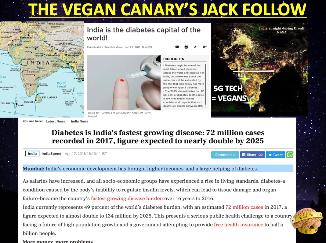
QT# 19: DIABETES IS A BLUE LIGHT HAZARD DISEASE.


In the 19th century, Dr. E. Babbit, M.D. proved that colored light was capable of healing through the effect on the autonomic nerve fibers in the skin and via the nerves from the eye to the brain. Dr. Spitler proved in the 1930’s that psychiatric illnesses could be cured or improved by using a visual colored monochromatic light source. It is now known that there are at least four effects from light. These are:

1. The optic nerve to the pituitary gland, temporal lobe, and occipital lobe of the brain. This information affects the conscious part of the brain without interpretation.

2. A second nerve bundle from the retina to the hypothalamus, which is a major control area for both the sympathetic and parasympathetic nerves. Dr. Fritz Hollwich, M.D. has shown that color affects neurotransmitter and hormone levels in the brain and spinal cord, which in turn affect the rest of metabolism and biochemistry. No one in the modern nutrition world seems aware of his work in blinded and sighted humans. (Vermont 2017 video)

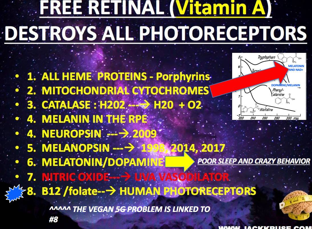
3. This path goes from the retina to the midbrain, and then to the superior cervical ganglion to the brainstem and then to the pineal gland. This area controls, among other things, our circadian (sleep/wake) cycle. This effect is mediated by melanopsin and retinol in humans in the eye, skin, subcutaneous fat mass, and the arterioles of the skin.

4. The last is a direct effect of the light upon particles that travel in the lymph, blood, and nerves. Researchers at the University of Vienna found that albumin is one of the particles able to be charged by sunlight.  It turns out albumin is made up of a large portion of aromatic amino acids that all absorb UV light in the blood plasma.  It also turns out the pH has a massive effect on how albumincan absorb UV light in the blood plasam.  The charges added to albumin are radically different if it is done by artificial light with blue light present.  After albumin has its topologic charge altered, it is then able to deliver this charge to tissues at distant locations (tissues) in the body.   Here is the original paper on how albumin works in the blood.  The higher one’s albumin ratio is the better they do long term.  This is especially true in most cancers.

Thanks to it, light at specific frequencies in the visible spectrum can act as a powerful tool to stimulate the biochemistry of the brain through the visual system by way of the retinal-hypothalamus brain connection.

INSULIN IS NOT JUST A PANCREATIC BASED HORMONE.

In 1922, Banting and Best found that insulin was the pancreatic hormone that regulates carbohydrate metabolism.  What many people do not know about the insulin story is that ophthalmologist, in 1950 Fritz Hollwich, conducted functional tests of carbohydrate control on blind subjects based upon the methods described by Staub and Traugott.  Their method involved a double alimentary glucose tolerance test given within 60-90 minutes.  They found in healthy people the second dose of grape sugar, which was given when the blood sugar was already falling had no effect or a slight effect on plasma glucose levels.

In his first run of experiments on ten blind subjects using the above methodology, Hollwich obtained negative results deviating from what earlier experiments showed in sighted people.  Hollwich demonstrated that light via the eye had an unknown effect on insulin physiology.  His work was confirmed in 1953 by Fuchs et al, and von Schumann (1953) and by Wassner in 1954.  Hollwich repeated his own experiments with larger numbers of blind patients in 1963, and again with the help of Diekhues in 1967.

After these finding in Europe, the insulin tolerance test of Radoslav became the gold standard. These experiments showed in all cases that in blind patients who received the insulin tolerance test, the blood sugar levels dropped far below the physiologic threshold they expected compared to sighted patients.  Hollwich was the first person in the world who showed that the results of both tests indicated a connection between blindness and a dysfunction of the hypophyseal portion of blood glucose regulation.  This finding is still not well known in modern diabetic research and diabetics with cataracts should be EXPECTED to have substantially different plasma glucose changes than patients without eye disease.

This is also true for diabetics with AMD.  Again none of this is found in the modern literature of diabetes because eye surgery has changed massively since Hollwich’s time.

Hollwich and Diekhues, in 1967 and 1971, did an amazing set of experiments on cataracts patients tested before and after eye surgery.  They showed a significant rise in blood sugar from 87.5mg to 98.5 % post surgery under a constant dietary regimen and light environment.  This variance in the postop Staub-Traugott tolerance test seen in the blind state before surgery and in the state of restored sight post-op can, with the food and light environment controlled served as definitive proof that ambient light’s entry into the eye has a massive influence on the regulation of glucose balance in humans.

Funny how you never hear any food gurus mention these experiments huh?  

This is pretty convenient for Big Pharma too who create drugs just that act on the gut hormone.  None of their solutions deal with the eye or skin.  This is why treating diabetics with just oral or IM injections fails to reverse anything.  I have a sense Big Pharma knows that.  


We now know, because of Hollwich that insulin is a solar hormone and has a diurnal rhythm independent from glucose intake. Those studies were done in 1974 and 1975 with radioimmune assays by Jarrett in 1974, Lakatuna et al in 1974 and Lestradet et al in 1974, Reinberg et al in 1974, and Thum in 1975. Thum’s paper in 1975, in particular, provides the possibility of assessing the precise means of light experiments with normal-sighted and blind subjects that should be done. None have because of the food guru perspective. Most are not even aware of this work in nutrition research because they only see what they want to see. What happens in the eye and skin when you eat is more important than what you eat. How is that for a truth bomb?


Most of the modern nutrition studies touted in the LCHF and paleo groups have no ambient light controls for the eye and Hollwich definitive proved in the 1960’s that Banting and Best initial observations of insulin’s effect is radically altered by ambient light via the eye or skin. We can add the skin to Hollwich’s work because in December 2017 we found it is in the skin and subcutaneous fat.


This is why most of the modern beliefs around carbohydrate metabolism are horribly flawed. I’ve been waiting 4 years for some of the gurus in here to go read his work and realize how much really is not known about ambient light’s effect via the eye and skin now that we found out melanopsin is in both tissues and melanopsin system radically effects the diurnal rhythms of insulin without ANY FOOD in the alimentary tract. Type 2 diabetes (T2D) is not purely a metabolic story tied to food, as you’ve been lead to believe. It has more to do with light via the eye and skin because of Hollwich’s experiments in the blind versus sighted humans from the 1960’s. It has been seriously upgraded by the news we found on melanopsin in the last 8 months. This was the topic of my Vermont 2018 talk. Most people believe diabetes is tied to food and a gut problem. It is not. When the circadian mechanism is off the eneterocytes do not turn over every 24-48 hours and this allows deuterium to enter the liver and this is what really causes diabetes problems most are familiar with. The process, however, begins with blue light exposure in the eye and skin, and this ruins the peripheral clock mechanism in the gut and liver as the picture below shows.


?The straw that breaks the camel’s back is epigenetic = fake blue light = melanopsin and Vitamin A problem = low melatonin and leads to poor mitochondrial DNA = problem in the eye and skin = a BLUE LIGHT HAZARD in the peripheral gut clocks in the enterocytes that spreads to the liver rapidly. That is really what diabetes is to a mitochondriac. That is what I showed in Vermont 2018.


?Eat SEASONALLY, but crap LIGHT environment = T2D.

?‍The wise N=1 ➡️ Reasonable seasonal carbs in the AM in summer, but get lots of sun with skin in the game + keep technology in check past 6 PM in any season. Your HbA1c never gets above 4.6 and you won’t need meds or physicians.

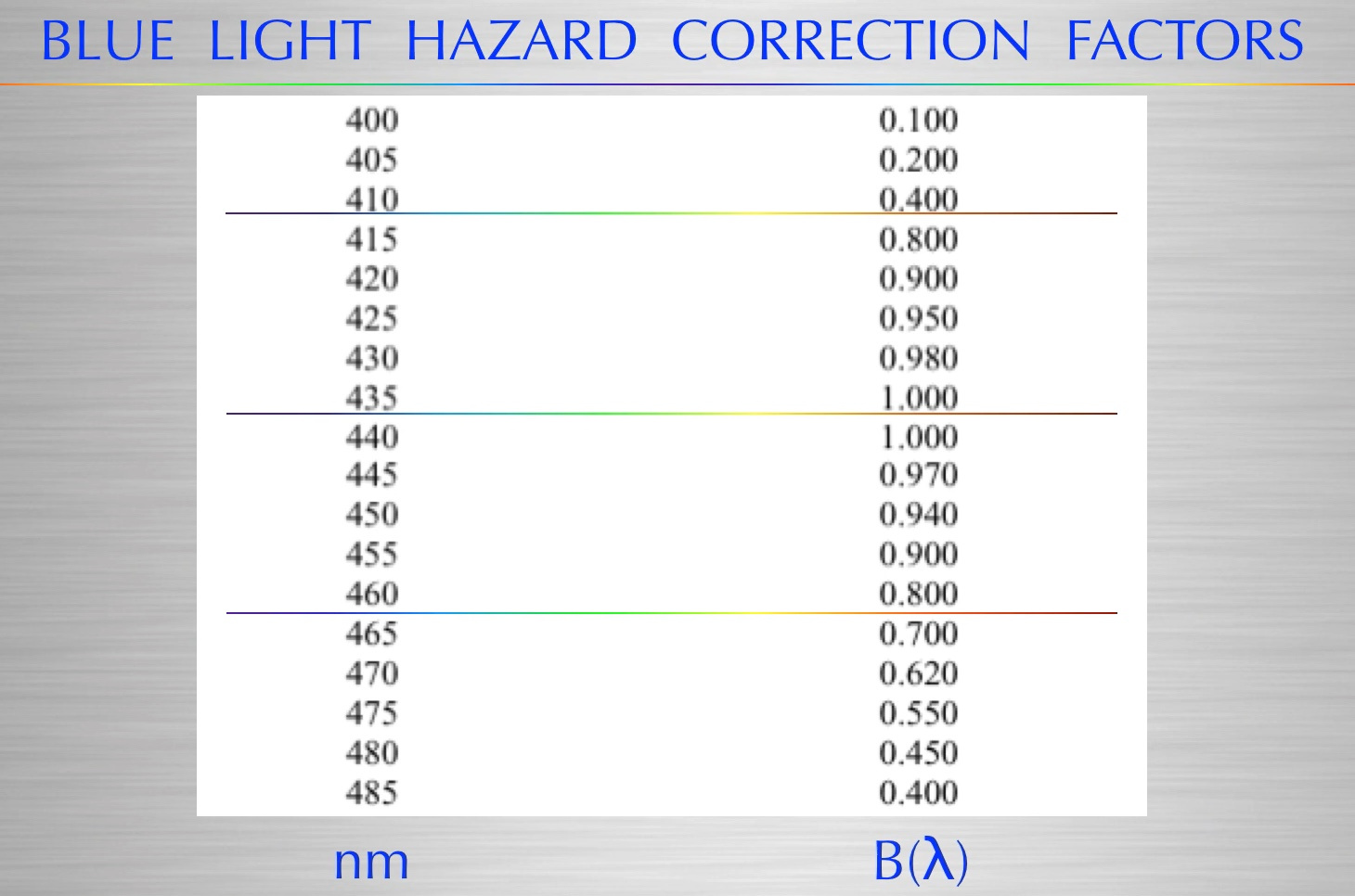
Artificial blue light  stimulates the anterior hypothalamus via the central reitnal pathways, by activating the PVN.  Normally sunlight with blue light and red in the AM can help activate the parasympathetic nervous system while stimulating the anterior pituitary hormones.  Subtracting out the red light and adding the blue is our modern problem.   This means that all colors in the bluish spectrum – from blue/green through blue to violet at 400nm is a problem for the gut because of melanopsin and the Vitamin A link to melatonin function.  Blue light via the eye or skin activates digestion and stimulate insulin secretion in the gut without the need for any food in the gut because of how melanopsin lowers melatonin by altering retinol function to ruin photoreceptor function that controls the human circadian mechanism.

VIDEO 1

VIDEO 2

CITES:

Hollwich, F.: The Influence of Ocular Light Perception on Metabolism in Man and in Animal. Berlin, 1985.

https://medicalxpress.com/news/2018-06-diabetes-results-breakdown-epigenetic.html

http://www.jbc.org/content/171/2/767.full.pdf

http://www.biotopics.co.uk/as/insulinproteinstructure.html

Szent-Gyorgyi, A.: Introduction to a Submolecular Biology. Academic Press: N. Y., 1960.

Wurtman, R.: The Effects of Light on the Human Body. In: Scientific American, July 1975, Vol. 233, Nr. 1, S. 68-79.

Gabel, S.: Information Processing in Rapid Eye Movement  Sleep: Possible Neurophysiological, Neuropsychological, and Clinical Correlates. In.: Journal of Nervous and Mental Disease, 175, 1987, S. 193-200.

Toupin, A.: Photic Avtivation and Experimental Data Concerning Colored Stimuli. In: Neurology (Minneap.), 16, 1966, S. 269

CPC: #29: Unexpected Physics in Biology is MUSIC to cells.

Circadian cycles allow for self organizing features and functions in cells. Effectively these cycles are the musics your cells are addicted too. Every AM you are designed to retune the system with the AM sunlight so the music of life contains to reverberate in you cells continuously without interruption.


Dr. Franz Halberg coined the term “Circadian Rhythm” in the 1950’s, as a result of his studies in what became Chronobiology.  He found our bodies have a repeating cycle, a rhythm lasting about (circa) a day (dian).  He measured these rhythms using the body’s core temperature and levels of activity and referred to the shorter daily waking and sleeping cycles as Ultradian Rhythms.  Sleep centers use EEGs (ElectroEncephaloGraphy) to record brainwave activity during our sleeping ultradian rhythms.  Dr. Halberg discovered the biological processes for producing essential proteins, which are the building blocks of our bodies, are only produced at the start of each ultradian cycle.  The most important ones are ubiquitin, methionine cycles, and proteins made from tryptophan.  Sunlight tunes them all in unique ways with photoentrainment.

The sun directly via its electromagnetic waves to tightly coil DNA/RNA while all versions nnEMF seem to uncoil DNA to affect its energy and information status.  We gained this idea in data on bacteriophages.  They pack their heads with DNA tightly under great force as the bottom left slide shows.


The area under curve in bottom right graph is a measure of the energy required to pack DNA inside the head of the phage.
Notice the energy increases greatly as more DNA is packed. High energy is due to DNA stiffness and its negative charge.

It appears circadian disruption uncoils DNA for transcription and this induces an electrical status change.  DNA becomes more positive in charge because electrons are lost to aqueous proteins that the ultradian cycles produce to change physiologic function of cells.  This appears to be the basic mechanisms of how things should operate to structure tissues and organize cells.

A priori optimum development in organisms, from single cell to multicellular organisms, or from skeletal cells to a skeleton or limbs exemplify Fibonacci patterns. These developmental patterns consist of two primary characteristics: (1) a number of the organisms structural arrangements may fall into the series 1, 1, 2, 3, 5, 8,…or (2) logarithmic growth based on the ratio of consecutive Fibonacci numbers (1.618033988…, also known as the golden ratio or φ). The growth patterns observed occur throughout nature in the arrangement of skin pores in tetrapods, the spiral shape of snails and sea shells, and the overall structure of plants. Fibonacci numbers occur in atoms and electrons (Huntley, 1969), the DNA molecule (Wahl, 1988), biological cell division (Spears & Bicknell-Johnson, 1998), models of growth and death (Hoggatt & Lind, 1969), bronchial airway segment bifurcations (Goldenberger, West, Dresselhaus, & Bhargava, 1985), experimental growth of tumor nodules (Prokopchuk, 1981), and many other aspects of human biology (e.g., position of facial features, body proportions).

Does musical composition exhibit this organizing pattern too?  Good music does.

Can we use it to help fine tune us when the melanopsin system is de-tuned by poor light choices?  It turns out we can.

Have you ever heard a smart phone interfere with audio equipment at a party or in your car?  This often happens with incoming calls in a car using satellite radio or music apps.  It sounds like bursts of noise when an active cell phone is near a radio or even an MP3 player or Public Address system. Now ask yourself, “How is it possible for something that doesn’t even have  a radio antenna in them to translate cellular signals into these audible bursts?” The answer is simple and central to understanding our current health problems in our electropolluted environments.


Cell phones, like nearly all wireless devices, use some form of frequency hopping to better utilize available radio spectrum, reduce interference and use lower power. The wireless carrier frequency is usually in the hundreds of megahertz to gigahertz range and at the low power emitted by cell phones the radio waves should pass harmlessly through us.  This is going to change and get substantially worse in most cities when 5G networks that are already built are allowed to broadcast.

So why does epidemiological evidence show a higher risk associated with exposure to these radio waves? The fact you can hear audible bursts with an audio amplifier that isn’t a radio receiver, or a receiver not even tuned to the same carrier frequency, gets to the crux of the electromagnetic problem mankind is facing now.

The culprit is the interval at which the frequency hopping occurs, and not the megahertz to gigahertz carrier frequency of the transmitted bursts. The frequency hopping interval, I am using ‘interval’ instead of ‘frequency’ to avoid confusion with the carrier frequencies, is in the audible range. Our bodies are sensitive to frequencies we can hear, there are frequencies that heal us by working with our circadian cycles and frequencies that make us ill before they kill us because we decouple these cycles from the proteins they make.

It’s as basic as how music affects us. When musical instruments are out of tune or out of harmony with each other the result is agitating noise. And when we hear such a cacophony of dissonance, we turn the music off or leave the performance.

Today, light waves are disrupting the circadian cycles most, but it appears that we might be able to use music to heal.

Self-organized criticality is also a way of understanding complex systems in nature. A power law is a mathematical relationship often referred to as the 1/f law (after the 1/f electronic noise of transistors) in which the distribution of power density at different frequencies (power spectral density) is inversely proportioned to the frequency. Before venturing further, let’s see what the 1/f distribution can tell us about music. This song happens to be one that entrances me when I write about quantum biology.

Fractal musical time is built around power laws buried in nature and sunlight. It is even in the human heartbeat and EEG.

Why do we love music? Have you ever asked yourself this? One reason we enjoy music lies in its balance of predictability and surprise, so researchers claim. They found a musical pitch (frequency) spectrum following a 1/f power law, which achieves this balance of predictability and surprise; but what about musical rhythm? Musical rhythms, especially those of Western classical music, are highly regular and predictable; are they? Daniel Levitin at McGill University Canada, Parag Chordia at Georgia Institute of Technology Atlanta and Vinod Menon at Stanford University California in the United States decided to put that to the test by analyzing the rhythm spectra in 1 788 movements from 558 compositions of Western classical music in their papers.

The rhythmic content of the compositions was systematically measured by noting the duration of the notes and of the rests, transforming the durations into Hz (cycle per second), plotting the data and finding the spectral exponent in the slope of the line obtained. They went through the works of 40 composers in 16 subgenres and found an overwhelming majority of rhythms following the 1/fa power law with a ranging from ~0.5 to ~1. An exponent of 0 would be pure white noise, completely unpredictable, whereas an exponent of 2 and above would be highly predictable. Notably, classical composers whose compositions are known to exhibit nearly identical 1/f pitch spectra demonstrated distinctive 1/f rhythm spectra: Beethoven’s rhythms were among the most predictable, and Mozart’s among the least, with Haydn in between. This song by Tool, above, was built around nature’s key frequency law. The difference in rhythmic predictability is such as to allow composers to identify their compositions uniquely and to distinguish them from works of their contemporaries. No one has made a song like a cadence in this modern piece. What does it say to you as you listen to it? For me the take home is simple: It is not the melody or voice that commands the story;

it is the ear that deciphers the code contained within. It is very similar to how a mitochondria deciphers the light waves that collide with it.

CITES:

1. Mathematical Models in Biology  By Leah Edelstein-Keshet 1988.

CPC #28 IS OBSTRUCTIVE SLEEP APNEA PROTECTIVE?

IS SLEEP APNEA PROTECTIVE AND NOT PATHOLOGIC?

I believe the answer is yes.  Why do I say this?

It appears sleep apnea protects defective mitochondria from high oxygen tensions. Want to know more watch my May 2018 webinar to understand the mitochondriac perspective.  I go further into detail in my August 2018 webinar that is being released today to my full members.

This means it is really not a disease……it is a reaction to low NAD+ and high ROS made in mitochondria to control the flow of oxygen just as a carburetor does in a car.

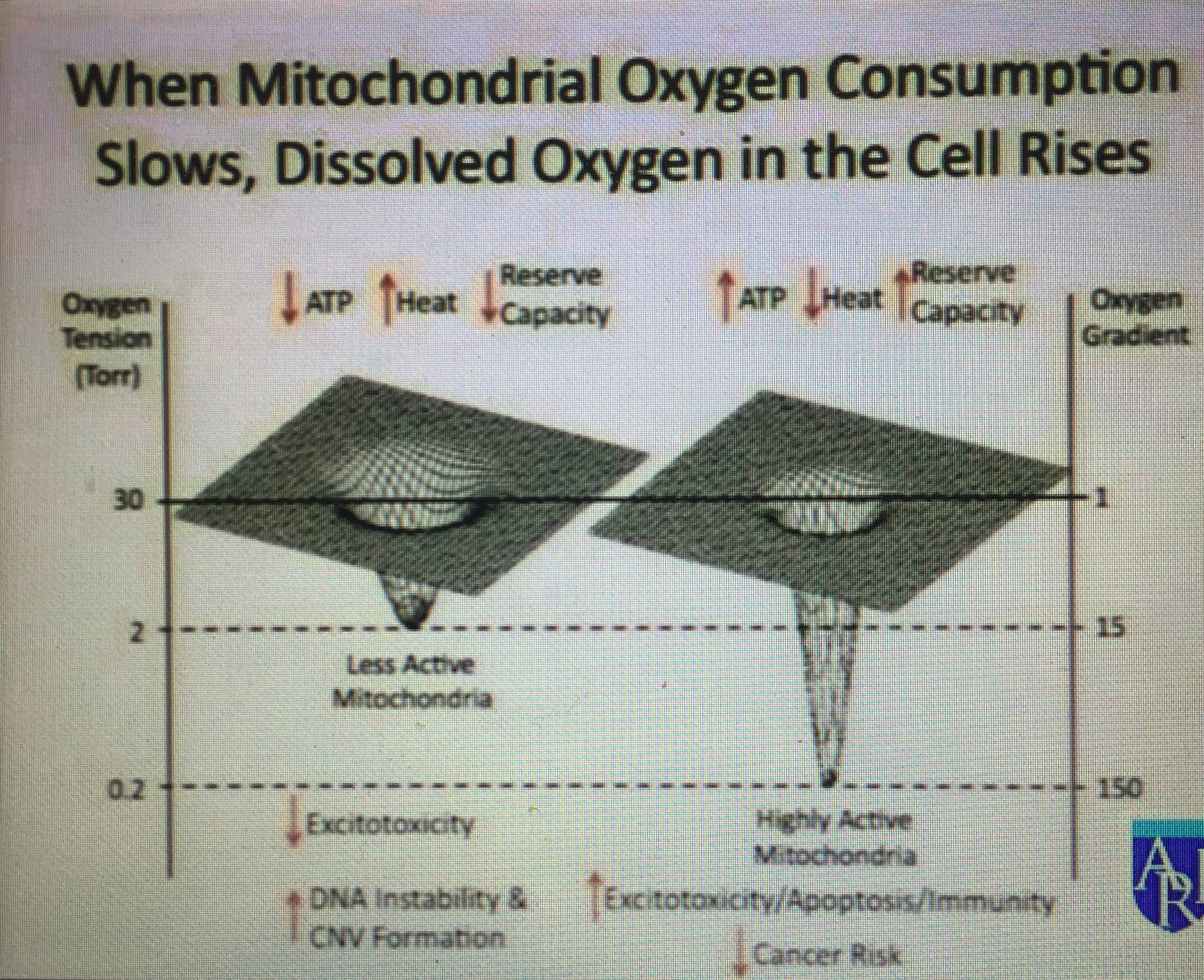
Over-eating is going to stress the ECT………that is ruined by the increased space between the cytochromes…….this implies oxygen levels have to be decreased when autophagy is ruined.  The biggest offender is blue light exposure via the eye and skin to cause this change in the melanopsin and Vitamin A couple.  Blue light exposure decouples Vitamin A and Vitamin D from one another as the slide below shows.


When ECT flow remains brisk and OSA is present, oncogenic risks is higher. Very few people with apnea seem to know that sleep apnea carries a pre-malignant risk with it because of this mitochondrial connection to defective apoptosis.


PREDICTION: This is what I think is going to happen to Jimmy Moore and his fat laden diet married to his high technology appetite. This lifestyle subtracts the sun because of his blue light addiction. This ruins the melanopsin/retinol mechanism that is protective to the metabolic rate variations in uncoupling proteins. These factors then lead to the key change in Kreb’s bicycle slowing both cycles kinetics causing methionine to rise.


When that happens autophagy and apoptosis are broken and these things all follow:

Cancer Changes:

1. that affect signaling of damage (the DNA damage response or DDR),
2. the extremely important WNT pathway that controls aspects of making RNA from DNA and especially proteins for apoptosis versus cell division. (The name WNT comes from pathways wingless in flies and int in mice)
3. enzymes called kinases that are part of vital pathways similar to WNT (MAPK, AKT),
4. the cytokine interferon that signals to the nucleus regulating and responding to making blood vessels, cell movement, defense against invasion, and cell death, and
5. inflammatory cytokines that stop apoptosis (nuclear factor-κB (NF-κB),


Cancer must inhibit apoptosis to occur. May 2018 webinar has it all laid out. Melanopsin optical signaling interruption in the eye and skin is capable of doing it ALL. The key feature in ALL CANCERS: ECT must be maintained while apoptosis inhibited.

OSA is a phenomenon that protects apoptosis from inactivation by keeping oxygen levels low. This protection scheme goes on until the deuterium entombment in the cell membranes of the PUFA’s of the mitochondria overwhelm Kreb’s bicycle and leads to COX-2 amplification.  This detail was covered in the May 2018 webinar and I went even deeper in the August 2018 webinar.  All cancers need high levels of oxygen to maintain high ECT speeds. The rising levels of methionine cause this.  This occurs because of the chronic slowing kinetics of the TCA and urea cycle eventually ruins the recycling of methionine from homocysteine.  This leads to blood vessels releasing angiogenesis factor to sprout new blood vessele to raise delievery of blood and oxygen.  Once this happens cancer is much more likely in a patient with OSA because it inhibits apoptosis.

This happens because oxygen is highly electronegative on the periodic table and it draws electrons from NADH in cytochrome fast. Another way the electromagnetic force is utilized in cristae.

Moreover,  sleep apnea sufferers struggle with spatial recognition and quantifying correct time.  This links it to a dopamine defect somewhere in the system.  This is also associated with a melanopsin defect in the eye or skin, as a result.  This implies sleep apnea is a protective effect cells use to buffer mitochondrial damage from melanopsin/retinol disruption.


You pay the price of both, with your choices made daily around the electromagnetic spectrum. You pay a biologic toll because of your choice, or you gain a biologic benefit because of your choice. At the outset, the dopamine levels are the same. After you decide and the results are in, you will see that the environment will add or subtract from your dopamine bank account. The presence of time alteration and sleep apnea are two signs you better change what your are doing. The result determines the reality you get. In this way, it should be clear how the environment dictates results we get.


AM sunlight balances the blue light of the morning. Blue light power increases during the day while red light remains constant in sunlight.

This implies blue light alerts us and is somehow related to time creation in the human brain.

Blue light opens the optical windows of the skin and eyes via the melanopsin effect in arterioles to other portions of light in our environment. When that is sunlight all is well. When that light is all blue light and no UV or IR light sleep apnea is coming faster to your life.


What does this all imply? Red light slows time. Cold slows time. Blue light creates and speds time perception up in the brain by altering the circadian clock functions. UV light rebuilds and replenishes time in a complex dance that begins in your eyes and skin because of melanopsins effect on Vitamin A = retinol. Therefore, excessive chronic blue light hazard speeds up time, the time mechanism in clock genes, and can give you mitochondrial diseases like cancer. When you are blue light toxic your life is lived like a blue straggler star. Quick and bright. Youre alert, and get sick quicker, and die sooner.


AM sunlight is the light switch that creates the ideal level of dopamine and melatonin assuming your melanopsin system is working properly and OPERATIONAL.  The switch is tripped by what happens first in the eye and then the skin by the balance between dopamine and melatonin created by the days first light.  Many papers attribute bipolar dysfunction to the lateral habenula in the brain.  People with bi-polar disorder often have sleep apnea too and have altered time sensation.  This area has a lot of melanin in it and has melanopsin inputs to it as well.  This region of the human receives input from the stria medullaris thalami.  This region links the central retinal pathways and the SKIN to the hypothalamus. The stria medullaris is a part of the epi-thalamus. It is a fiber bundle containing afferent fibers from the septal nuclei, lateral pre-optic-hypothalamic region, and anterior thalamic nuclei that connect to the habenula.  The habenula sends many outputs to several midbrain regions involved in releasing neurotransmitters, such as dopamine, norepinephrine, and serotonin.


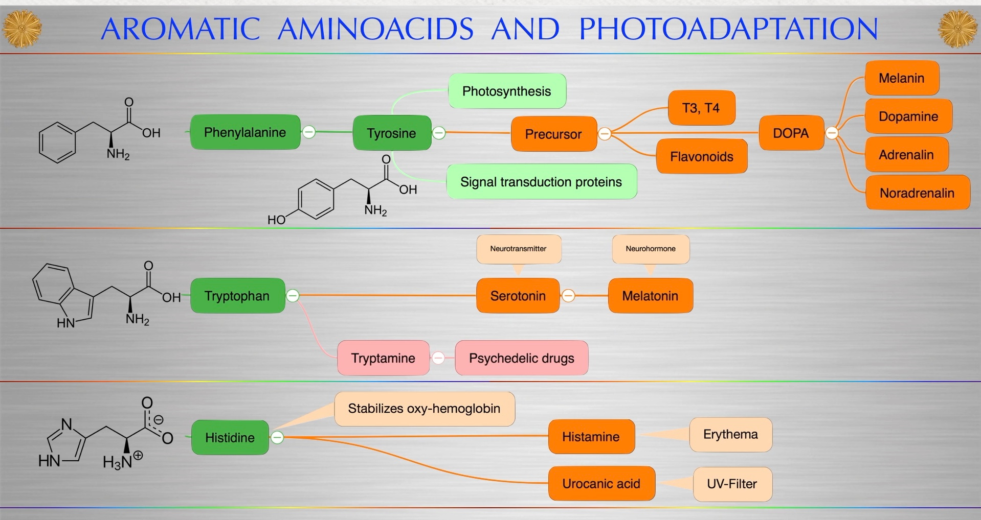
All of these chemicals are made from aromatic amino acids that need to be programmed by AM sunlight.  All three of these neurotransmitters are made initially in the eye and then the skin from sunlight and aromatic amino acids.

Obstructive sleep apnea (OSA) patients show multiple neurobehavioral difficulties, including deficits in attention, psychomotor and executive functioning, memory, and affect (Beebe et al., 2003). However, the principal deficits in the syndrome involve the loss of motor control over the upper airway muscles, which fail to discharge in a timely fashion, when they should dilate the airway during diaphragmatic descent, and a loss of control over autonomic motor activity, with chronic, exaggerated sympathetic discharge from the PVN (Somers et al., 1995), and inappropriate lagged or muted dynamic sympathetic responses to blood pressure or ventilatory challenges.  This puts the problem in the brainstem in the dopamine tracts.

The habenula regulates the activity of midbrain centers, including the dopaminergic ventral tegmental area (VTA).  This area is huge in Parkinson’s disease development from melanopsin uncoupling on the surfaces of the eye and skin.  Phenylalanine and tyrosine can both be converted into these three neurotransmitters easily because both can make dopa. L-DOPA, is synthesized in the brain and kidneys of humans.  Look at the picture above where DOPA is made = tyrosine.

Dopamine is also synthesized in plants and most animals. The brain includes several distinct dopamine pathways, one of which plays a major role in reward-motivated behavior. Most people do not even realize with a dopamine defect, control of blood glucose is lost too. This is why people with apnea often have diabetes.

This becomes a key feature in the melanopsin damage seen in all forms of diabetes.  This one is best understood but researchers have done a poor job realizing how these pathways all begin with light signals in the retina/skin/blood vessels for proper physiologic functioning.  This is why bi-polar disease, diabetes, and parkinson’s disease and SLEEP APNEA stump modern medicine.


AM sunlight also stimulates POMC which makes beta-endorphin to make us seek AM light.  A lack of beta-endorphin sets up humans for an opiate crisis.  Modern humans have lost this GUIDING solar signal because of how they live their life and what light they live under most commonly.  Most types of rewards increase the level of dopamine in the brain.  In some diseases, like schizophrenia, too much dopamine at the wrong time ruins your ability to maintain cognition in time and space and leads to a crazy life.  This occurs in schizophrenia.  Without solar guidance, many modern humans seek opiate rewards in chemical ways to offset the deficit.  This leads many nights living humans to use many addictive drugs to increase dopamine neuronal activity to offset the loss of sunlight. Other brain dopamine pathways are involved in motor control and in controlling the release of various hormones.  These get damaged by chronic sunlight loss or too much blue light at the wrong time post sunset. It can even be a problem during the day if done chronically by a job and a computer screen or a phone to a teenager.  These pathways and cell groups form a dopamine system which is neuromodulatory and controlled by the circadian system by the melanopsin/retinol interactive controller.  This is the topic of my August 2018 webinar and it is why my Patrons are getting this blog right now at the same time.  Patrons it is time you sign up for your real Black Swan training here:  HYPERLINK

Outside the central nervous system, dopamine functions primarily as a local chemical messenger and it needs the direction of sunlight.  How do we get light signals deep into the body this way?  Hemoglobin in the eye and skin is how it begins.  It is loaded with iron porphyrins which absorb 250 – 600 nm.  The water that surrounds it can absorb all the way to 3100nm.


This light is ferried to cells to program the dopamine and melatonin release locally ot change the metabolic rate of mtDNA by altering the UCP2 permeability of deuterium. The same is true of melatonin. In blood vessels, dopamine inhibits norepinephrine release and acts as a vasodilator with Nitric oxide (UV-A); melanopsin does the same thing using other frequencies of sunlight. In the kidneys, dopamine increases sodium excretion and urine output; This changes the EZ size in blood plasma. In the pancreas, it reduces insulin production (diabetes link); in the digestive system, it reduces gastrointestinal motility and protects the intestinal mucosa gut barrier (leaky gut link); and in the immune system, it reduces the activity of lymphocytes which is important in autoimmunity.

I said June 2, 2018 in Vermont that is not one human chronic disease not LINKED to the melanopsin/retinol mechanism. Here is the slide I used.


This post is a dip of the toe in why I might be correct in my reasoning. With the exception of the blood vessels, dopamine in each of these peripheral systems is synthesized locally and exerts its effects near the cells that release it. All of these link back to sunlight to the most fundamental level and no one but Black Swans realize it.

The mitochondriac perspective always explains things we observe over the things modern medicine espouses.


The more I study the history of intellectuals in medicine, the more they seem like a wrecking crew, dismantling civilization bit by bit — replacing what works in nature with what sounds good and repackaging it as evidence-based therapies.

Nothing replaces this……….and it is free.


Start using it if you have sleep apnea because that symptom is your colony of mitochondria telling you there is a problem.

CITES:

https://www.ncbi.nlm.nih.gov/pmc/articles/PMC3930100/

QT#17: SWINGING ON THE SPIRAL: TIME


Few of us realize the liquid crystals in us create time using light.  Time and space are modes by which we think and experience reality but neither are conditions in which we live on Earth.  We live by swinging on the spiral arm of a galaxy constantly moving through space .

Ever since the derivation of the theory of relativity, physicists have thought they have done reasonably well in understanding time; however, biologists are still waiting for a comprehensive theory of timing in living systems:  they remain waiting for a corpus of “laws” describing how cells and organisms can precisely initiate and terminate processes at specified times because they are focused on the nuclear genome and not the mitochondrial genome.  Time perception is critical to mitochondrial biology but it is not especially important to the nuclear genome because the energy provided by the mitochondrial genome allows time to manifest in living things. This deficiency in biology is particularly acute in developmental biology, where complex mechanisms of various paces and durations must be orchestrated to solve huge developmental problems such as the one faced by the fertilized egg: how to become an organism using energy/information and electric and magnetic field to direct and power morphogenesis.

Animal development is, in fact, nothing but time. From the cell cycle to the beating of the heart, our own lives are composed of a multitude of microscopic and molecular oscillations. For developmental biology, the study of causal relationships implies the examination of two time points: inducing the cause and looking at the effect. In QED, cause and effect is blurred because of the uncertainity principle.  In biology, cause and effect “appear” to be linked because of how time is perceived.  We are generally ignorant of the temporal rules governing this transformation. Light and aromatic amino acids form those foundations.  Whereas the rules of a Swiss watch involve a timing mechanism that uses a uniform unvarying tempo to translate rapid oscillations (seconds) into longer periods of time (hours), the rules of the developmental clock of animals are much more complex because they use light and light is non linear in the violet part of the visible spectrum. The animal clock is made up of a variety of counting mechanisms that follow varied temporal rules at different frequencies and often run in parallel without any “apparent” interaction with each other, because their linkages are via molecular resonance. The goal of the developmental clock is not simply to mark off time, but to integrate and unify the myriad temporal signals received from throughout the organism.

However, the frequencies and amplitudes of most, if not all, developmental clocks are robustly determined within the same species, suggesting that these are genetically encoded devices being linked and controlled by resonance instead of being produced by the system itself, as an “emergent feature.”  I have always believed time is an emergent property of life because time emerges from the complexity of light the force carrier of the electromagnetic force. Today, the “old paradigm of biologic time (Franz Hallberg) is dominant in the published literature, but as Max Panck said, things change in science one funeral at a time.  Morever, if the “belief” is true that time is encoded in our DNA (it’s not), what are the genetic balance wheel, hairspring, and gears, and what are the specialized tools that are needed to elucidate the intricate workings of the genome? There are many possibilities to consider here, but I will discuss two.


The first one concerns clocks generated by mechanisms in “trans”, generally based on the postulate made in the early 1950s by Alan Turing (mathematician, philosopher, and inventor of the concept of the computer), who argued that the diffusion of substances with activation, repression, and retroactive potentials can generate intrinsic oscillations in space and time. The literature has begun to decipher some of these ideas and more are likely to come in the near future.

The second possibility involves mechanisms in “cis”, using the linearity of our genetic material itself, our chromosomes, to measure time; an admittedly elegant solution, but one that severely lacks both experimental and theoretical support. Chronobiology is stuck on these two options because they do not realize that time is a superposition of many possibilities. This is why they ignore the mitochondrial genome. This genome creates many possibilities when small changes occur to it. When we disrupt it with anesthesia timing is completely altered, we observe it and we measure it, but we do not understand the implications for man or his diseases. This will change with the coming age of quantum biology. The old guard expected some help from the human genome sequencing and related high-throughput technologies, but they got stonewalled because it destroyed constructs that we inherited from Watson and Crick. I reject most of their thesis about biology.


Modern epigenetics data has blown up much of Darwin’s thesis from 1859 but few biologist will admit it today because it is akin to removing the binding from a book.  All organizing principle are afloat now in molecular genetics because of the new insights of epigenetics.  So is science and life.  Every end is a new beginning in understanding and learning with respect to nature’s wisdom.  Darwin’s supporters today are still trying to hold the theory together but the concept of timing and energy is 100% tied to energy collection and generation and distribution in the cell. They have no Earthly idea that energy and information processing are coupled by light.

There is little doubt that the mechanisms in ‘trans’ will be advantageously studied in this context. Time-lapse proteomics and metabolomics, adapted to minute amounts of material such as cohorts of less than a thousand cells, would certainly show significant recurrences and, perhaps, allow the temporal algorithms underlying developmental clocks to be uncovered.  But trends in protein or metabolic movement can just describe the cellular processes and it won’t explain it.  The explaination will take a mind filled with innovated thoughts to link them back to the fabric of nature’s laws.  Today’s developmental chronobiologists believe that a reductionist approach to clock mechanisms will give us the key answers.  In fact, we just gave three people a Nobel Prize recently for discovering the biologic molecules involved in the circadian mechanism.  For example, the tightly regulated timing in the successive appearance of whiskers on the nose of a rodent would likely be deciphered if we could microdissect the whisker field like a chess grid, and submit each square to full protein content analysis.

On the other hand, the challenge in identifying ‘cis’ processes would be to use several species showing graded variations in clock parameters, such as in the frequencies of the same clock, and to find correlations with the “cis organization” of DNA segments through a large-scale approach. Such correlations, if any, could reflect the existence of linear time-measuring devices, from the mere duration of transcription to more complex processes such as progressive transitions in the chromatin state. However, that is easier to say than to do; a genuine “casse-tête” for bioinformaticians.


I believe based upon what we know today about epigenetics, it is seriously pretentious to try to extract the fourth dimension, time, out of our DNA/RNA structure when we still lack complete understanding of the other three dimensions of our world. But the human quantum computer does not need complete data sets to jump to conclusions. That is what innovation and imagination are useful for in complex problem solving. This science is the privilege and the difficulty of working with embryos: The spatial construction can be understood only in the light of time. The irony of this idea, is that light is what creates time.


This concept continues to trip up biologists because they are ignorant of the queerness of the physics of light. Similarly, evolutionary genomics will likely teach us what happened during phylogenetic times, and our ontogenic clocks may be revealed by global comparative analyses.


Chronomics is on the way, but only the unripe mind will think it is the be all end all in the discussion of what time really is.

Time allows us to see what we have become, even when we miss it. The liquid crystals inside of us build this library. Time is erased for some of us, because we cannot use sunlight and water to build the crystals properly to manage our time. Sometimes we erase people and family, and walk away from the shadows creeping up on us. Sometimes you must give up on people, not because you do not care, because they do not care enough for themself by making the correct choices.

Spiral out…………..

QT #16: WHERE DOES ELF-UV COME FROM?


So do we glow when sunlight hits the skin? Yes, we do. What kind of light do we glow with? ELF-UV light manifests in our blood plasma = do we gain or lose electrons when this occurs? We lose electrons and give them to other anions in our blood. Sulfated amino acids are added to many proteins and lipids in our blood under the power of sunlight. Sunlight increases sulfhydryl groups which are the major anions in our blood plasma.


Our tissues glow because when an electron is delocalized light is given off and that light produces change without atomic change because it changes the topologic charge of atoms in our skin proteins because UV light causes a loss of electrons in out tissues.  This mechanism is what causes release nitric oxide from the arterioles in our skin to change the topology or femtochemistry of the surface of the skin when it is irradiated by IRA and UVA light.  The change in femtochemistry results in attochemical changes in deuterium in our blood plasma.

Full spectrum sun contain UVA/B light which is capable of altering the topologic charge of nitric oxide (NO).

UVA light releases the most NO into our blood.  IRA light is how the exclusion zone is built initially in sunlight in our plasma.  Both of these frequencies penetrate below the epidermis to where the arterioles of our skin.

The key thing to comprehend is there is no way to satisfy the octet rule for both atoms in NO because of their valence shell electron state. The structure is a double bond with three non-bonding electrons on N and four on O but really one of the electrons on N is not localized to N but the entire molecule.

Formal charge = electrons in ground state – (non-bonding electrons + 0.5 * bonding electrons)

Formal charge for N = 5 – ( 3 + 4 * 0.5) = 0 (there are two bonds so four electrons are involved in bonding)

Formal charge for O = 6 – (4+ 4 * 0.5) = 0

Sunlight alters that balance of charge on our surfaces.  What does it do?

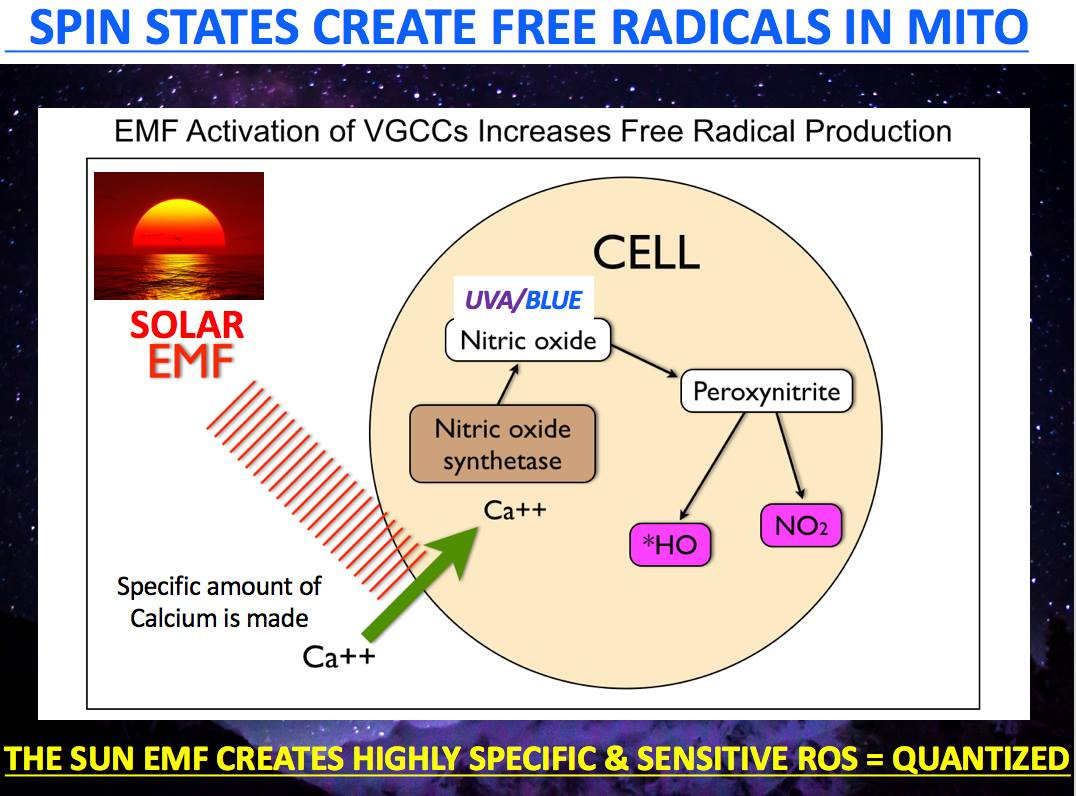
It creates the triplet state of free radicals because of the effect on electron quantum spin.  Every surface in our body has this mechanism available if the sun is allowed to interact with it.  If the sun is absent this will not occur and it will lead to collateral damages.   Where else is electron spin altered?   MITOCHONDRIA.  This is where the critical ROS and RNS signals are made.  These signals affect a third cycle in us that controls the sulfated amino acids.  This is the sulfated amino acid cycle contains cysteine methionine and homocysteine.  This cycle is altered because of the changes in free radical creation by the light our skin does or does not experience.

Full spectrum solar exposure of the skin increases sulfhydryl groups in the blood plasma.  This increases the anions present in our blood.  Sulfur amino acids are a kind of amino acids which contain sulfhydryl groups, and they play a crucial role in protein structure, metabolism, immunity, and oxidation. Recent studies have demonstrated the oxidation resistance effect of methionine and cysteine, two of the most representative sulfur amino acids, and their metabolites.

Methionine and cysteine are extremely sensitive to almost all forms of reactive oxygen species MADE IN THE MATRIX, which makes them antioxidative and critical redox switchs that tell us about TCA and urea cycle kinetics.  They can both be used to tell us when the matrix is slowed by deuteration.

Moreover, methionine and cysteine are precursors of S-adenosylmethionine, hydrogen sulfide, taurine, homocysteine,  and glutathione. These products are reported to alleviate oxidant stress induced by various oxidants and protect the tissue from the damage. However, the deficiency and excess of methionine and cysteine in our diet can affect the normal growth and longevity of animals.


It is well known that oxidation from free radicals of the cysteine thiol groups occurs in all stressors in humans.  This results in defective glucocorticoid receptor (GR in pic above)  function and it affect ligand and DNA binding in cells.  This implies that in a 5G world where the toplogy of the skin will be affected, cysteine and methionine hacks will be quite important to understand and use when one gets afflicted with electropollution type diseases like autoimmunity, sepsis, and cancer.   To understand how these sulfated amino acids operate you must understand how the sun works in your skin.  I discussed this in the Vermont 2018 talk now available for purchase to Patrons and the public.

Vitamin D3 supplement pills don’t and can never give you triplet state being described here because you need the light stimulus on the skin to get it.  You also need skin in the game to generate the light pressure on your skin and arterioles to enable the chiral effect.   Sunlight exposure of the skin, works in unison to sulfates the cholesterol anion substrates in the skin and blood from the sulfated amino acids so that cholesterol can become Vitamin D3 via various steps between the skin, kidney and liver under the power of UVA and UVB light.


KEY POINT ALERT:

UVA light helps in performing this task because of the action of activated NO in the skin.  NO inactivates cytochrome 4 while IRA light allows the ATPase to spin freely with fewer electrons needed from foods/fat due to the action of NO on ECT.  UVA light inhibits or slows electron chain transport flow on the inner mitochondrial membrane.   With this knowledge are you still afraid of black lights indoors?  You should not be.  It might be a great hack for you depending upon your N = 1 context.


Nitric oxide (nitrogen oxide, nitrogen monoxide) is a molecular, chemical compound with a chemical formula of NO that is a colorless gas under standard conditions. Nitric oxide is a free radical—i.e., its bonding structure includes an unpaired electron with a specific electronic spin state. It is a practically important intermediate in the chemical industry. In addition, some are unavoidably produced during combustion of fossil fuels in power plants and automobile engines, with an excess of NO being created when there is present more air or higher temperatures than needed for efficient and complete combustion of the fuel. More oxygen and higher temperatures in a cell favor H+ bonding.


They lower deuterium bonding in the skin. This is very important because it is the key mechanism of how and why deuterium is found in the high concentration of the blood plasma in the irradiated skin by the sun. In skin thermal temps are raised most by UV/IR light and oxygen is increased by UV light striking the skin alone.


Not only does skin make it with the powerful sun but so does the atmosphere in a quantum fashion. It is also produced naturally by the extremely high air temperatures produced along the path of lightning in thunderstorms.

Now stop here.  Did I not tell you in the past on previous blogs that the inner mitochondrial membrane can generate the same voltage as a bolt of lightning in the “What powers life and death blog years ago?”  Yep.

30 million volts is possible.

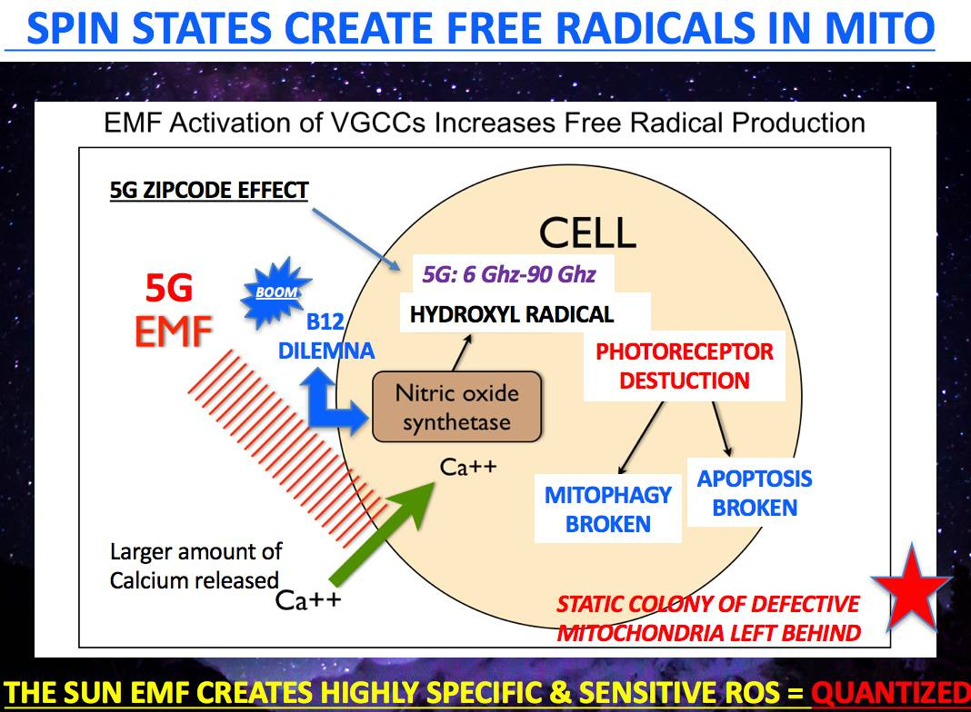
It was found that NO acts through the stimulation of the soluble guanylate cyclase, which is a heterodimeric enzyme with subsequent formation of cyclic-GMP. Cyclic-GMP activates protein kinase G, which causes reuptake of Ca2+ and lowers the intracellular calcium by the opening of calcium-activated potassium channels. This is highly protective in high nnEMF environments. 5G will block this effect in the skin. 5G will also destroy the covalent bond between melanopsin and Vitamin A.  This will demolish the entire circadian mechanism in all the peripheral clock genes.

This is why 5G risks will ramp up mitochondrial diseases linked to the melanopsin/retinol system.  Melanopsin also uses calcium as a secondary messenger.  This creates another risk of nnEMF in destruction of the melanopsin mechanism.  nnEMF alters calcium flows and this is the signal that makes free radicals in the matrix for proper quantized signaling.


The fall in concentration of Ca2+ ensures that the myosin light-chain kinase (MLCK) can no longer phosphorylate the myosin molecule, thereby stopping the cross-bridge cycle and leading to relaxation of the smooth muscle cell in our skin arterioles allowing more RBC’s loaded with porphyrins to continue absorbing UVA light to keep this action of NO working properly to keep deuterium inside of our arteries where it belongs.  Deuterium concentration in the blood plasma is how the body normally creates ELF-UV within our blood plasma to activate cell water to become an exclusion zone.  This is a topologic charge effect that must occur endogenously.

Nitric oxide, is also known as the ‘endothelium-derived relaxing factor’, or ‘EDRF’, is biosynthesized endogenously from L-arginine (urea cycle), oxygen, and NADPH by various nitric oxide synthase (NOS) enzymes. Reduction of inorganic nitrate may also serve to make nitric oxide in our gut. The endothelium of blood vessels uses nitric oxide to signal the surrounding smooth muscle to relax, thus resulting in vasodilation and increasing blood flow so we can collect a lot of UV light from skin irradiation. How does hemoglobin fit into this blood UV story?

Two important biological reaction mechanisms of nitric oxide are S-nitrosation of thiols and nitrosylation of transition metal ions. S-nitrosation involves the (reversible) conversion of thiol groups, including cysteine residues in proteins, to form S-nitrosothiols (RSNOs). S-Nitrosation is a mechanism for dynamic, post-translational regulation of most or all major classes of protein in all living things. This is why the ubiquitination series has so many blogs and threw so many new details at you.  They were foundational for this blog.

Sunlight controls protein turnover via ubiquitin marking and nitrogen flow in cells using this mechanism.  Methionine is needed for this step no matter the protein being made by a cell.  The second mechanism, nitrosylation, involves the binding of NO to a transition metal ion like iron or copper in your cells.  In CPC #25 you saw how this process is broken in sepsis.

Methylene blue, IV Vitamin C, and glucocorticoids in low doses can improve the function of sulfated amino acids in people with mitochondrial damage who do not get enough sunlight.  The sulfated amino acids are the critical optical switches in many neurotransmitter and glucocorticoid receptors.  They are also critical in DNA function.  This has been shown in many diseases like sepsis in many papers on PubMed.  Sulfur and iron clusters are found in the mouth of every cytochrome protein. Did you also know that cytochrome C oxidase is also a heme protein that has 4 red light chromophores embedded in its atomic structure?   This is why IRA light exposure is a critical part of solar exposure and control of apoptosis.

In this nitrosylation function, NO is referred to as a nitrosyl ligand. Typical cases involve the nitrosylation of heme proteins like cytochromes, thereby disabling the normal enzymatic activity of the enzyme when it becomes necessary in cold environments where sunlight is poor.  This was the key point I made in the Cold Thermogenesis 6 blog years ago.

Nitrosylated ferrous iron is particularly stable, as the binding of the nitrosyl ligand to ferrous iron (Fe(II)) is very strong. Hemoglobin is a prominent example of a heme protein that may be modified by the NO free radical by both pathways: NO may attach directly to the heme in the nitrosylation reaction, and independently form S-nitrosothiols by S-nitrosation of the thiol moieties. There are other heme proteins you will soon learn about that refine this process even further.

Hemoglobin absorbs light at 280 nm (UVC) and 3 other IR frequencies. Maybe now you can see why they link to NO function via the 42% red light in our sun.  The addition of sunlight and seasonal ketosis keeps the TCA and urea cycle clear of a lot of RNS and ROS from broken kinetics in both cycles due to a loss of control of hydrogen isoforms from cell membrane damage.

The sun is designed to increase the mammalian battery using UV/IR light and cold for humans with bad environments.  The Quantlet was designed with this idea in mind.  The best idea is not to rely on any man made device, but to rely on sunlight with a seasonal ketosis regimen in humans to make metabolic cell water devoid of deuterium in the matrix.  Deuterium needs to be kept in the blood to make UV light in us endogenously that programs the proteins in us because it passes its information and energy to water that surrounds every protein in us.


 CITES:

Dr. Kruse talk June 2, 2018 Shelburne Farms.  

QT#14: METHIONINE AND TRYPTOPHAN ARE TIME CRYSTALS

video
play-sharp-fill

WHAT IS THE QUANTUM MECHANISM FOR EVERY AUTOIMMUNE CONDITION ON EARTH?  

Why did this blog start with a Tryptophan operon lecture?  Tryptophan and Methionine are the only two amino acids that have SINGLE codons in the human DNA code.  This makes both of them unique compared to the other known 500 amino acids.  Our DNA only codes from 20 amino acids.  Methionine has to be obtained from the environment.  This makes it more unique.  What does methionine do  in humans that make it UNIQUE?  Our start codon to begin all protein synthesis always codes for methionine in HUMANS.  This means the methionine cycles are a pretty big controller of something important.    Have you ever wonder what they control and why they exist?  What if I told you methionine and tryptophan were two-time crystals that tick everytime sunlight in a season interacts with us.  One is essential and made only in the environment and the other is made by us and it forms one key chemical that controls our mitochondrial DNA.

You think that might be important?  

What do codons and ubiquitin marking  have in common?  What is a codon?  It is the code for an amino acid to be translated when it is low or needed.  After it is made it can be activated by light, electrons, or by protons.  

Are all amino acid codons the same?   No, they are not.  

In fact, not all 64 codons of the genetic code specify a single amino acid during translation. In fact, one amino acid can be encoded by more than one mRNA codon-triplet. Arginine and leucine are encoded by 6 triplets, isoleucine by 3, methionine and tryptophan by ONLY 1, and all other amino acids by 4 or 2 codons.  So is this why methionine and tryptophan are special?  Why is tryptophan coded only by the bases UGG in DNA?

 Science knows that the common 20 amino acids are coded by several kinds of codons,  except for methionine.  It is coded by AUG.  You know there is something else interesting about methionine, don’t you?    

Methionine is the amino acid a cell sends to the ribosome to begin to signal to start a protein translation process. This means methionine is a gatekeeper for protein translation.  Might it be because every peptide bond in proteins costs us 5 ATP?  I think so.  Methionine action on protein translation appears to be quantized to energy flux in the cell.  This means the light signals from antenna arrays in our cells are what link to the timing of how the methionine codon operates.    

This means the more ELF-UV light released in a cell means more protein translation.  

Since methionine is a sulfated amino acid that is essential it cannot be made by our cells.  It has to come from the environment, so that means methionine is capable of telling the cell something about the environment.  

What is this information that it seeks to “know”?   

If more than one amino acid can decide a start point for proteins, the coding sequence would be interrupted in unwanted locations and this would affect its semiconductive diode abilities.  The exclusivity of methionine tells us that nature wants no electromagnetic waves interrupting this wireless communication process.  The fidelity of this signal must be important for some reason.

We know that there are several exceptions in the standard genetic code for codons but over the 3.8 billion of years of evolution there is only one codon as methionine code for tryptophan.  That is extraordinary fact.  It is as extraordinary as DHA never being replaced in 600 million years of eukaryotic evolution in the nervous system and in cell membranes.  It is so extraordinary that there must be a missing role in biology that has yet to be discovered about why this is true.  This blog is about that reason.

DHA, methionine and trptophan are the key gears that make up the circadian mechanism in every eukaryote on Earth.    It forms the basis of the circadian mechanism in complex life in eukaryotes and it links to deuterium biology in our circulatory system.  The topology of our skin sits between these two areas.  The skin is loaded with electromagnetic antenna arrays.  Our apocrine glands and the melanopsin/retinol arrays are the most important antenna’s man has.   

I posted this very question on my FB page ten days before I spoke in Vermont in June 2018 and was quite surprised one of my misfits got the answer to the question fairly close, and it was clear he was making the connections a black swan mitochondriac needs to optimize health and reverse disease using quantum biologic principles.  I said in Vermont in 2018 all diseases were linked to blue light toxicity right at the beginning of the talk.  That is a big statement.  Here is another big piece of data to back up my current beliefs.  

Luke Marshall remarked that ubiquitin marking increases in a 5G blue-lit,  nnEMF world due to mitochondrial matrix deuterium accumulation.  This subsequently leads to dehydration in Krebs bicycle and no DDW is made.  As a result, the respiratory proteins in the cytochromes spread out causing electron transport to become inefficient, and this cause a loss of effectiveness of autophagy.  If it persists ECT can collapse and fail, and sleep is destroyed and melatonin drops.  If apoptosis is intact it can take out the bad engine if it too is not an efficient process because of the circadian mismatch, then our tissues become overloaded with bad engines.  As a result,  we no longer make DDW in the mitochondrial matrix and we cannot fat burn or use amino acids properly.  The kinetics are what is off and this affects how a time crystal operates with light pulses.   

Tryptophan is one of those amino acids that makes melatonin.  Melatonin is the neurohormone that repairs or replaces mitchondrial DNA.  If tryptophan is not properly activated and programmed by the environment, melatonin will not be created.  If melatonin is not created properly we lose control of the only two self regulatory programs in mitochondria.  Those two programs are apoptosis and autophagy.  This blog is about that amino acid and how it operates in a quantized cell.  You already learned about methionine in this series.  When we can’t make water in the matrix our metabolic pathways are not surrounded by the water they need to operate like a semiconductive diode. Blue light antenna’s in our skin control melatonin levels.  The light antenna in us is blue and it is linked to Vitamin A.  Vitamin A is linked to Vitamin D and both are linked to the DHA receptor.  This is how the peripheral clock genes are linked to melatonin.  As Vitamin A drops in the plasma so does melatonin levels.  Melatonin repairs the peripheral clock gene mechanism in humans.


As a result,  our proteins become dehydrated and lose their topologic charge, this causes a size and shape changes in many small signaling proteins in mitochondria that control apoptosis and autophagy.  The defective protein in these self regulatory programs  then gets marked for a turnover by ubiquitin. The protein that does this is ubiquitin.  I have written an entire series on this protein.  This protein is also controlled by the peripheral circadian mechanism.


Now you can see how it fits your surfaces in your eye, skin, lung, and gut to operate.  Typtophan makes melatonin.  With respect to tryptophan or any amino acid, a specific gene will need to be expressed in order to replace the protein, the start point is always methionine. As ubiquitin marking increases so does the costly process of protein synthesis, all protein synthesis begins with start codon methionine. 


This is a big deal in the gut.  Enterocytes use the peripheral circadian clock genes to get rid of their deuterium laden cells every 24-48 hours to keep functioning normally.  If this process is slowed or absent because circadian timing is off, it leads to the accumulation of lipids in the substance of the liver.  Fatty liver is a circadian disease linked to the inappropriate collection of deuterium in the liver from a malfunctioning portal circulation due to the defects in the sloughing of enterocytes.


Leaking gut is not what most think it is.   Methionine is a hydrophobic sulfur-based amino acid, cysteine is synthesized from methionine and methionine is recycled from homocysteine.  So if homocysteine is raised you know you have a circadian mismatch present.  It likely goes back to blue light toxicity at a surface.  


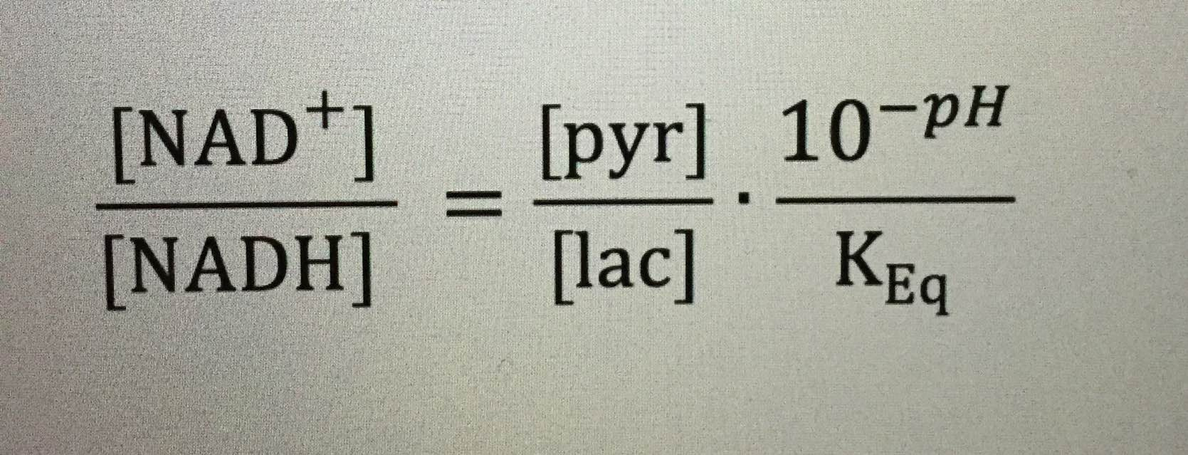
Luke went on to speculate that  “Methionine is hydrophobic but it has the potential to act as a hydrogen bond acceptor. It seems like there is going to be a difference in disulfide and hydrogen bonding with methionine compared to cysteine, which then affects deuterium concentration.”   Luke is a mitochondriac paying attention to my work.  Now that you know methionine is the start codon in the tryptophan operon in the video above let me show you more connections you might not have made yet.  The tryptophan operon links all the way to cytochrome 1 (NAD+/NADH) of mitochondria with damaged autophagy and apoptosis.  Cytochrome 1 efficiency SETS one boundary of the redox potential in the mitochondrion as the picture below shows.  This means it is critical to understand WELL if you are going to get better.


In mammals, NAD+ is synthesized from four different biosynthetic precursors in two distinct pathways. De novo synthesis (the deamidated pathway) uses the amino acid tryptophan, which is metabolized to form biosynthetic intermediates. These intermediates ultimately generate nicotinamide (the pyridine moiety of NAD+) and then form NAD+. Dietary vitamin B3 compounds, including nicotinic acid, nicotinamide and nicotinamide riboside, serve as NAD+ biosynthetic precursors and are salvaged from the diet (the amidated pathway) to generate cellular NAD+.

NAD+ is where “carbohydrates electrons” usually enter mitochondria at cytochrome number 1.  How many people do you think know that Typtophan can be a “bisexual amino acid”?  Tryptophan is a deep UV fluorophore amino acid (240-300 nm) is the basis for construction of NAD+ which is a 340 nm fluorophore protein?  

240 nm light is DEEP SHORT WAVE UV Light.  Where does that light come from folks?  Look at the picture below?  Can it come from the sun?  


It cannot.  This means that there is a pathway in the body that MAKES UVC light to program typtophan to make melatonin and NAD+ in some way.

That was the topic of my talk in Vermont.

Tryptophan catabolism in humans it is very difficult to comprehend for most but I will give it a try. 

It turns out, one major pathway results in the formation of acetoacetyl CoA, the precursor of ketone bodies – acetoacetate, 3-hydroxybutyrate, and acetone.  Tryptophan is activated and excited by light from 240-300nm.  This means it is an aromatic amino acid that has a phenol ring.  This is UVC light.   UVC light is noted to be TOXIC to all life by NASA.  This should confuse you.


People believe UVC sterilizes objects and planets.

Why would a living quantum system use UVC light to do something in their blood?

Did you know humans have 150 ppm of deuterium in their blood?

Did you know ketone bodies are deuterium depleted?

Did you know that they also block the NLP3 inflammasomes?

How is this process related to disease reversals and sunlight?  

Might tryptophan be an optical switch or time crystal that allows the body a way to “know” what season it really is because each season has its own electromagnetic fingerprint?  Yep.


This raises the question what is an inflammasome?    

Inflammasomes are multi-protein signaling complexes that trigger the activation of inflammatory caspases and the maturation of interleukin-1β. Among various inflammasome complexes, the NLRP3 inflammasome is best characterized and has been linked with various human autoinflammatory and autoimmune diseases.  

If you were fortunate to see my Vermont 2018 talk on how the skin, you’d begin to see how big a deal this really is.  

The NLRP3 inflammasome is activated in the cytosol where the matrix of mitochondria makes deuterium depleted water at Krebs bicycle.  This is where the TCA and urea cycle meet because fumerase adds water to both cycles anionic substrates.  

This is how information from SUNLIGHT is transferred to metabolic pathways to know what season it is, and understand how light and oxygen tensions determine the choices that a cell must make.


Black Swan Mitochondriacs are people who are highly-driven have what’s called a high “need for achievement.” If these people value something, they go after it with relentlessness.  Nothing else matters but THEIR health.  The better a mitochondriac knows nature they better they can “know” themselves.  As a result, the better their relationship is with the rest of the world.  Any time there is mitochondrial damage oxygen becomes toxic and ROS rises and cells become UNABLE to use the TCA and urea cycle and have to default to the older evolutionary pathways present in all cells before the Cambrian explosion, namely glycolysis and the PPP.  Glucose/fructose in foods isn’t toxic to any human or animal.  It is the damage to their mitochondrial that gives silly talking confident monkeys that sugars might be.  It is a horrible narrative done by poor thinking because the silly talking monkeys got tired of thinking and got lazy with their logic. 


Knowing about biochemistry and not connecting it to anything else in nature, like the sun’s biophysical levers, in a cell, is akin to buying a guitar that Prince once owned and then expecting that you could play Purple Rain as he did in life.


The implications of all the biophysics in this blog should be obvious.  When deuterium leeches from cell membranes in us and becomes prominent in places it should not be (matrix),  biochemistry in the (TCA/urea cycle) is no longer cyclical and its KINETICS are ruined.  Kinetics ruins the peripheral clock timing mechanism.  These cycles begin to run like a linear chemical reaction driven by an equilibrium bias and it is slow. This lowers oxygen levels and the cell MUST use glycolysis and the PPP to avoid oxygen toxicity.  This pushes a cell closer toward an equilibrium bias, which is another way a mitochondriac use to describe that journey to illness and eventual death.


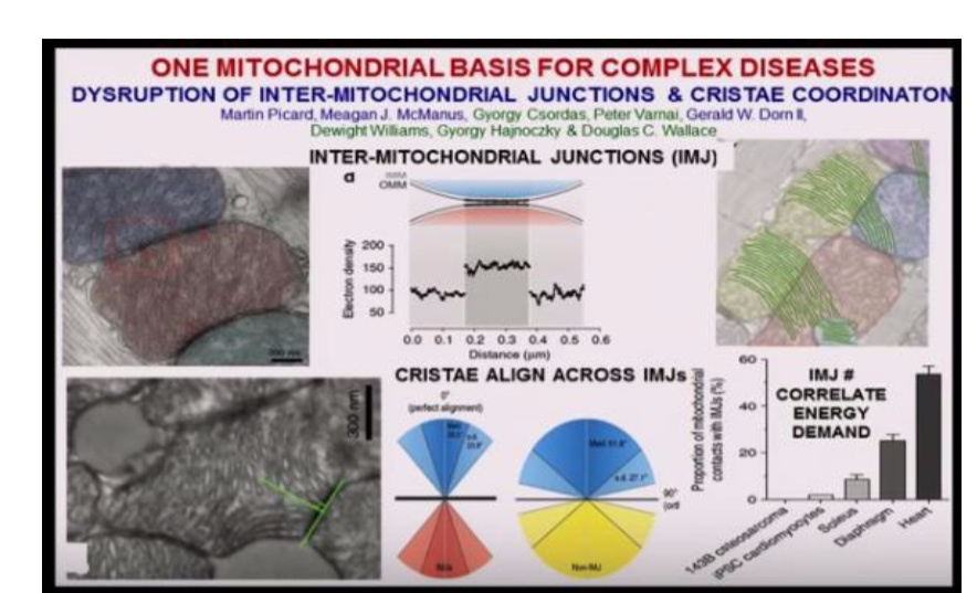
To a mitochondriac, death is the slowest form of death we create by controlling oxygen and light like a biophysical carburetor to keep a cell far from equilibrium.  Now think about what this means for deuterium in Kreb’s bicycle, and what Dr. Doug Wallace has found at the IMJ’s (above bottom right of pic).  IMJ’s do what they do for energy because there are built using the information quanta in sunlight.  It is about as counterintuitive as one can imagine.  But it is what life is built around.  These ideas are the results of deep thinking and what the Quantum Thermodynamics blog series is all about.  These blog ideas can do wonders for any silly talking primate when they pay attention to nature’s directions and not other silly talking primates.

Hopefully, with all these lessons in the QT series, you’re beginning to also understand how the connections of light and oxygen links to the physiologic function of melanopsin, blue light, and the Vitamin A cycle.


This allows you to see how the pieces fit and understand how they can fall apart because of the action at the NLP3 inflammasome.  When Vitamin A goes awry you lose the circadian mechanism and autoimmune conditions become more likely because you cannot create full spectrum shortwave UVC light using deuterium in your blood plasma.  

If you cannot do this, can you make a large EZ?  NOPE 

If UVC is absent can you make melatonin from tryptophan?  NOPE

You remember tryptophan use UVC to activated huh?  

It is kinda weird that the spectrum of activation of Tryptophan spans UVC and UVB light isn’t it?  

Could this be the way an amino acid that makes serotonin and melatonin to control mtDNA seasonally?  Yep.  

This is black swan mitochondriac stuff here.  This is how the pieces fit and I figured it out only after seeing them fall apart in thousands of my patients over the last 25 years.  

Tryptophan is difficult to make sense out of without understanding the biophysics of UVC and UVB light.  Deuterium makes them inside of you using the light that strikes your skin.  

Do I believe UVC is toxic to life in 2018?   

No, I no longer do and I think NASA and biology are dead wrong about UVC light.  I think some UVC falls to earth and the little that does is critical inside the tropics to work with Lo haplotype and black skin.  Why do I believe this.  Look at the picture of all the aromatic amino acids below……….they all use UVC light to operate. 


So you might be asking yourself, OK Jack,…….tryptophan making ketone bodies explains how it works in winter when no environmental UVB light is present.  Tryptophan is a seasonal EMF time crystal signaler. 


It means the phenol rings of this aromatic amino acids (ring above) never gets excited by sunlight, therefore, it forms a ketone body that is deuterium depleted for proper use of fat burning in the TCA cycle when it is cold.  

How does it operate in spring and summer?  

Is this the reason why serotonin is also so misunderstood by clinicians and researchers?  Yes, it is.  Serotonin is a quantized semiconductive protein that can act as a neurotransmitter, a protein, and an optical switch for metabolic pathways because it is only made and excited when a certain light frequency range in the solar spectrum that VARIES as the power density of seasons vary, therefore those frequencies must be present in the environment to program this amino acid.  


Tryptophan catabolism includes the generation of the glucogenic amino acid alanine, which allows tryptophan to be classified as a ‘glucogenic and ketogenic’ amino acid, hence why I called it bisexual above. It turns out serotonin is excited and made in summer because UVB returns to Earth to cause the presence or UVC light in the blood plasma from the pinch of deuterium in the skin I mentioned in the Vermont 2018 talk.


This version of serotonin has its version or tryptophan fully excited by short wave UVC light from 240-300nm.  This means the photonic programmed amino acid can act very differently in tissues because all aqueous proteins/lipids respond using a molecular resonance phenomenon.  What is the purpose of photosynthetic reactions in animals?


It turns water into the fuel that cells need and require to operate their proteins.  Water that is made by the mitochondria and surrounds proteins is a repository of electromagnetic waves.  Sunlight is the key driver of animal photosynthesis in humans.

This is why tryptophan and serotonin confuse the non-mitochondriac.   Now for some hardcore science to show you just how confusing tryptophan is to understand without knowing about the biophysical levers that control it.

TIME CRYSTAL WORK BY THE ELECTROMAGENTIC PULSES OF SUNLIGHT.

They are broken by the blue light and 5G pulses of man’s light and we do not realize it.


BIOLOGY GEEKS:

The tryptophan catabolic pathway starts with either tryptophan 2,3-dioxygenase or indoleamine 2,3-dioxygenase 1 which open the indole ring to form N-formyl-kynurenine. N -formyl-kynurenine is then converted to kynurenine by the enzyme, arylformamidase. It is also called kynurenine formamidase in the literature.

Kynurenine then undergoes a series of catabolic reactions resulting in complete oxidation to acetoacetyl-CoA. The first is catalyzed by kynurenine hydroxylase (also called kynurenine 3-monooxygenase) forming 3-hydroxykynurenine. The next step in the pathway is catalyzed by kynureninase and produces 3-hydroxyanthranilic acid and the amino acid alanine hence the classification of tryptophan as a ‘glucogenic and ketogenic’ amino acid. The next step in the pathway is the conversion of the 3-hydroxyanthranilic acid to 2-amino-3-carboxymuconate-6-semialdehyde (ACMS) and quinolinic acid by the enzyme 3-hydroxyanthranilate 3,4-dioxygenase. ACMS can be metabolized via the action of the enzyme ACMS decarboxylase followed by 10 other enzymes to acetoacetyl-CoA, the end product of complete tryptophan catabolism.  All the enzymes use proton tunneling to operate.  The light that controls this is beyond the normal red light that controls photosynthesis. 


 There is so much escitation that ocurs beyond the 250-780nm solar spectrum that life uses in mitochondria. The 1538.5-3100nn range deals with proton movements in enzymes and the matrix.

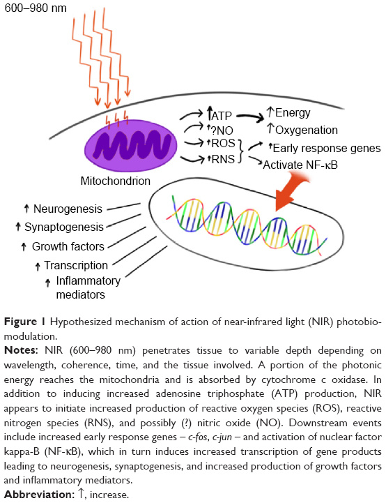
Through cytochrome c oxidase, IR-A light gives a ‘molecular kick’ to the mitochondria from the 42% of the red light in the sun’s rays that are present from sunrise to sunset; this informs or tells (information quanta) the cell to turn on a large number of antioxidant and energy-boosting genes only when ATP is being made by the combination of IRA and UV light we see in parts of a day.
Water viscosity changes with the red light in sunlight to facilitate the ATPase to work at 100% photothermal efficiency. That boost is greater when UVA and/or UVB light are present because of both of those UV frequencies BOTH slow ECT at different places on the inner mitochondrial membrane from cytochrome 1-4 by a quantum stimulus/design.


Cytochrome c oxidase has 4 red windows at 620, 680, 760, and 820nm of light. Those are some frequencies to consider. Hemoglobin has a sharp cut off at 600 nm. Both are heme proteins which act as circadian time crystals with radically different optical windows. Cytochrome c oxidase is present in mitochondria and RBC have a ton of hemoglobin in their cells which is HAVE NO mitochondria by design because of these light mechanisms. RBC’s are designed to ferry 280-600 nm (IRA and UVA light) 600nm-1000nm red light would cover the activation of fibroblasts to make collagen to stimulate repairs mechanisms. This means humans have a deep ability to go behind the red region to make energy from light that will re-write the biology books.


This should alert you to just how many paths are possible to alter tryptophan function.  This explains why serotonin and melatonin, both created from tryptophan can have so many varied functions because this amino acid can be programmed by excited electrons, ground state electrons, or protons that activated to transfer information between 900nm-3100nm light.  

The second notable feature of red light beyond the normal photosynthetic window is the energy buildup in water. This creates order and a separating charge both require energy.  These components in nature have plenty of precedent.  They seem to be the boost that helps begin the initial steps of charge separtion in photosynthesis on Earth.  That initial energy boost comes from red light from the sun. Spectral measurements showed that all wavelengths explored, from 300 to 5000 nm, contribute, with the most effective lying between 1500-3000nm in the infrared region beyond cytochrome c optical window.

This a small example of how cells are capable of using quantum superposition in an aqueous solution, from the mitochondriac perspective.  Conventional biology has no chance of comprehending this because they do not understand how the biophysical levers change water’s physics.  

Why is this BIG to the mitochondriac?  

All diseases and aging are associated with low NAD+ levels at cytochrome 1.  This occurs when blue light interacts with Vitamin A and melanopsin to lower melatonin levels to cause mitochondrial damage and elevates heteroplasmy rates and human disease show up in doctors offices and patients lives.


Did you get it?  

Guess how tryptophan fits also  fits into this developing story in the matrix??

Aside from its role as an amino acid in protein biosynthesis, tryptophan also serves as a precursor for the synthesis of the neurotransmitters serotonin and melatonin and kynurenine also serves as an important intermediate in the pathway for the synthesis of the nicotinamide adenine dinucleotide co-factors, NAD+ and NADP+.

IT FORMS THE KEY MECHANISM TO IMPROVE REDOX POTENTIAL IN THE MATRIX

NAD+ is the key electron acceptor of cytochrome 1 of the TCA cycle.

NADP+ is the key electron acceptor in the Pentose phosphate pathway.  The TCA cycle is used for biosynthesis when oxygen is plentiful.  The PPP is used for biosynthesis when oxygen levels are poor.  

BOOM!

This is the black swan mitochondriac level of understanding modern medicine needs to help the public.


I just showed you a lot.  Ask some people you care about to join me on Patreon if you want to help them become a black swan.

CITES:

1.  Amino acid synthesis and metabolism – http://themedicalbiochemistrypage.org/amino-acid-metabolism.php

2.  Urea cycle – http://www.ncbi.nlm.nih.gov/books/NBK27982/

3.  Miles B, Amino acid degradation – https://www.google.com.au/?gws_rd=ssl#q=Miles+Amino+acid+catabolism

4. http://www.soc-bdr.org/rds/authors/unit_tables_conversions_and_genetic_dictionaries/genetic_code_tables/

5. https://www.imperial.ac.uk/news/186732/new-type-photosynthesis-discovered/

6. mdpi.com/2310-2861/3/4/43/pdf

QT#13: METHIONINE 101

Nobody sees the links of nature in metabolism until they do………….

I think I showed that in the Vermont 2018 talk.  This series is showing you a side of biology foreign to the most learned eyes of life.  Today the onslaught continues to show you how nature uses an essential amino acid as an exogenous time crystal in our tissues.

While amino acids are the building blocks of proteins, different amino acids also participate in a wide variety of biological processes. For example, amino acids supply carbon and nitrogen molecules for biosynthesis, feed substrates to maintain TCA cycle activity for ATP generation, and provide reducing equivalents to bolster anti-stress capacity for redox homeostasis. Therefore, all organisms have developed strategies to cope with metabolic stress and challenges posed by the deprivation of amino acids. In mammalian cells, there are at least two major adaptive mechanisms that sense and respond to fluctuations in amino acids levels. Mammalian target of rapamycin (mTOR) is a conserved Serine/Threonine kinase that senses amino acid availability to regulate cell growth and autophagy. Another important sensor is the GCN2 (general control nonderepressible 2) kinase that regulates protein translation initiation in amino acid–starved cells by detecting uncharged tRNAs. These two kinases are highly conserved from yeast to mammalian cells and play major roles in the control of protein translation, transcriptional programs, and regulation of adaptive responses during amino acid starvation.

Not all amino acids are equivalent in their physiologic abilities.  This tells the mitochondriac they all have different levels of information processing.  It also implies that metabolism is not just about energy.  It is about how tissues learn using information in the movement of protons and electrons in the cycle amino acids operate in.  The major ones are the TCA, urea, and the methionine cycle.  This one is indirectly linked to the kinetics of the the TCA and urea cycle.

It turns out methinone can and has the ability to control autophagy/mitophagy in cells.  Autophagy is one of the self regulatory programs mitochondria uses to recycle themselves and cells that have sustained damage that is fixable.  This means it can be both an antiaging strategy and one that can back fire and cause cancer.  How so?

To understand the perspective you first have to see how the information is transferred from the sun to cells.  In order for mammalian cells to live and function, amino acids are required for protein synthesis and the generation of metabolic intermediates. An imbalance or deficiency of amino acids often triggers an “amino acid response” (AAR) to allow cells to adapt to their environment.

Reseachers have found a unique and dramatic gene expression program that occurred only when cells were deprived of methionine, but not any other amino acid. They also found that these methionine-specific changes depended on changes in histone modifications and an intact creatine biosynthesis pathway (PPP).

So I am being clear here, restricting methinone seems to make us live longer in several studies, while too much methinonine seems to set us up for mitochondrial diseases like cancer and heart disease.  Is this point of view axiomatically true or is the system built dyamically by light?  I bet you can already guess at the answer.

Always, the eye sees more than the mind can comprehend, and we go through life self-blinded to much that lies before us. We want a simple world, but we live in a magnificently complex one, and rather than open ourselves to it, we perceive the world through filters that make it less daunting. This is one of the greatest errors I see every day on social media in people trying to recover from an illness.


How does a single amino acid affect cells in such a profound way? It turns out methionine has direct effects on the surface of DNA via histone proteins. These proteins are the first step in controlling the beginning steps of all protein translation. This means methionine affects the surface topology of DNA. Topology concerns itself with how the surface electric charge of “something”, changes the physics of what is possible below that surface layer. How does topology work? Well, the surface electric charge changes the size and shape of things below the skin level. This new branch of science shows you how surface sunlight can change the optical window of cells to change the biochemistry possible below. Methinine is a key gear in the circadian clock mechanism in the SCN and in all peripheral clock genes. It works with melanopsin and retinol to control this process in us. The implications are far bigger than most of you can imagine. This paper gives you a hint at what many have missed. I fully expected this because of the previous work of DelGuidice and Preparta in 2000, and the follow up work of Pollack in exclusion zone water in the last decade. But this paper was the game changer because it was the final piece to make sense of a lot of loose ends.


The electrical change of water is influenced dramatically by the amount of full spectrum sunlight the skin gets.

Therefore, topology is critical in two steps in cell biology, which the organism needs to exert extreme control. Extreme control implies serious communication strategies. This is how the nonlinear aspects of UV light are put to use in a cell. This is where information quanta mechanism comes into play. Methionine deprivation reduced the degree to which histone proteins were indirectly modified by methionine via histone methylation. Methylation has three hydrogen atoms on one carbon. The isoform of those hydrogens are critical in understanding the critical role of methionine in the control patterns of autophagy and apoptosis. Let me illustrate the point with an example.

Methionine is an essential amino acid humans cannot make so it has to be made by photosynthesis in the food web on Earth.


Sun exposure increase the sulfhydryl groups in the blood plasma naturally with no added ENERGY needed.  Sulfhydryl groups are the major anion of our blood under solar exposure.  This increases the number anions present in our blood.  Sulfur amino acids are a kind of amino acids which contain sulfhydryl groups, and they play a crucial role in protein structure, metabolism, immunity, and oxidation. Recent studies have demonstrated the oxidation resistance effect of methionine and cysteine, two of the most representative sulfur amino acids, and their metabolites. Methionine and cysteine are extremely sensitive to almost all forms of reactive oxygen species MADE IN THE MATRIX of a mitochondrion, which makes them antioxidative.

Moreover, methionine and cysteine are precursors of S-adenosylmethionine, hydrogen sulfide, taurine, homocysteine,  and glutathione in all cells in the body. These products are reported to alleviate oxidant stress induced by various oxidants and protect the tissue from the damage. However, the deficiency and excess of methionine and cysteine in diet, can dramatically affect the normal growth of animals. The slide below shows this effect.  The stress can come from any stimulus.  Light stimulus however, has the largest non linear effect.  This means a small change in our ambient environment can lead to massive changes in our skin’s topology.  Do not forget this lesson.  You’ll need it later on to understand a prediction I made to a patients in this blog.

Methionine is known to change the color of an animals coat as seasons change.  In fact, when I learned about this I did hacks to grey my own coat and reverse it using methionine hacks I came up with over the last 5 years.


It is well known that oxidation from free radicals of the cysteine thiol groups occurs in all stressful stimuli in humans.  Thi sincreases the firing rate of the PVN while reducing outflow from the vagus nerve in th ebrainstem nuclei.    This results in defective glucocorticoid receptor function and this dramatically affects the reactivity of ligand and DNA binding in cells.  When stress occurs it knocks off the retinol loosely bound to melanopsin on the skin.  This lowers the Vitamin A levels in the plasma and that information is immediately transferred to the CSF because all CSF is made as an ultrafiltrate of our blood plasma.  The blood gets irradiated in our retina and and in our skin where this melanopsin/vitamin A couple reside.  Since this bond is a loose covalent bond it is easlily broken.  This is common in human primates but it varies greatly in species closely related to us.  It is 180 different than what we see in noctural mammals.  They have a very storng covalent bond between melanopsin and retinol. Melanopsin is a known blue light detector.  Blue light has enough power to break that weak bond.

Since the human bond is weak, this implies that in a 5G world where the toplogy of the skin will be affected by massive electric currents, Vitamin will drop in the plasma rapidly.  What are the collateral effects?  As Vitamin A drops so will the ability to make Vitamin D from our skin.  Why?  both vitamins are yoked in humans.  What else will happen?  Cysteine and methionine cycles will no longer operate as they were designed.  Both of their cycles are tied to the kinetics of the TCA and urea cycle in the matrix of humans.   This means doing hacks on the sulfated amino acids will be quite important to understand and use when one gets afflicted with electropollution type diseases like autoimmunity, sepsis, and cancer.   I have been doing these hacks now for the last 7 years.

Today’s riddle: Biohacking tip #250 from my new unpublished book. Why do sulfated amino acids have one requirement if it is natural and made under the power of photosynthetic processes, but if you make it in a lab, like a supplement maker would have to do, human need double the amount to get the same effect?  Might it be the information quanta carried by H+ in methionine somehow controls the metabolic pathways?  Could this amino acid be linked to the kinetic flow into and out of the urea and TCA cycle?  We know when the TCA and urea cycle kinetic slow methionine levels seem to rise.  Can H+ in an amino acid or the methyl groups it controls offer this level of physiologic protection? Yep.

There is now a general consensus concerning normal sulfur amino acid (SAA) requirements. WHO recommendations amount to 13 mg/kg per 24 h in healthy adults. This amount is roughly doubled in artificial nutrition regimens. This highlight that things made in a lab are not equivalent to those amino acids in the sun.  This is bad news for the food gurus and supplement makers.

You need to eat way more fake stuff to get the same benefit from food done under the power of the sun.  Eating more unnatural food supplements actually lowers longevity, while increasing mTOR signaling.  This leads to more disease, not less.  You can use your hair color and your blood smears to do these hacks.  Your perspective gets skewed when your vision is not the same as nature’s method of information transfer using light or water in your body.

Methionine is a sulfated amino acid and its concentration varies with solar cycles.  It is found in seafood, eggs, parmesan cheese and brazil nuts.  Methionine is a precursor of homocysteine. So when we eat too much protein with methionine in it, homocysteine can rise if it is not recycled quickly enough by the action of the kinetics in the urea and TCA cycle.  When homocysteine rises it is “big tell” to the wise clinician that deuterium leak from a cell membrane is happening at fumerase in mitochondria.  Blu elight exposure of the skin or eye causes our cell membranes to break down and liberate PUFA’s.  What holds the PUFA together strongly in our cells?  Deuterium does, with it massive kinetic isotope effect.  Deuterium gets out of the blood into tissues using sunlight.


So too much of a good thing can lead to problems too even in a state of decent health.  This is why in the CPC #25 blog Dr. Marik said most people with severe sepsis seemed to always have “co-morbid disease” as a precursor for sepsis.  That tells you they had higher than normal heteroplasmy before they got septic.

Heteroplasmy is a synonym for deuterium slowed TCA and urea cycle kinetics.   The deprivation of most amino acids, except glycine, is capable of triggering a robust and mostly conserved Amino Acid Response.

Quite unexpectedly, the data shows that methionine deprivation triggers the most dramatic and extensive gene expression changes in the nuclear genome in HUMANS.  

This information is quickly relayed to the mitochondria via the tensegrity system of the cell.  

HOW?

Blood plasma.  


UV-A light increases NO release from the arterioles in our skin to vasodilate the microcirculation.  This dramatically changes the hydrogen bonding network of water below.  That water touches every cell membrane in our body by way of the blood.  Cell membranes connect to the outer mitochondrial membrane by actin filaments that have integrins connected to them. These connect the nucleus to the mitochondria in cytosolic water.  These components are the tensegrity components of your cell. Integrins are designs to tighten or loosen the system to alter size and shape of everything in a cell. What controls the tensegrity system is the redox potential in the cell.

The generation of free radicals is a synonym for the redox potential energy within the cell. If you cannot make them, you can not have ideal health or cellular signaling. This is tied to the amount of electrons and the TYPE protons in the system that can work at large distances apart in different chemicals like oxygen, nitric oxide, and hydrogen sulfide.

Modern biology would have us believe each biologic process is structured and orderly on its surface, but the truth is they are all driven by random molecular motions (entropy).  Free radical are proxy makers for entropy that orders the tensegrity system in the cell.  

This is why tensegrity is ultimately tied to the redox potential inside the cell that is controlled by the amount of UV /IR light added to the skin, eye, gut, and lung to control the tensegrity system of the cell. 

This is why photons are the force carrier of the electromagnetic force of the environment of the cell.  The electromagnetic force can only control charged particles, and only electrons and protons carry negative and positive charges.

This is why the sun controls the topologic electric charges in the skin that sets the entire program of life in motion.  

CAN YOU IMAGINE THE EFFECT OF TATTOOS ON THIS?

Somebody wise in the Vermont audience asked me this question.  I could not give the detailed answer then, but I can now.  

Did you know methionine is capable of turning OFF autophagy in humans because of its nuclear genomic effect on epigenetics?  It alters histone methylation which changes how proton tunneling can affect certain parts of the double helix.

Methionine can raise homocysteine and is the most notable cause for concern in people with broken kinetics of the Kreb or urea cycles.  When these cycle are broken ( by deuterium KIE) it leads to unique changes in the flow of amino acids through these cycles. Let me give you a few examples.

Among the twenty-two standard amino acids in humans, nine are considered “essential” because the human body must obtain these from diet. For different types of cells, there also might be different dependencies based on their genetic makeup and metabolic flexibility as well as the microenvironmental stresses of these environments that could link to cancer. There has been much interest in identifying nutrient addictions of cancer cells with the hope for new therapeutic opportunities. The best one is the used of DDW which seems to increase TBW turnover to free up the kinetics of hydrogen flow in all these cycles.

For example, acute lymphocytic leukemia (ALL) cells are deficient in the asparagine pathway and require large amounts of exogenous asparagine. Therefore, asparaginase, through its ability to deplete extracellular asparagine, has become a cornerstone in the treatment of ALL cancers. Glutamine addiction is found in basal-type breast cancer cells and cancer cells with activated Myc and Ras. Certain melanoma cells have a leucine addiction caused by defective adaptive autophagy due to a LACK of solar programming with information quanta.

Methionine is unique because it is an amino acid processed outside the urea and TCA cycle.  It has its own cycle for function.  Its amount is quantized to the kinetics of the TCA and urea cycle.  Methionine participates in multiple cellular metabolic pathways, including the salvage pathway, the SAM recycling pathway, the trans-sulfuration pathway for cysteine biosynthesis, polyamine synthesis, and creatine biosynthesis. Methionine, indirectly via SAM, also donates methyl groups for protein methylation, which can result in epigenetic changes when the proteins being methylated are histones.

Alterations in the TCA and urea cycles indirecly affect the movements of hydrogen in mitochondria via methionine creation.  Remember this series is teaching you that particles with a half-integer quantum spin state, like H+ are the particles that nature uses to transfer information from the sun to the living system.

This happens in mitochondrial diseases as I covered in the May 2018 webinar and it is why mTOR activation in cancer shows up in so many papers. The researchers have no idea why this occurs because they do no understand how light controls metabolism.


This blog aims to end that mystery today.  Acute high doses of methionine can lead to acute increases in plasma homocysteine, which can be used as an index of the susceptibility to cardiovascular disease and cancer.  It has even caused death.  High levels of methionine in the plasma is an awesome marker for a physiologic log jam at fumerase at the junction of the urea and TCA cycles at Kreb’s bicycle.  This is why I had to teach you about them in the last 3 blogs and June webinar.  That webinar is must see!

Mitochondriacs should pay attention to these linked actions to understand nature’s goals.

Is this how autophagy can be ruined before apoptosis to lead to organ failure before death?? I think so.  I think aging is a loss of information control within the methionine cycle.  This makes methionine a marker for heteroplasmy status,  and this is why I warned you to use the calcium index score as a marker of mitochondrial biology.  High homocysteine is often associated with excessive calcium deposition in the arterial walls.


In fact, sufficiently high doses of methionine can actually result in death when heteroplasmy rates are high or redox potential in mitochondria are low. I believe this is caused by rapid slowing of ECT flows in the heart without the ability to make ATP using the red light of the sun. More on that later in the series.

When the urea cycle slows the TCA cycle it can slow ECT and kill ya like cyanide can. How is that for a shocking insight? Longer-term studies in adults have indicated no adverse consequences of moderate fluctuations in dietary methionine intake, but intakes higher than 5 times the normal amount resulted in elevated homocysteine levels. These effects of methionine on homocysteine and vascular function are moderated by supplements, like the water soluble vitamins B-6, B-12, C, and folic acid. I believe these B vitamins all facilitate proton tunneling (H+ again) to improve information transfer in enzymes.


When present in sufficiently high levels, methionine can act as an atherogen and a metabotoxin. An atherogen is a compound that when present at chronically high levels causes atherosclerosis and cardiovascular disease by lowering of nitric oxide concentration in the microcirculation as we age or by an elevated concentration of methionine BECAUSE OF A LACK OF SUNLIGHT on the skin. Remember UVA sunlight makes nitric oxide folks. You must have skin in the game to win this battle as the pic from Vermont 2018 shows.


A metabotoxin is an endogenously produced metabolite that causes adverse health effects at chronically high levels. These can be caused by nonlinear changes at fumerase explaining further how DDW works in cancers, arterial disease, and heart disease. We know that heart failure occurs in diastole via autophagic failure. All the pieces fit, because as a neurosurgeon, I have seen them fall apart in thousdands of my patients.


Methionine is also an important part of angiogenesis, the growth of new blood vessels. This brings more oxygen to tissues when they may not need it.  This was a key point made in the May 2018 webinar.  This is another key webinar for this series of blogs!   

This can be an advantage in wellness but it can be a death sentence in a pre oncologic state as the May 2018 webinar showed. Why? The pathologic Warburg shift requires cancer cells to shut down apoptosis while increasing ECT flow. How does it happen? Methionine’s kinetic cycling is broken in the sulfated amino acid cycle in the blood anytime the kinetics of the TCA and urea cycle also slow.  This happens for many reasons.  When the urea cycle slows, BUN rises and protein becomes a metabolic toxin.  This is why mTOR activation looks bad in many papers.  Doctors like Ron Rosedale make this error all the time because he is undereducated about light.  So do supplement makers.  This is why Mark Sisson and Asprey protein powders can destroy people.  The internet is littered with many examples of this but no one asks the right questions why this happens. Now you know the answer.

This causes it to rise because of its indirect linkage of the TCA to urea cycle via fumerase and betaine.  Betaine?  Yes.  It is another water soluble B vitamin linked to methionine clock gears.  When Kreb’s bicycle is altered it affects the methionine cycle in cells too.   How?

In another pathway, only in the liver, betaine (TMG) is the source of the methyl group transferred to homocysteine.  TMG = tri methyl glycine.   Selenium deficiency increases transsulfuration of homocysteine, and decreases global DNA methylation.  This is why seafood is so powerful outside of its DHA effect.  It provides a robust amount of selenium and methionine in nature’s dosing.

As urea rises, the liver and kidney both become fatty (deuterium effect).  The tri methyl glycine (TMG) de-fats both organs because TMG osmotically protects cells from urea and high electrolyte concentrations.  This is why so many diabetics have ammonia and urea measure at high levels.  When this occurs their cognition drops.  Most of the people on the internet complaining of cognitive haze have this DEFECT.   Their livers are loaded with deuterium, which is fully capable of ruining urea cycle kinetics.

Part of the methionine cycle is the creation of betaine which helps protect us from urea cycle dysfunction. This is how the cycle communicate via the movements of hydrogen (H+ again folks!).

This is also why glycine is useful in broken metabolisms.  This is why the survivor soup blog was written.

TMG converts homocysteine to methionine in the liver, whereas folate-dependent remethylation of homocysteine takes place in all cells. People with poor solar exposure have poor folate levels and poor B12 levels.  This is why diabetes is linked to blue light toxicity in the eye and skin and real lack of sunlight exposure.  Strong light and TMG strongly reduces plasma homocysteine in subjects with low serum folate, and has a weaker effect when folate is high, whereas Vitamin B6 has little or no effect on plasma homocysteine levels.

Glycine makes betaine and it can create DDW in the urea cycle to lower methionine levels and improve autophagy.  If you get in the sun you also improve apoptosis and your disease risk shrinks.  Normally the sunlight exposure on our skin make DDW in mitochondria and this is why we sweat out water loaded with deuterium from skin surfaces.  This is how we work.

Diabetics have altered sweating so they can never get rid of their excess deuteium via the sweat.  This is why sweating was a key feature in the Leptin Rx.  Go back and look.


In people with circadian mismatch diseases due to blue light exposure, this causes methionine to build up and stimulate angiogenesis. This is why I wrote the CAC blog for you.  More oxygen helps bad cells become cancer rapidly via the ideas in the May 2018 webinar.  I hope you are making these connections rapidly now.

When the urea cycle kinetics slow down what happens to methionine? It rises as a substrate in the cytosol and plasma. This implies underutilization or overconsumption of methionine can cause cancer. Why? Is this a proton spin story Jack? Yes it is.

How? It turns out the methyl group donor in DNA methylation, is related to cancer growth in a number of studies. What turns the process off? AM sunlight when IR-a leads to UV-A light.  This is why you saw the slide below 6 months ago appear on my social media pages.


PUTTING IT ALL TOGETHER:

Methionine is an essential amino acid.  Humans do not make it, they must eat it.  It can be recycled from homocysteine and cysteine.  Methionine is coded for by the initiation codon, meaning it indicates the start of the coding region and is the first amino acid produced in a nascent polypeptide during mRNA translation. This means the hydrogen that it is linked to must come from methionine recycling.  This is where the information is hidden by nature my fellow mitochondriacs to control autophagy.  Fasting lowers methionine so this is why fasting is linked to improved autophagy if done in the SUN and not indoors under blue light.  Exercise also depletes us of methionine, assuming the urea and TCA cycles are opertional.  If they are not exercise can hurt us.

Methionine is one of only two amino acids encoded by a single codon (AUG). Anyone want to guess why?  It is so important there cannot be multiple nuclear signals for it because of hydrogen.

The methionine codon AUG is also the most common start codon in humans. A “Start” codon is message for a ribosome that signals the initiation of protein translation from mRNA when the AUG codon is in a Kozak consensus sequence. This is critical in understanding how the Warburg shift goes from normal to pathologic in cancers and autoimmune conditions.

Why is it the key signal in the cytosol at Kreb’s bicycle when diseases is present?

Almost all the substrates in the TCA and urea cycle are ANIONS. Methionine and SAMe are CATIONS. This changes the topologic charge in matrix water.  Remember how I started this blog?   This is a big freaking deal folks because everything in the urea and TCA are anions.  This offers another level of control to methionine for the circadian mechanism at our surfaces.

The methionine-derivative S-adenosyl methionine (SAM) is a cofactor that serves mainly as a methyl donor in biochemistry. This is where functional docs stop their knowledge. You need to be aware of it.

The issue of a changing toplogic charge is huge in information quanta transfer and they do not know it. They have zero understanding of biophysics of the matrix. SAM is composed of an adenosyl molecule (via 5′ carbon substrate built by the PPP) attached to the sulfur of methionine, therefore making it a sulfonium CATION. This happens because the three substituents hydrogen all positive charge. Those hydrogens cannot be the deuterium isotope of hydrogen. The PPP controls the hydrogen flows in the cell. The sulfur atom acts as a soft Lewis acid (i.e., donor/electrophile) which allows the S-methyl group to be transferred to an oxygen, nitrogen, or aromatic system in the urea or TCA cycle, often with the aid of other cofactors such as cobalamin (vitamin B12 in humans).

Metabolism is not JUST about energy folks. It concerns itself more with information quanta, in case you are not getting the message from this blog.

Think of each thing you learned in this blog that happens in nature in a day as critcal information your cells need………it is neither good or bad when it is missing its natural context……..then it is just information buried in quanta.  Once you understand the context properly, then use the information to adjust your life choices.   Consider that before becoming more aware you wouldn’t have even noticed how bright the lights you live under really are, nor made ANY connection between the artificial lights (and EMF) and the quality of your sleep that night. When you think like a mitochondriac you begin to see how ironic nature is.  It’s funny how what looks like a setback (sunburn) might even be considered a step forward when it is done by the sun.  “Blue burns” indoors are deadly information to cells.

DDW makes sure excess methionine becomes glutathione and is not able to stimulate angiogenesis to deliver more oxygen to mitochondria. Both glycine, serine, and DDW can lower homocysteine levels because of the effect on methionine. Sunlight also helps make DDW and this is why it raise redox better than anything I have found.


People will be shocked to hear this but artifical light bulb flicker also induces a PVN stress response via the eye and skin because of melanopsin and the Vitamin A link.  In fact, I think it might be behind the cancer epidemic for several reasons.  In 1951 Fisher and Fisher found that flickering man made light altered eosinophil counts in man.  In fact, they made the case that looking at the peripheral blood smear could tell a clinician about the state of the retina.  In 1960, Ponte did experiments on the eye using flicker and conformed their work.  I used this data to diagnose a skin cancer in the skin of a tattoo’d patient who had developed an eosinopenic drop in her blood over 5 years that her primary care doctors missed.  It turns out the flickering light on a computer screen shined on a tattoo might be a quite way to get a skin or blood cancer.  Why?

The stronger the flickering effect is in the blue range, the stronger is the eosinopenic effect on the peripheral blood smear.  It turns out that an increase in the lux number of man made light, causes the eosinopenic effect to occur earlier and more distinctly.  This affects the skins circadian mechanism in a bad way as you’ll soon see.  How long have we known about this effect?  In 1956, Doe et al followe ddiurnal fluctuations in urine cortisol levels and found a distinct diurnal eosiniophilic curve.  This linked light function on the eye and skin to the immune system controling arm in the adrenal gland. Later studies done by researchers showed this effect came from the macula or the retina.  So sitting in front of a flickering screen is a great way to get skin cancer and blood cancers.  Both are rising as technology spending has soared.  This is why you need RaOptics and Iris software on your computer.

The flicker effect cause PVN stress to max out in the human diencephalon to destroy the brainstem PVN and the adrenal cortex at once. People do not know the science of light well enough to know why people are killing themselves with human PROGRESS. Mitochondriacs do.

STRESS AND TRAUMA and methionine.

The more stress response one gets from any cause, this increases PVN activation in the hypothalamus, and as a result the more methionine you’ll need to combat 5G stress.  Any type of stressor increases our need of methionine.  This is true of trauma, sepsis, nnEMF exposures, and even greying of your hair!  In hacks when my hair would grey hair I knew to eat more eggs, seafood, and Parmesan cheese to augment the methionine effect.  How did I learn all these links?  It came from a patient who had a pretty amazing tattoo effect linked to a mercury stress as her redox fell because she worked at night in the IT department of a hospital in the basement.  I want to share it with you because it mimics the effects in CPC #25 blog and it shows you the deep wisdom in the May 2018 webinar.


The tattoo lesson on methionine: More on how your surfaces can be affected by a lack of sunlight and methionine in your skin. People with poor kinetics in the TCA and urea cycles begin to have a raised level of methionine in their plasma.  We can confirm this with their homocysteine levels.  They should be rising based upon the data in this blog.  This tells us their is a kinetic isotope effect of deuterium in one or both these cycles between some of the substrates.  If methionine is raised because of this effect,  it stimulates angiogenesis in the surrounding tissues and this supports a pathologic Warburg shift in a stressed tissue.  Why was this skin in this IT worker stressed?  She was around nnEMF 24/7 and the tattoo was old.  She had it when she was healthy and had no issues with it until her environment changed.  She got the tattoo when she was younger and never had a problem with it until she worked in the basement of the hospital at night around electropollution.   After 3 years the red ink of the tattoo began to look inflammaed and infected.  Many high quality red inks have cinnabar in them.  Cinnabar is a safe mercury compound used for centuries in tattoo art.  Why did it cause an issue here in our modern world?  The blue light she was around caused a mercury resonace phenomena in her skin in the red ink that raised the local heteroplasmy rates in the red parts of the tattoo.  Remember blue light penetrates deeper than we think to where the ink is located.  This acted like a photochemical stressor.  She also wore a Apple Iwatch on this arm.  As the methionine went up angiogenesis made cancer in the red ink region likely because of her co-morbid nnEMF risk; this lowered her redox state because of her occupational risks.  Incidentally, she reported insomnia.  She was vegan too.  This was a key symptom in knowing her autophagy efficiency was dysfunctional.  The veganism was a clue to why her homocysteine was elevated.  I thought the risk was high that apoptosis was also dysfunctional because of all her risks.  I was worried this was an unusual cancer and not a reaction to her ink.   Her vitamin D level was 18 and her LDL cholesterol rate was over 300 and her homocysteine was 284.  Vegans are well known to have high homocysteine levels (HC), unless they are in strong sunlight,  so I knew she was having trouble recycling HC to methionine and this is why I was worried about this being a possible tattoo ink cancer from an acute skin stress in somebody with bad TCA/urea kinetics.

In December of 2014, she decided to have it looked at.  A biopsy came back as an unusual type of skin cancer by the dermatologist. This is just another perspective of how getting a tattoo in your past, when you were healthy, can be used as a hack of a dropping redox because of a changing environment.  The teaching point here is that the stimulus of cancer in this person was the blue light and nnEMF she faced in her job.  This caused a stress reaction in her skin, and her eyes and subsequently altered melanopsin cycles in the tattoo and this broke the kinetics in her Kreb and urea cycles in the red inked skin.

This slowed the urea and TCA cycle kinetics allowing methionine to build up and destroy autophagy for years and cause increasing angiogenesis in the red skin.  This angiogenesis inhibited apoptosis setting the stage for cancer via the Warburg Shift we spoke about in the May 2018 webinar.   Everything was fine for her up until apoptosis was destroyed in this skin because this area NEVER got any sunlight because of her shift work was in a basement without windows loaded with nnEMF.  The tattoo was also most covered by her clothing.

She effectively lived under blue lights 24/7 for the last 8 years of her life.  What was the result? Cancer inside the boundaries of the red ink where the mercury was and resonanted with the blue light she was exposed too.  If that skin had of gotten a minimum of UVA or UVB light I bet this would have never happened.  This case is a text book teaching case in how blue light stress of the skin and eye can destroy autophagy and apoptosis and can lead to a cancer in red ink on the skin.  It is pretty amazing.

Cinnabar is a compound that has mercury in it. It is perfectly safe unless your kinetics of the TCA and urea cycle become dysfunction by deuterons in our metabolic pathways. The crazier part of the story is that if she had of eaten bone broths with serine and glycine or more foods with methionine (seafood) she might have avoided the surgery altogether because the cancer might have been prevented.

Did you know that foods with serine, selenium, and methionine can clear mercury from our body quickly?  It is true.  This is the kind of redox/detox mitochondriacs advocate.  This is how we get grey hair too folks.  All enable us to clear heavy metals quickly.  She was a vegan refused to eat seafood which contains all of these natural heavy metal chelators.  Seafood and or sunlight might have saved her from the cancer or the surgery.  She was not interested in my ideas about this.  It amazed me how dietary dogma can keep a person from curing themselves with the wisdom of nature.

Mitochondriacs need to make sure if you get a tattoo that you think of all of the things that can go wrong and how your body could be potentially disfigured from it. But you can also use the tattoo as a redox monitor too as this case shows.  You can also use your tattoo’s as a canary in the coal mine for the efficiency of autophagy and apoptosis if you choose to keep it. If this person was Patreon member or a member of my website she could have gotten the wisdom that the use of DDW, seafood, survivor soup, eggs, and parmesan cheese might have been wiser maneuver before undergoing surgery for an inducible cancer.

Amazing how nature works when you understand her.

It also appears that any augmentation of glycine metabolism is beneficial because it lowers methionine in dysfunctional mitochondria.  This is why bone broths are so helpful to those mitochondrial diseases.   This is why I gave you the survivor soup blog already.  Now you can see how the piece fit in the Quilt of life.


Have a look at this video on youtube.

Given what I said above, what do you think of this as a way to celebrate beating cancer?

In my opinion, his cancer will be back soon………..This begins with your relationship with yourself. If you do not love yourself entirely and actively ensure your own needs are met, you will find it difficult to do the same for others.
Know this: however you treat yourself is how you will treat others. This is why, ironically, the most selfless thing you can do is to be self-centered (albeit not selfish).

The take home of this monster:

Have you ever wonder why chemist just don’t get quantum biology? The proteins life plays on are the stage upon which the drama of life unfolds. Proteins are carbon polymers of electrons waiting to interact with sunlight. The actors on “this stage” can be none other than small and highly mobile units such as electrons and protons.  Chemists equations don’t see these small things in their metabolic pathways…….but you can bet your ass they happen in every living thing on Earth. This is why life exceeds a biochemistry mindset.

EVERY CELL HAS A CLOCK infront of every human gene………to protect. blue light destroys their operation because of the linkage of Vitamin A to melanopsin and its weak covalent bond.  This is why we are all getting sick today.

Vermont 2018 WISDOM:
Truths are approximations. All of them because they are relative to the physics of the present moment.
Most think of sunlight as energy and as something that we can see.
Sunlight is energy but is MOSTLY information and it ONLY speaks in the language that our body completely understands. That language is called the entire visible part of the spectrum of light.
Artificial light = foreign language for cells
Artificial frequencies = foreign language for cells
…………..
Photon — travels at the speed of light
information tells photons where to go on the back of H+ and electrons —– faster than light via Fermat’s law and the use of entanglement.  Mitochondria decipher the information and spit out free radicals depending upon the information you give them.

This is why you need to be a mitochondriac.  Join my membership and learn all this for yourself.  I go where others cannot because they do not know this territory exists.

CITES

https://www.sciencealert.com/your-body-has-trillions-of-clocks-in-its-cells

https://www.benbest.com/health/Meth.html

QT#12: KREBS BICYCLE 3: The retina

video
play-sharp-fill


Most people who have watched my Vermont 2017 youtube have come away astonished at the insights I put together there using the science published in the last 100 years.  This blog might be more shocking to those who fully grasped the complexity of the physiology of the retina.  I briefly mentioned in the Vermont 2017 talk the role of Muller cells and RBC’s in the retina.  What I did not then, is that both of those cells in the human retina operate using a non-pathologic Warburg metabolism.  That talk gave an extreme level of quantum biology and was done to show you how obesity begins in the eye.  Obesity is extended when the signals in the skin are also usurped by a blue light environment.  I made a prediction in my own hacks 13 years ago that if the CT protocol and Leptin Rx were true, that a melanopsin like photoreceptor had to be present in the skin, its arterioles, and subcutaneous fat mass to explain obesity in humans fully.  Little did I know in 2017 right after my Vermont talk that paper got published and my educated guess was correct.

I did not mention this in Vermont 2017, because the next step in your quantum education links directly to what happens in the skin.  This is why I was provocative with the audience there last year and I suggested to the Dave Hollenbeck that if he wants to see some more amazing quantum biology he should have me back in Vermont in 2018 to talk about how the skin operates with the peripheral clocks in every organ in humans.   It turns out he took me up on the challenge.  I went there on June 2, 2018 and unleashed my ideas on the topic.  So my members and Patrons get the early heads up on what innovations are coming in this talk in Vermont.   Seeing the details on the slides I have made, in person, might just blow your mind.

The teleological explanation for the presence of the Warburg effect in the mammalian retina is simple to understand once you have a basic idea of what Krebs bicycle really is.  When proteins synthesis is massively upregulated by ubiquitin, the cell cannot rely on the steady removal of anions from the TCA or urea cycle to fuel biosynthesis because of slowed down kinetics of either cycle.  This creates a logjam for the cell with respect to the cell cycle.  The collateral effects are felt in the matrix, with respect to hydrogen movements, because of the need for of H+ to run the ATPase to power both cycles.  So what does a cell do when redox drops and the TCA and urea cycle spin rates are lowered for any reason?

It relies on the older evolutionary pathways, glycolysis with the help of H+ to transfer information over energy to get the job done.  Each C-N  bond in proteins costs 5 ATP.  This is very costly on an energy basis.  Information costs are even greater.  This means biosynthesis is an energy hog for the cell.  Does the retina use the TCA or urea cycle exclusively for biosynthesis?  No, they do not.  This should surprise people considering the metabolic rate of the retina.   So how does this process occur in the retina?    The retina is a tissue with a high oxygen demand in the back of the eye but a poor supply in the front of the eye.

The anterior chamber of the eye has to rely on getting oxygen from diffusion through the cornea from the air because the cornea and lens must remain transparent for vision and no blood vessels can impede the passage of light.  If the cells of the eye used TCA intermediates exclusively for biosynthesis, considering this oxygen issue,  it would create a chronic proliferative state in areas of the eye and this would impede its function.  This defeats the purpose and the physiologic requirements of the central retinal pathways, pituitary, and hypothalamus.  All of these places in the brain that are light relay stations are quite small in size.  The reason these parts of the brain are small in stature is that they deal almost exclusively in information quanta processing over a growth and proliferation state.

This implies that the retina must run two different metabolisms to get the job done in vision.  Is this true?  Yes, it is.  It turns out the adult mammalian retina is non-proliferative because of how it is built to interact with light, it shares similar biosynthesis requirements to neoplastic tissue because of the prodigious turnover of the opsin proteins in the disc membranes of the photoreceptor outer segments.  This explains why certain cells of the retina use the Pasteur effect.  You had a blog on that effect recently here at Patreon.  Each mammalian rod outer segment consists of a stack of ∼1500 distinct discs enclosed by the plasma membrane. Approximately 60% of the dry weight of the disc membrane is protein, of which, opsin comprises 90% of the protein content.  Remember all opsins are “loosely” covalently bound to Vitamin A to operate in humans.  Hence, the retinal-bound opsin, rhodopsin forms a large structural component of the rod disc membrane. These components are even larger in the outer retina where melanopsin controls the quantum release of melatonin release from the pineal after 4 hours of dark.  This is the major quantum mechanism that controls mitophagy and autophagy signaling.

All G-coupled receptors are linked to the circadian mechanism in humans.  Rhodopsin is a G protein-coupled receptor comprising 348 amino acids, with a rich glycine and serine component.  This is the very same serine and glycine amino acid components that link them to the efficiency in the PPP (and glycolysis).  The PPP is the major way we regenerate the major reductive moiety called NADPH you learned about years ago in the EMF 4 blog post.

Tissues with poor oxygneation cannot use the TCA or urea cycle.  This is critical in tissues that cannot use the TCA or urea cycles because this is a backdoor way for the cell to augment informational quanta transfer using H+ pulled from the serine cleavage system.   RBC’s, Muller cells and every single embryonal stem cell uses glycolysis and the PPP by nature’s design.  None of them have high oxygen contents.  This keeps a lid on their growth rates.   There is NOTHING pathologic about these cells that have to rely on gluconeogenesis.  The key point in understanding is that when cells use gluconeogenesis for biosynthesis exclusively they MUST use glycolysis and the PPP for biosynthesis.  What determines their use?   The amount of oxygen tensions in the tissues is deterministic for the pathway a cell uses.  Glycolysis and the PPP are dominant when oxygen tensions are low!

This is where you may remember David Sinclair paper about pseudohypoxia, NAD+ and aging I mentioned in December of 2013 on the blog.  Sinclair was talking about cells losing the ability to use the TCA and urea cycle as they age so they all seemed to have a common phenotype of having low NAD+ and pseudohypoxia.  He called it a cardinal feature of aging and disease.  It turns out he miss read the tea leaves.  As cells age, they become less able to use the TCA and urea cycle because autophagy loses its efficiency, due to a loss of redox power.  When autophagy becomes faulty, sleep is poor as a marker, and as a result, a cell loses control of atoms of deuterium buried in the PUFA in the mitochondrial membranes.   You heard about this mechanism in the May 2018 webinar.  This is a huge big deal folks.  Why?  What are the collateral effects of this mechanism?   Is this how cells make and create the ELF-UV light they use for a mitotic division?  Yes.  Is this the basis of the research of Fritz Popp work on ELF-UV light.  Yes.


Richard Young did the seminal experiments on the retina.  The deuterium critics have no clue how this complex dance operates because they do not understand how information is transferred via the quantum spin state of protons and electrons to Orbital Angular Momentum (OAM).  Moreover, they have no idea what the OAM is used to do in your cells or blood.  The next few ideas link my Vermont 2017 talk to the one I gave on the skin last week.  This is a story of how deuterium does things people never anticipated to help mitochondria and cells self-regulate their own behavior under the power of the Auger effect (UV light).  UV light is critical in controlling ECT flow in mitochondria several ways as this series has shown.

Now you do and I am showing you how it links to the physiology that controls melatonin creation and releases in the mammalian retina.  In fact, this quantum mechanism is so powerful that Mother Nature decided to use it kingdom-wide in our clade and on every surface of their body plans.

The rhodopsin turnover rate in the human retina parallels the degree of aerobic glycolysis found in many different species in mammals.  The rhodopsin turnover rate matches the Vitamin A and D cycles in humans almost perfectly.  I showed a slide of this in Vermont.  This is why unsupplemented Vitamin D levels are great proxies for defects in Vitamin A cycles in the brain and retina.  This makes your unsupplemented Vitamin D3 level a great proxy for what is really going on in your skin and eyes mitochondrial matrix.  The next part of the puzzle is not well known.

Photoreceptors have a relatively low rate of turnover in lower vertebrates, and it is temperature dependent (another clue to the H+/D effect).  Recall that higher temps favor H+ bonding in aqueous environments versus D bonding in water.  As temperature increases, the non-pathologic Warburg effect becomes temperature-dependent.   In mammals the story is exactly the opposite.  The rate of turnover in the mammalian retina is astounding and this is why mammals became warm-blooded.  They used heat release from their mitochondria to precisely control the flow of H+/D in the matrix and cytosol to control growth and metabolism of the retina just by using light and temperature and oxygen concentrations in the eye.  This is the basis of the CT 4 and 6 blogs written over a decade now.   All of these reasons are why I made the educated guess years ago that melanopsin had to be present in the skin/fat of mammals.  In December of 2017, I was proven correct.

Since melanopsin is present in the eye, arteries of the skin and fat mass this becomes a critical point in understanding how the skin becomes the gatekeeper of deuterium flows from the blood plasma versus the tissue.  Remember phototaxis is well known in the leaves of plants.  Most third graders know that leaves grow toward light and can change their angle of inclination of growth toward sunlight based upon the type of sunlight present.  Very few people realize that the RBC’s in mammals do the very same thing as leaves.  This maybe why aneurysms form in arteries because  RBC function differently when they are out of sunlight.  Arteries should have laminar flow in the center of a vessel and why blood flow is more turbulent towards the artery wall. Sunlight causes the entire arteriole to migrate toward the sunlight of UVA light.   Sunlight can directly affect the RBC’s in the center of your blood vessels to act just as leaves do to collect IRA and UVA light as a form of animal phototaxis.

Recall UV spectrum in sunlight heats mammalian skin up the most significantly.  IR light in sunlight is considered cold light energy on a relative basis but it is the initial light of the sun that builds the EZ in tissues and our blood.  Remember, via chemistry’s laws that heating favors H+ in hydrogen bonding over deuterium bonding, so organisms can use their body heat as a fractionation method to control growth in tissues.  This helps you understand further why mammals evolved warm bloodedness.  Heat is a powerful physical tool in controlling the flow of different isotopes of hydrogen in your cells. It turns out UV light raises the temperature more than any other light waves in sunlight.


Having the innate ability to uncoupling your protons gradients helps keeps deuterium in the blood plasma because blood moves so rapidly, it can diffuse its heat like a radiator, but tissues do not have the same ability. This relative difference is all that is needed to control where isotopes of hydrogen should be found. When deuterium is trapped in the arterioles of the skin is can be pressurized by sunlight in the UV range and the chiral heat effect to pinch deuterium in the blood vessels coming to the surface, while at deeper levels H+ accumulation is favored in our tissues. Compression of H+ or deuterium allows the body to make a continuous UV spectrum from the isoform of hydrogen trapped in both tissues.

The UV spectrum is not equivalent because of the chiral heat effect. In the blood, the spectrum of light extends deep into the UVC range, and this frequency matches the optical absorption spectrum of the EZ in the blood. Recall this is built by the water and IRA light initially from the sun. When this phenomenon occurs in the blood plasma, the EZ can grow further to absorb light rays into the UVC range to further energize blood plasma to carry more light energy and information in the ophthalmic artery distribution of the retina. The flow of information is more important in tissues that cannot use the TCA or urea cycle. This helps the areas of the retina that have poor blood flow. This circulatory pinch of hydrogen allows deuterium and hydrogen to become a creator of full spectrum UV spectrum light creators via an unusual nuclear effect specific to hydrogen on the periodic table. This photic phenomenon extends the size of the EZ in the blood and vascular space to become something more than just a battery or capacitor as Pollack has shown in his studies on water.


The NIR light addition to blood allows water in our bodies to transform into an optical resonator or a mirror. This mirror can then be used to optical resonator to create tiny lasers at our skin surface or in our ophthalmic artery which feeds the retina. That UV light created by this mechanism is then used to regenerate melatonin, dopamine, and build many more substances in quantum thermodynamic fashion. This system is amazing when you see how the quantum system is organized to support the semiconductive circuits of the central retinal pathways I laid out in last years Vermont talk. How it links to the skin is also quite remarkable. You might want to watch that Vermont video 2018 when it goes live here on Patreon.

We can see the wisdom of information quanta by nature when we carefully examine the anatomy of the photoreceptors in our eyes. I showed some of Dr. Wunsch slides in Vermont in 2017 but I could not get into this level of complexity with that audience because they were not facile with the science and they were not mitochondriacs who understood what Krebs bicycle was. I am hoping to change that this year. You have to understand how quantum thermodynamics differs from classical thermodynamics to understand the process fully.

In the retina information from light is the key resource that determines the anatomy. The organization of the retinal cells involves the highly compartmentalized cellular configurations, with the confinement of mitochondria to the inner segment, absent from the outer segment. The dense aggregation of mitochondria in the ellipsoid region of the inner segment reflects the considerable reliance of this portion on oxidative energy from the TCA/urea cycle. Evidence that supports these ideas was the finding of high concentration of malate dehydrogenase (an enzyme involved in the TCA cycle) in the primate photoreceptors inner segments. The malate result revealed a level 30 times higher than the outer segment of the photoreceptor. Malate needs fumerase to be completely free of deuterium to add water to it to support the functioning of the inner retina. This tells the wise person nature had to build a light-based control system to control the flow of H+/D if any retina was to operate with sunlight. I began to put all the pieces together 15 years ago. Now you can see information processing in the eye and skin really begins with H+ and deuterium fractionation occurring immediately in the surfaces of the retina and skin. This helps explain why nature really built the retina in the way she did. For those of you who don’t know the retina appears to build inside out. This has confused biologists for ages because none of them understand light well. The same idea is true in the skin’s physiology.


Since this was true in the retina, I knew that it had to share metabolism with several pathways that worked with different oxygen levels. I hypothesized that we should see data that lactate would be higher in areas where the PPP and glycolysis are used in the retina. I believed strongly in 2003 the same thing had to be true in the skin because of their shared embryology. Do we have any evidence of this? Yes we do. Contrary to conventional wisdom, the measured fractions of lactate dehydrogenase (LDH) used in glycolysis was significantly LOWER in the inner segment where the cones are located as compared with the outer segments of the retina where melanopsin and Vitamin A are used. This tells you cone vision needs lowered oxygen tensions because the light they work with is less powered than the blue light that melanopsin works with.


This is why the fovea/macula has a POOR blood supply.  I should those pictures last year in Vermont (above).  How does the retina accomplish these gymnastics?  It did exactly what the RBC did in evolution.  It eliminated mitochondria from the cone cells while restricting blood flow to make sure no TCA/urea cycle metabolism could occur there.  Anyplace mitochondria are, deuterium must be rare, and vice versa.  The exclusion of mitochondria from the photoreceptor outer segment necessitates its reliance on glycolysis/PPP for cellular energy production, while still permitting information transfer from sunlight via protons with the H+ isoform in the retina and into the small structures of the brain mentioned above.  There is a deep lesson here for the mitochondriac about how and why the retina where built as they were to work with hydrogen to make UV light inside our body to drive photonic programming of physiology.   This is how information quantum is created and moved in the central retinal pathways to affect your brain and CSF pathways to build the world wide web in your head called your mind.

CITES:

https://onlinelibrary.wiley.com/doi/pdf/10.1111/ceo.12462

https://elifesciences.org/articles/29217

Dr. Jack Kruse Vermont 2017 Youtube talk
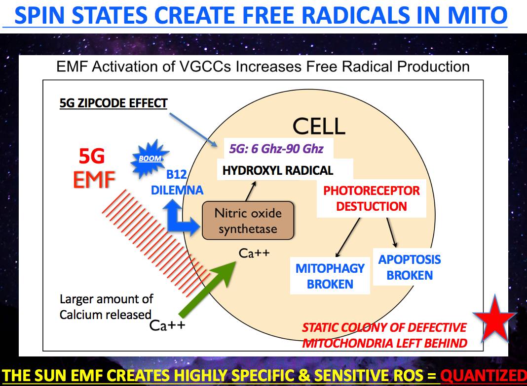
Dr. Jack Kruse Vermont 2018 talk

QUANTUM THERMODYNAMICS #11: KREBS

This blog is coupled with the June 2018 webinar.

KREB’S BICYCLE WISDOM:  You should never rely on thirst to dictate water consumption because it lags the real effect. Calculating your total body water deficit is another way to try to measure how badly your engine (TCA/urea cycle)  is working.  Total body water is a function of metabolic rate and state of the mitochondrial matrix.  We covered this in the April 2013 webinar.  By the time thirst kicks in, your serum osmolarity is already impaired. Dehydration is a major cause of daytime fatigue as well, and dehydration slows your metabolism by 2-3%. As time goes on this imbalance can steepen dramatically in a person in an altered environment.  In fact, just a 2% drop in total body water can cause neurologic changes to show up.  How do I know this?  I am a neurosurgeon,  and we see these swings all the time in trauma cases and brain tumors that are associated with syndromes called SIADH, cerebral salt wasting syndrome, and diabetes insipidus.  They are related to vasopressin.  Now think about what I mentioned in the April 2013 webinar for our members.  Are you beginning to connect any dots that dehydration and nnEMF may be linked?  Could this be why modern humans are afflicted by the opiate crisis?  Look at the picture below.  To understand how we work you need to see how the pieces fit.
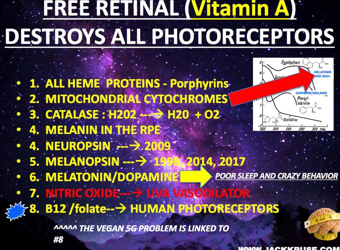
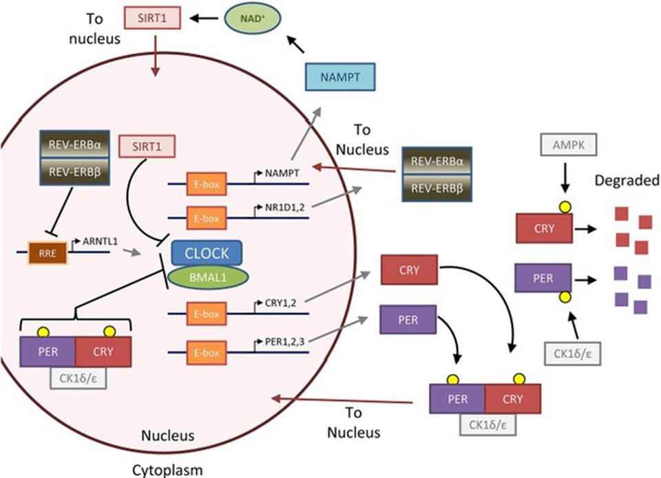
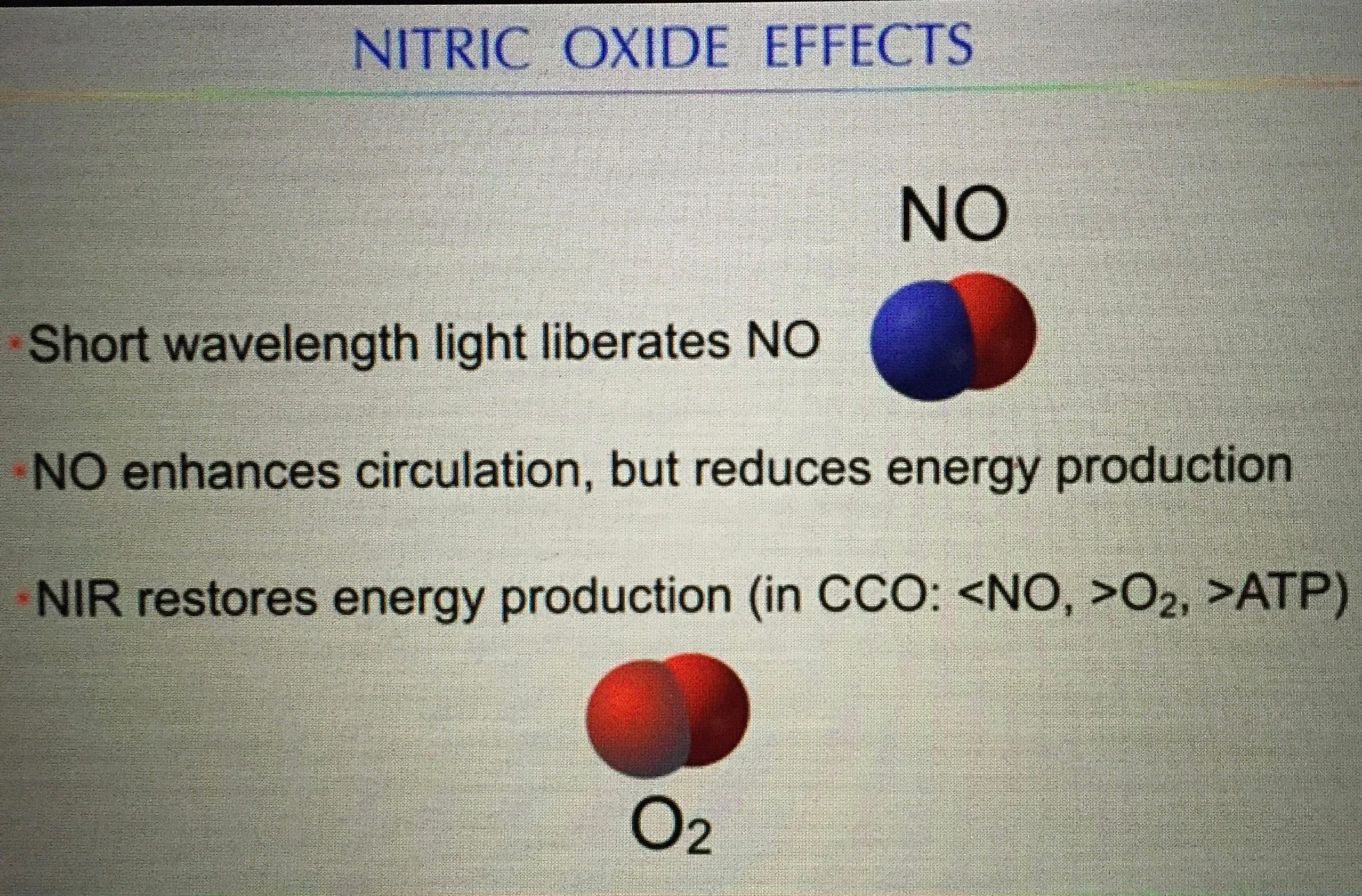


Beta-endorphin is made via POMC and solar exposure of the eye as I showed in my Vermont 2017 video on youtube. Morphine is an exogenous opioid that is used for people in pain. People deficient in solar exposure in the IRA, UVA, and UVB ranges have more pain and require more opioid drugs than those who get outside with the skin and eyes exposed to sunlight more often. This implies morphine dosing is not properly quantized to the light we live under and may lead to collateral effects that could cause bad signaling and lead to addiction. How is this information transferred from the sun to tissues in our brain to lead these diseases? It occurs on waterways in the brain called CSF water networks. The human brain is surrounded by a sea of CSF made almost entirely of water and salt. It happens to be loaded with H+ isoform of hydrogen made from the the blood plasma by the choroid plexus.


The latest piece of data I unleashed in Vermont 2018 (above) is that when the electric topologic charge of water is altered we can absorb more UV light in water.  This has big implications for the creation of melatonin from tryptophan in our bodies to optimize autophagy to restore proper functioning of mitochondria as we sleep.  What happens when we live a life based around modern blue light and not the sun?

Mitochondria function declines.

We get the opiate crisis and we get serious changes that occur in the blood plasma that lead to problems that do not allow proper signaling between mitochondria and nuclear DNA.  As a result, the mtDNA cannot keep the nuclear DNA quiet, and as a result, ubiquitination rates rise.  This means the body needs more methionine,  since it is the start signal for mRNA translation in ALL EUKARYOTES on Earth for the last 600 million years.  It is a real big deal as people found out in my Vermont 2018 talk.

Synthetic opioids affect a posterior pituitary hormone called vasopressin.  Vasopressin (VP), or antidiuretic hormone, is secreted in response to either increases in plasma osmolality (very sensitive stimulus) or to decreases in plasma volume (less sensitive stimulus). As such, larger numbers in osmolality indicate a greater concentration of solutes in the blood plasma.  Normal human reference range of osmolality in plasma is about 275-299 milli-osmoles per kilogram.

Calculated osmolarity really is tellng us about salt content of the blood, glucose content and the urea content of the blood = 2 Na + Glucose + Urea ( all in mmol/L).  Urea is often found as BUN on a lab.  This is why I am always so interested in people’s BUN/creatine ratios.  It tells me a lot about how their urea cycle and blood plasma are operating.

In normal people, increased osmolality in the blood which will stimulate secretion of antidiuretic hormone (ADH is vasopressin). This will result in increased water reabsorption, more concentrated urine and less concentrated blood plasma.  Morphine tolerance is mediated by complex formation between the vasopressin 1b receptor, β-arrestin-2, and the μ-opioid receptor as the picture below shows.


Why is this important?  All opiates affect vasopressin release from the posterior pituitary.  It is tightly controlled by light and dark cycles.

Consider the following:  Codeine is an opioid analgesic used for moderate to severe pain. Its analgesic effect is dependent on the conversion to morphine and morphine-6-glucuronide because the binding affinity of codeine to μ (mu) opioid receptors is 200-fold less than that of morphine.  This means use of morphine really cause massive changes to serum osmolality of our CSF and blood.  When salt is present in water is absorbs more UV light spectrum.  This affects the size of the exclusion zone in the blood plasma.  As my members found out in Vermont 2018 this has MASSIVE implications for wellness.   When opiates are used in patients this is why the gut function is so disrupted.  The change in salt content, ruins the gut peripheral clocks that are supposed to turnover every two days.  This is why they cause constipation and lethargy are frequent side effects of opiates.

The side effect profile, such as nausea, vomiting, hypotension, tachycardia-bradycardia, confusion, imbalance, headache, dizziness, fatigue, urticaria, ureteral spasm, reduction in micturation, and the very rarely seen tonic-clonic seizures and respiratory depression is quite wide because of this effect of UV light on water in your blood.

However, some of the symptoms observed during the use of codeine/morphine such as nausea, vomiting, headache, fatigue, confusion, and seizures can also be the symptoms of hyponatremia too.  Hyponatremia is a lack of salt (sodium) in the blood plasma.  This affects the release of vasopressin from the posterior pituitary where there is no blood brain barrier.  Neurosurgeons have to deal with a condition called SIADH.  This is a disorder of water networks in the blood and CSF that can be deadly.

The syndrome of inappropriate antidiuretic hormone (SIADH) was first described in 1957 by Schwartz and colleagues in two lung cancer cases that showed urinary sodium loss. You learned in the May 2018 webinar that apoptosis has to be inhibted and ECT speeds have to be kept elevated by raising oxygen levels distal to the ATPase to pull electrons along.

Cancer cells have to use oxygen to do this because cytochrome 1 is usually destroyed from blockades to repair the NAD+ levels there.  When NAD+ levels drop cells have to reduce oxygen levels to protect themselves from a highly proloiferative state.  When all the back systems to make NAD+ are inactivated, cells will use oxygen to increase ECT speeds and this pushes us closer to cancer.  This is why SIADH was first seen in two cancer cases in the 1950’s.  The last back up system for cytochrome one was shown in Vermont.  Tryptophan catabolism is capable of making NAD+ but it needs sunlight and a fully functional Kreb’s bicycle to do it.  In cancer, both of these are lost.  The implications of the Vermont 2018 will continue to reverberate for a long time for you members now.

SIADH is characterized by hyponatremia, inappropriately increased urine osmolality, increased urinary sodium losses, and decreased serum osmolality in a euvolemic patient without edema.  Excess water retention in the ECF space is the main problem in SIADH and therefore dilutional hyponatremia develops.  The water retained by the blood plasma is not the same water that a mitochondria makes or that you drink.  This should get you thinking about why the opiate crisis is linked to technology and and a lack of AM sun.   It turns out nnEMF and a lack of sun mimic the effects of opiates by depleting us of salt.  Excess glucose in the blood does the same thing.  Tecnology causes amplification of blood glucose because of the effect on AMPk pathways as Nora Volkow showed in 2011 with cell phone use.

WHAT ABOUT THE WATER WE DRINK?

Does it affect our CSF and blood and the metabolic pathways a cell can use??

Yes in many ways.

The water you drink should be un-fluoridated and COLD!  If you have a mitochondrial redox issue making it deuterium depleted helps.   You might be beginning to understand how the quilt was built now.  Limiting fluoride increases the size of the EZ in water networks in us.  Water then is cold carries more oxygen and electrons in it to enlarge the EZ in water.  DDW can former larger exclusion zones and build redox power quickly in sunlight.  What is the effect of oxygen in a cell in this case?  Look at the picture below carefully.   Oxygen changes mitochondrial function based upon the redox state of the mitochondria.  This is why I told you redox power is the key to understanding how we work and not food.


Mitochondria need oxygen and electrons when the redox potential is high so they can operate the TCA and urea cycle well. When the redox potential is lowered for any reason, mitophagy is impaired, and oxygen becomes a toxin because more ROS is made because slowed ECT speeds leave too much oxygen in places in the matrix too make too many free radicals. Too many free radicals lead to improper optical signaling in cells. This is one way we deplete our stem cell supply. This is another aspect of why cancer is a killer. If you cannot recover apoptosis and ECT speeds with sunlight and the creation of water in your mitochondria you inhibit apoptosis while you ALSO depleting your stem cells depots of there life force.

This causes us to use up the enzymes catalase and glutathione as a buffer inside our cells. Glutathione is made from 66% sulfated amino acids. Cysteine is the most RARE amino acid. Sunlight makes sulfhydryl groups in our blood as I showed in Vermont in 2017 and 2018 talks. This makes the anions in your blood that help you create UV light from deuterium to create melatonin to optimize mitophagy.

This means that TCA and urea cycle function at Kreb’s bicycle can indirectly affect the sulfur amino acid pathways in cells as a collateral downstram effect. Why is this a big deal? Methionine is the key sulfated amino acid that control protein synthesis in all eukaryotes. Our cells cannot make it, it is essential and means we need to eat it in pork, eggs, brazil nuts and parmesan cheese. This amino acids is an exogenous time crystal that controls mRNA and all protein translation. It only has one codon, AUG, like tryptophan does (ACC).

The evidence that sulfated amino acid cycles are broken in your mitochondria is a rising homocysteine level in your plasma. This is why the 5G and CAC blog exists. When HC rises and BUN rises, arteries calcify. What else happens? If the BUN/creatine ratio is up at the same time too, it is a bigger deal. This tells us you cannot make water in either the TCA or urea cycle. Dr. Boros got on the stage in Vermont and told everyone the key purpose of mitochondria is not energy production it is the production of water. I agree with this because the hydrogen proton ater network is how sunlight moves information in matrix water in cells.

When these things occur, the wise thing to do in this case is not take exogenous methyl groups, as the functional medicine doctors push, it is to increase your methionine intake with a high fat diet, while drinking water with a deuterium content that is below 135ppm and DOING IT IN THE SUN!!!!

This radically improves things along the inner mitochondrial membrane by lowering ROS and increasing endogenous glutathione production.


Water is the stage life dances on. Water is a repository of electromagnetic radiations which carry energy and information from sunlight. Water is designed to work wirelessly with the sun, but its abilities can be usurped by man-made EMF’s. They are capable of changing our osmolality. In a blue-lit microwaved world, the size of the EZ water is the ultimate Faraday cage for mitochondria in a cell provided it made from H+ bonding. I mentioned the chiral heat effect in Vermont 2018 and you’ll soon have a blog on it here too.

Water allows the proper quantum dance to happen in all complex life, not just us humans. We are just beginning to unfold its mysteries and why it is vital to all life on this planet. Beta oxidation in mitochondria not only makes CO2 but it creates water!!! This isoform of hydrogen in water is the stage life is built upon because it can move fast under the power of red light from the sun. 42% of it is RED between 600-3100 nm.


Water is what makes all plants grow via photosynthesis (PS). PS only works with sunlight 400-780 nm. PS does not use UV light. All animals in the eukaryotic tree need water to survive. When water falls from the sky as rain, it is distilled to a degree; at least it used to be before our atmosphere was polluted by modern chemicals and EMF’s in the ionosphere. (geoengineering)


If you use the photoelectric effect of the sun to enlarge the charge in cell water, while simultaneously grounding to Earth,  your needs and want for water will begin to approach your aquatic ancestors and your brain will repair and re-grow faster.   Why?  It can assimilate more UV light from the sun to expand your neocortex while optimizing the chemistry of your CSF all at once.

This is quite important in diseases like autism and Alzheimer’s disease.  Signs of leptin sensitivity begin to appear and labs start to improve.  Eventually, your thinking improves and your life re-evolves.  I left some key details out in this blog post on purpose.  Some of my members saw Vermont unfold live.  They will get the implications of this webinar fully.  The rest of you will have to wait until the video edit is complete.

As future series goes on you will see where this is all headed (quantum thermodynamics).  It is time you begin to fill them in with the other lessons you have learned from me over the last decade. The April 2013 and April 2018 webinar is a big clue in this path to Optimal that the story links to hydrogen is an old one for me.  You might have never realized it back then because you needed all the lessons you now have at your disposal as a member.

GSH is glutathione in the picture below.  Glutathione is made from sulfated amino acids like methionine, cysteine, and homocysteine which also operate in a cycle linked to the kinetics of the TCA and urea cycle.  All these cycles are linked to oxygen consumption in a cell.  This goes right back to how the redox potential is built in a cell.  On the stage in Vermont 2018 I said all one needs to really know about biochemistry is that the TCA and urea cycle need high oxygen levels and DDW in the matrix to work.  If either one is broken for any reason the older pathways of glycolysis and the PPP have to be used. These two pathways evolved before the Cambrian explosion and the change of our sun’s light.  This is why photosynthesis only uses the visible part of the spectrum above 400nm.

You can see the highest redox power in mitochondria are present when GSH and NADPH and NADH are working well kinetically between the TCA and urea cycle.  This is why understanding Krebs bicycle is mandatory for any mitochondriac in training.  Note where the redox levels of cytochrome C is.  This cytochrome protein controls apoptosis.  When it drops below 200 Mv you are at risk for serious mitochondrial diseases.  Note, you do not need a large redox potential to make ATP.  This is why red light and photobiolodulation can help a badly functioning mitochondria.  I actually gave you this information in the Energy and Epigenetics series but your understanding was not yet at the point where you could handle the quantum science to make sense of it.  Now that has changed.


THE MITOCHONDRIAC WISDOM THAT IS COUNTERINTUITIVE to THE food guru perspective: We saw that on display with Dr Zach Bush. His theory on the microbiome was demolished in front of the audience eyes. I never intended it to happen but Jeff Leach gave me the ammo and I was not about to waste it as a teaching point.

Cells that don’t or can’t use the TCA and urea cycle; these cells tend to have lower metabolic rates by design in nature. This happens because deuterium leaching from broken cell membranes gets into Kreb’s bicycle where it should not be. Autophagy/mitophagy controls the cell membrane turnover and it too is linked to the circadian mechanism linked to melanopsin and Vitamin A coupling to melatonin creation in our skin and eye.

Cells that are unable to use the TCA and urea cycle tend not to grow well. This is benefit in stem cells, retina, and the brain. This is why some of them use these older pathways. Cells that don’t do this by design, like the enterocytes, are usually forced into apoptosis because of deuterium assimilation from foods or from defective mitochondrial membranes adjacent to Kreb’s bicycle are no longer controlled by the tight regulation of autophagy and apoptosis. This is why nature built our gut cells to be replaced ever 48 hours and why our brain cells are rarely replaced unless need be.

Cells that cannot recycle mitochondria are afflicted with defective mitochondria and they cannot use mitophagy to repair themselves, but they usually can use apoptosis. This is the surest sign of the presence of a circadian mismatch from blue light toxicity via the skin and eye surfaces. After my 2018 Vermont talk you should be seeing how the pieces fit because now you’re becoming an acute observer of the pieces fall apart.

This is the wisdom built into the QT #1 and 2 blog posts. Cells that use the TCA and urea cycles well tend to have the highest metabolic rates and must have an intolerance for deuterium in their matrix.

Oxygen consumption marries to the speed of ECT and the creation of free radical signaling in the matrix. The more oxygen that a mitochondria can handle = a higher ROS. That is not always a good thing if the process is not quantized properly. When your skin and eye are illuminated by blue light this is the key way ROS is made in modern humans.


As free radical signaling rises (ROS) this implies that MORE ELF-UV bio-photons are being released from the matrix. This is a key sign of a falling redox state.

WHY DO SICK CELLS RELEASE MORE ELF-UV LIGHT?

Fritz Popp showed this effect in all living cells. He had no idea where the ELF-UV light came from. In Vermont I told you where it HAS TO COME from.


This ELF-UV light is likely coming from the matrix where trapped deuterium is compressed by the electric and magnetic fields produced by the mitochondrial matrix.  BOOM.   This deuterium is tightly bound to the anion substrates of the TCA and urea cycle.  When deuterium is compressed in the presence of anions not moving in the matrix it creates a nuclear pressure on deuterium causing it to release full spectrum UV light.  This was the excessive light that Popp found in cancer cells. I have a sense this is the same thing that a black hole does too in nature.

Might it be that the body is regulating metabolic rate in various tissues by controling deuterium concentrations to marry the specific physiologic demands of tissues kinetically at “Kreb’s bicycle”?  Is this done for a biophysical reason? Yep, I think so.  When redox power drops a cell become less able to use the TCA or urea cycle and wants to LIMIT oxygen presence to cap proliferation.

Many diseases uses this as a safety break like AMD, retinal bleeds, strokes, and sleep apnea.  To gain this protection, these cells MUST  default to older evolutionary pathways present before the Cambrian explosion that use lower oxygen levels.  Today many think cancer has to use glucose/glutamine exclusively because the paradigm is missing the pieces I gave in Vermont 2018. That is how big a deal this is folks.

Remember the TCA and urea cycle are both places loaded with anion substrates naturally.  As they cycle, this creates a need for the matrix to collect cations in the form of H+ over 1,000 fold.


Might this be why a cell uses deuterium as an optical switch in the matrix to control the kinetic rates of both cycles? Yep. This is why tissues can have a variable metabolic rate, and it control is tied to the circadian control of proton uncoupling. This is why the brain and gut have very different ways to handle deuterium to control the process. I hinted at this in Vermont with this slide below.


The regulation of anaplerosis and cataplerosis in the TCA and urea cycle depends upon the metabolic and physiologic state and the specific tissue/organ involved. Many people forget this basic aspect of biochemical physiology. They incorrectly believe pathway biochemistry is always the same and does not dynamically change its function as the redox potential and oxygen levels change. IT IS JUST NOT TRUE. Biochemical pathways are thermoplastic by design to control the fractionation of deuterium and hydrogen at surfaces in our body. Mitochondria liberate heat, in the form of infrared light, and this heat also has a massive effect on keeping deuterium in the blood plasma and out of tissues.

In this way a mitochondria mimics what a black hole is believed to do when it recycles matter. Black holes consume all matter that gets close to them, while emitting full spectrum light at the poles. Black holes seem to be recycle plants for matter. Mitochondria spit out UV and IR light using the same quantum principle. They appear to recycling matter from food to create light. This light is captured and slowed by cells to make new matter in cells. This was the key point in Vermont 2017. As light slows down we can make new things with mass. This is why aromatic amino acids are playgrounds for shortwave UV light and water is a red light chromophore.

For example, during starvation, cataplerosis occurs via phosphoenolpyruvate to support gluconeogenesis. This process is regulatory in the liver, whereas in the kidney anaplerosis occurs via the uptake of glutamine. I have a deep sense that the H+/deuterium fractionation present or absent in the matrix is the key optical switch in tissues controling matter creation. In a nondividing cell, there are 6600 H+ ions for every atom of deuterium in the matrix, yet the human blood is jammed packed with it (150ppm).


Have you ever thought to ask yourself why nature would do something that appears counterintuitive? Moreover, why the healthy cell would do this? Is this how growth occurs? To go even deeper into your curiosity, consider the H+/Na antiporter in human cells.


In the normal cell cycle when a cell wants to divide the mechanisms is thus: The cytosol must first become the adult size, and it is the size that gets the cell to the G1 step in the cell cycle. In this phase, H+ is pumped out of the cell and nucleus while “some deuterium” is allowed to enter to create a swelling stimulus to cause chromosome division. This means that in a cancer state we should see swelling in the cytoplasms water supply before mitosis. Is this observation true? Yes, it is. Look at the picture below.


In fact, the inventor of MRI, Roy Damadian was the first person to understand that the cytoplasm of cells might hold the clue to cancer in 1971 I wrote about this long ago when I was teaching you about Gilbert Ling.

What Damadian and Ling did not know is the swelling in cancer was due to a higher concentration of D2O over H20 in the cytosol because of poor kinetic flux of the entrapment of deuterium. D20 has a way higher viscosity than H20.

A higher viscosity will not let the chromosomes pull apart easily in mitosis of the cell cycle. When this occurs, it alters the signals for interphase, which are precisely QUANTIZED by the release of a specific spectrum of ELF-UV from matrix deuterium. Not all cells use the same frequency of light to cause mitosis. I think ever cell line has a special spectral line to cause normal growth.

The pressure and heat released by a mitochondria affects the amount deuterium a cell lets into its matrix. This is what creates a unique specific light signal to control cell growth in different tissues. It turns out the small amount of deuterium let into the matrix by the Na+-H+ antiporters generate just the right amount of ELF-UV light using uncoupling proteins to get the cell to begin the division process to separate chromosomes without aneuploidy.

UV light assimilation in water is affected by its salinity and charge.

This is why the salt content of the blood links to hydrogen flows in our body. As I showed in Vermont it also links to the amount of UV light our blood can assimilate from the sun.

This is why I went into the story about SIADH/vasopressin earlier. You likely forgot I mentioned vasopressin in the CT 4 and 6 blogs years ago. Now you can see I have been slowly unfolding this story for years. It is quite complex but nature makes the application of her rules easy as the slide shows.


I am hoping you can see now why my complexity leads to simplicity by using nature’s laws as poetry to get my points across coherently. I don’t want you to fall prey to poor thinking as some have in the past.


These thing all affect how the Na+-H+ antipoters operate in mitochondria to control deuterium fractions everywhere in our body to control how the TCA and urea cycles operate.

In normal cells, when the H+/Na+ antiporter is blocked, just adding deuterium depleted water added to the culture medium, stops mitosis very quickly.  This is how DDW works in cancer.   Think about all the cells that are normally obligate glycolytic cells in the body I mentioned in QT #9.  They all work this way to control proliferation in the retina and brain.

They cannot work this way when deuterium is not in the correct spot in a cell.   This is why I mentioned to you months ago in Q & A’s to think about the cellular environment in relation to H+ and deuterium fractions.  Deuterium has a positive and negative regulatory role in the cell.  Light and temperature controls the flow of hydrogen in us.  When information is lost inside the cell, the ability to control the flow H+ and deuterium is also lost.  This means you have no way to gain shortwave UV light to drive proper optical signaling via the blood plasma.

That concept was covered long ago in Quantum biology 1, 3, and 4 blogs, but I bet you forgot them.  I think you might see another side of this coin when you go back and re-read those blogs today that might make you more curious.  The mitochondrial ROS/RNS cycles were built for cells who respond EFFECTIVELY when both autophagy and apoptosis are fully operational.  When they are non functional, then ROS and RNS become deadly.  They are only this way when the redox power of the mitochondria is below -200mV as the picture above showed you.

In stem cells, when we activate glycolysis and the PPP in the presence of pseudohypoxia to use autophagy over apoptosis to control quiescence.  This is how senescent proliferative cells are kept dormant.  In cancer, the opposite situation manifests, where the TCA and urea cycle are hindered by deuterium clogging the anion gears of both cycles making water that has a high viscosity because of a higher fraction of D2O.  This forces the cell on a CHRONIC basis to use glycolysis and the PPP for biosynthesis.

The change in oxygen tensions alters the time window for oncogenesis.  These are the key steps that must occur for oncogenic transformation because acute and chronic hypoxia are handled differently in cells.

Chronic hypoxia leads to the formation of angiogenic growth factors released from our blood vessel linings, that increase blood vessel formation to bring new vessels and oxygen to the impaired cells and this fosters cancerous growth.

Guess what causes those angiogenic factors to be released?   Methionine does.  This makes methionine our exogenous time crystal.  Methionine rises in blood when the TCA and urea cycle are defective.  This is how cancer begins.  The cytosol and matrix are filled D20 over H20 and this is the key difference seen in cancer cells and obligate glycolytic cells.  When deuterium is squeezed in the matrix it makes massive amounts of ELF-UV to stimulate uncontrolled mitosis and biosynthesis.  This drives cancer growth.   This is why tumor cells all have huge ROS/RNS free radicals associated with them because cells cannot make ELF-UV light without oxygen and ROS as Roeland van Wijk shows in the first 100 pages of his book.  You might want to go back now and read it with your new understanding now.


At the same time, cancer cells all must inhibit apoptosis, while having impaired autophagy and fast ECT speeds to become immortal like embryonic stem cells (ESC) are to use the Warburg shift to survive. Now think about the group of normal cells who do not use the TCA/urea cycle in normal cells. The homology is remarkable, when you see the real purpose of biochemical pathways for the first time.

The reason why researchers cannot solve cancer should now be obvious. They see the biochemical changes and cannot see the biophysical levers because they do not understand hydrogen biophysics between the matrix and our blood.

Cancer cells do not use the TCA/urea cycle, because they cannot, due to the KIE of deuterium as Ray Damadian showed when he invented the MRI machine in the 1970’s. Moreover, the cancer phenotype shows they only use glucose or glutamine to fuel biosynthesis because they have too because the TCA and urea cycle have poor kinetics.

They must use glycolysis and the PPP because the redox power is below -200mV or oxygen levels are poor. So eating glucose and protein with cancer is not as bad as today’s LCHF cancer experts think. It becomes bad when you are out of the sun and lose control of how deuterium is supposed to move from the blood to tissues. It is needed until you can recover the cytosol water fractions using DDW to clear the cytosol and matrix of deuterium laced water. This is why cancer cells and normal cells look as they do (pic below). Cytosol in normal cells is smaller because it has less deuterium in it.


Guess what the link between both cycles is? Fumerase. It is where DDW is made by both cycles to add DDW to both anions. Since water has a different viscosity and ability capture UV light to lower the surface tension of the anions substrates to move freely to make the process run smoothly to control growth. (reminder pic below from Vermont 2018)


It has been well established in the literature that cell swelling triggers its proliferation.  This manifests when autophagy is impaired and the we call this a rising state of heteroplasmy.  It is also quite clear that the literature  has numerous reports as they cytosol and cell shrinks this promotes its apoptosis and cancer’s do not manifest when this happens.

There should be no more mystery why cancer occurs or what one should do to control it and reverse it.  The key is recapturing both autophagy and apoptosis using circadian signaling to avoid the situation and prevent the disease before they manifests.  Mitochondriac wisdom 101.

ARE YOU FEELING ME NOW?

CITES:

Volpe DA, McMahon Tobin GA, Mellon RD, Katki AG, Parker RJ, Colatsky T, et al. Uniform assessment and ranking of opioid Mu receptor binding constants for selected opioid drugs. Regul Toxicol Pharmacol. 2011;59:385–90

https://www.ncbi.nlm.nih.gov/pubmed/16984813

https://www.omicsonline.org/open-access/the-dysfunction-of-metabolic-controlling-of-cell-hydration-precedes-warburg-phenomenon-in-carcinogenesis-jbb.10000e59.pdf

WHAT WILL I PROVE BEYOND A SHADOW OF A DOUBT IN VERMONT 2018?

video
play-sharp-fill
I have decided to something I have never done.  I will give the punch line of the talk in the first ten minutes and tell you what the end point is……..and then I will show you piece by piece how they fit and how they fall apart.  I plan on beginning my talk with the video above and ending it with the video at the end of this blog.  The words of the videos should be your focus and not the song, albeit the songs are awesome. 
If you are going to Vermont, you might want to read this before you see my talk live.  
For those of you not attending, you’ll have a chance to see the video here on Patreon when it is ready. 
NAD+ links directly to tryptophan and the circadian mechanism foundational to human life. 
Ask yourself what do codons and ubiquitin marking have in common?
The answer is circadian biology.  Did you know that tryptophan and methionine are the basis of the circadian mechanism in humans?  
They are our key time crystals in our eye, skin, gut, and lung.  They are the key gears in the eye clock I discussed last year in the Vermont video but nobody realized it.  They will this year when I show them last years slides and they realize I was forshadowing the coming story on deuterium and UV light.  
Look at step 8 above………I hope you remember that NAD+ is the key fluorophore protein of cytochrome 1 in every mitochondria in every cell of your body. 
When the circadian mechanism is disrupted by any stressor,  protein synthesis is increased and often not yoked to proper solar signals.  Since we now know melanopsin is now present in eye, skin, and dermal fat it is becoming clear ubiquitin is linked to the melanopsin system.
That opsin is linked, via sunlight to cytochrome 1, NADH/NAD+ couple.  Tryptophan has a unique absorption spectrum that hints it is a seasonal time crystal and it can be used by the endogenous timing mechanism by the addition of information and energy from photons, electrons, or by protons.  Did you know tryptophan makes NAD+?  I bet you did not.
These two aspects of subatomic particles program the phenol ring of tryptophan differently in seasons because season have a different electromagnetic footprint codified by photosynthesis in food and this is deciphered by mitochondria.  This is why tryptophan can be both ketogenic and glucogenic and it has but one codon because it decreases the signal to noise ratio to one.  
This links its production directly to solar cycles via photosynthesis depending on how it was created by the sun.  Methionine is the “exogenous time crystal” because it is also unique with one codon.  Methionine and tryptophan are the only two amino acids with one codon in eukaryotes.  Did you know this?  
This fact in our kingdom of life has been true for 600 million years of evolutionary history.  What else in our kingdom has not changed in 600 million years?  DHA has never been replaced once in the same time frame even though there are other PUFA’s more easily made by cells.  Ask yourself why this is the case?  Why would mother nature do this?  Now recall that the central retinal pathways in humans have more DHA in them than any other part of the brain.  You think there is a reason behind that tied to this story in the skin?  There is.
Methionine is an essential amino acid that cannot be made but has to be eaten, therefore, it gives energy and information to the mitochondria from the environment the animal is inhabiting. That signal must be yoked to the incoming light signals via the eye and skin to be ciphered.  The mtDNA deciphers the message to pass it on to the nucleus to change epigenetic signaling via the addition or subtraction of electrons, protons, and photons to histones and methyl groups to affect chromatin binding and DNA kinetics and the Auger effect in cells. 
Tryptophan is used to make melatonin and it controls mitochondrial DNA.  These are main clock gears of the circadian mechanism.  This topic will be explained in detail in Vermont 2018 on Saturday, June 2.  Strap in.  I’ve got a lot of people to prove wrong and this is going to be epic.
THE ENDING VIDEO FOR THE VERMONT TALK WILL GIVE YOU SOME INSIGHT TO THE SLIDES I HAVE MADE