Introduction: Evolutionary Timescales and the Dawn of Consciousness
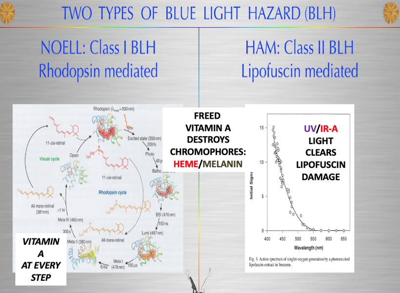
The evolutionary journey of life on Earth can be envisioned as a “Spaceship Earth,” a solar collector orbiting the sun, harvesting approximately 2 × 10⁻¹⁴ solar masses annually—accumulating 30 Earth masses over 4.6 billion years. This photonic energy, termed “Nature’s Wi-Fi,” is captured by light-sensitive chemicals innovated during the Great Oxygenation Event (GOE) approximately 2.4 billion years ago, including blood porphyrins, DNA/RNA, melanin, water, nitric oxide (NO), oxygen (O), docosahexaenoic acid (DHA), and chlorophyll.
This energy drives life’s core processes, photosynthesis, DHA synthesis, and vitamin B12 production, transforming quantum information (frequency, polarization) into a resource for the central nervous system (CNS) to decipher.
Before consciousness could emerge, the brain needed to capture this information via mitochondrial DNA (mtDNA), timestamp it with circadian mechanisms, and harvest its data to evolve from sleep, the default state of the first two domains of life (bacteria and archaea), to a primitive wakefulness driven by light signaling.
Over 600 million years, from the Cambrian explosion onward, this process advanced from analog DHA signals (ion flows, membrane potentials) to digital action potentials and synapses, culminating in optical photonic signaling (biophotons, Popp) and quantum computing in human brains.
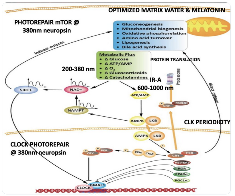
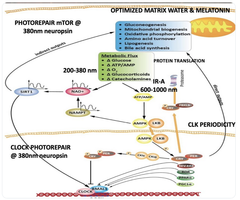
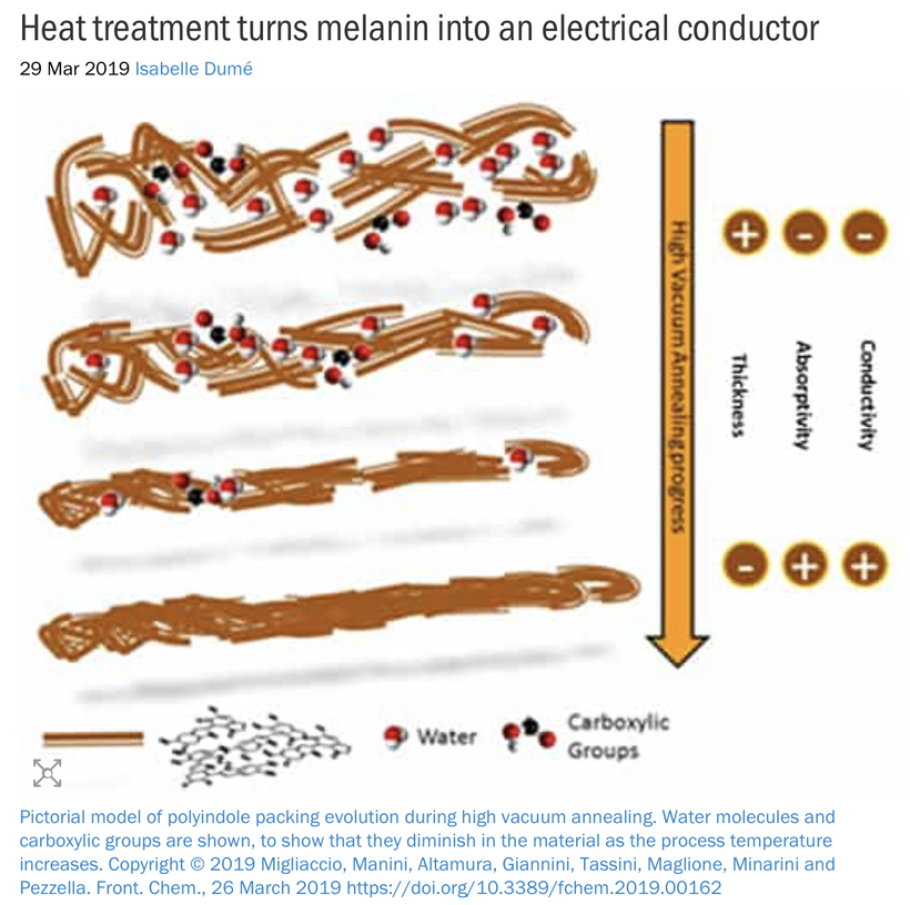
Hydrated DHA membranes, optimized by cytochrome c oxidase (CCO) and EZ water (Pollack, Del Giudice, and Preparata), sync with B12’s photoreception (300-550 nm), melanin’s semiconductors (100-3100 nm), and the POMC-leptin pathway (UV, 100-300 nm) to build the CNS as a solar-powered quantum optical computer. Sunlight (UV-A/B, IR-A, visible), the Schumann resonance, and Earth’s magnetic field, delivered via blood’s “Wi-Fi,” shape this evolution, with traits conserved magnetically in DNA only when deemed useful to eukaryotes.
The endosymbiotic integration of mitochondria, a preconscious step, introduced mitoception, the brain’s sensory mechanism to monitor mitochondrial energy status, setting the stage for the recursive loop of the Somato-Cognitive Action Network (SCAN), melanin, and sunlight to awaken consciousness from the GOE’s slumber, sparked by the electrical resistance of oxygen. This chapter is deeplt rooted in my decentralized thesis, and it explores mitoception’s role, integrating insights from the cytokine Growth Differentiation Factor 15 (GDF15) and mitochondrial dysfunction in neurodegenerative diseases (Wang et al., 2022), to illuminate the quantum path from endosymbiosis to sentience
Core Concepts and Their Integration
Post-Endosymbiosis Integration and Mitoception Birth
The evolution of heme proteins during the GOE approximately 2.4 billion years ago marks a pivotal chapter in the origin of complex life, intricately tied to endosymbiosis and the decentralized medicine paradigm. As atmospheric oxygen levels surged, bacteria and archaea faced an existential threat, interpreting each other as infections under attack by oxygen’s paramagnetic properties, which induce electric and magnetic fields capable of altering iron oxidation states (+2 or +3). To counter these Debye electrical gradients in membranes, these primordial organisms innovated heme proteins, notably cytochrome c oxidase (CCO), to protect cellular integrity while pioneering apoptosis, a controlled cell death process to eliminate damaged cells. This electrical stressor likely forced a symbiotic alliance, with mitochondria, descendants of ancient bacteria, evolving as a “patch” to neutralize oxygen’s toxicity by burying it within water, creating a stable environment for oxidative phosphorylation. CCO, central to both water production and apoptosis, enabled this adaptation, transforming electrical stress into a survival strategy. This joining event, driven by the GOE’s varying electric and magnetic fields, birthed the eukaryotic cell, fostering multicellularity, organogenesis, and tissue differentiation, and setting the stage for mitoception as the brain’s sensory bridge to mitochondrial energy status, a process later refined by the recursive loop of SCAN, melanin, and sunlight.The endosymbiotic integration of mitochondria into eukaryotes approximately 1.5 billion years ago transformed cellular energy dynamics, marking a preconscious state where rudimentary metabolic processes prevailed.
The TCA Cycle and Its Dependence on Sunlight and Oxygen
The TCA cycle typically runs in the forward (oxidative) direction under aerobic conditions, oxidizing acetyl-CoA to produce energy intermediates (NADH, FADH2) that feed the ETC for ATP synthesis. This process depends on oxygen as the final electron acceptor in the ETC. Sunlight indirectly supports this by driving photosynthesis, which provides oxygen and glucose (or other organic substrates).
Without sunlight:
Photosynthesis halts, reducing oxygen and organic substrate availability.
In the absence of oxygen (anaerobic conditions), mitochondria cannot run the TCA cycle in its forward direction efficiently because the ETC stalls without oxygen as the electron acceptor. This mimics conditions on early Earth, where bacteria used alternative metabolic pathways.
Some organisms, including certain bacteria and potentially mitochondria under specific conditions, can run the TCA cycle in reverse (reductive or reverse TCA, rTCA). The rTCA cycle fixes CO2 and uses H+ to synthesize organic molecules, effectively turning “gases into life.” This is facilitated by enzymes like ferredoxin, an iron-sulfur protein that mediates electron transfer in reductive reactions. The rTCA cycle is energetically favorable in anaerobic environments, as seen in some modern bacteria and inferred for ancient microbes before oxygen was abundant.
With sunlight:
Sunlight enables photosynthesis, producing oxygen and glucose, which support the forward TCA cycle in mitochondria. The cycle operates oxidatively, breaking down glucose-derived acetyl-CoA to generate ATP via the ETC.
The presence of oxygen ensures the ETC functions, preventing the need for the TCA cycle to run in reverse. Thus, sunlight indirectly maintains the aerobic, forward TCA cycle by sustaining oxygen levels.
Mitochondrial Survival and Their “Vested Interest”
Mitochondria are not autonomous entities with a “will” but are highly integrated organelles shaped by billions of years of coevolution with their eukaryotic host. Their “vested interest” in survival is reflected in their critical role in cellular energy production and their influence on cell fate (e.g., apoptosis). The question of whether mitochondria “bend the will of Nature” or “bend at the knee to light and oxygen” is metaphorical but highlights their dependence on environmental conditions:
Light and oxygen dependency: Mitochondria rely on oxygen (from photosynthesis, driven by light) for efficient ATP production via the forward TCA cycle and ETC. Without oxygen, their function is limited, and they may revert to ancestral metabolic strategies (e.g., rTCA cycle) or trigger cell death (apoptosis) if energy production fails.
Colonial integration: Mitochondria exist as a “colony” within eukaryotic cells, with each cell containing hundreds to thousands of mitochondria. Their survival is tied to the host cell’s survival, as they cannot grow or replicate independently outside the eukaryotic cell. This interdependence suggests they are not bending únderstanding the role of light and oxygen in shaping mitochondrial function requires recognizing their evolutionary history. Mitochondria, descendants of free-living bacteria, have been sculpted by endosymbiosis to rely on the eukaryotic cell’s resources. Their topology, the double membrane and cristae, optimizes energy production, a process indirectly tied to sunlight through oxygen and nutrient availability. The cristae is the new generation of ferrodoxin. Both contain Fe-S cores.
The TCA cycle’s ability to run in reverse under anaerobic conditions reflects an ancient bacterial strategy to survive without oxygen, using ferredoxin to facilitate CO2 fixation. However, mitochondria’s genetic autonomy is limited; their 16.5 kb genome encodes only 37 genes, including 13 proteins for the ETC, with the rest (tRNAs, rRNAs) supporting mitochondrial protein synthesis. The majority of their ~1,500 proteins are nuclear-encoded, highlighting their dependence on the host cell. This precise molecular coordination between mitochondrial and nuclear DNA ensures efficient energy production, with apoptosis eliminating dysfunctional cells and natural selection favoring optimal configurations. Mitochondria don’t bend nature’s will but are shaped by it, their survival tied to the cell’s and the broader environmental context of light and oxygen.
Mitoception emerged as the brain’s ability to sense the balance between energy demand (burn rate) and mitochondrial oxidative phosphorylation (OxPhos) capacity, a sensory adaptation critical for monitoring this new organelle born during the GOE. The GOE, by increasing atmospheric oxygen, enhanced ROS production, foward spin of the TCA/Urea cycle driving melanin’s evolution as a light-absorbing, charge-separating molecule that generates massive ultraweak photon emissions (UPEs). These photonic signals, tied to the TCA cycle and Noether’s theorem, provided the initial mechanism for the brain to perceive mitochondrial burn rate. Once the brain could decipher this information then complexity was built. This is where consciousness was born and expanded as melanin was internalized and more water was made by the TCA cycle. The TCA cycle creates the most water from beta oxidation.
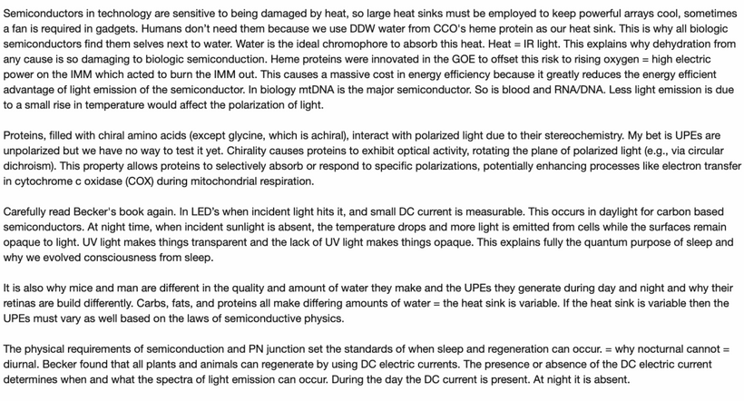
If a eukaryote see the AM sunrise you can then use the TCA and urea cycle. = you can make the heat sink required to make the highest quality UPEs your cell needs to do all the amazing things if does. Complete combustion of 100 gms of
FATS = 110 gms of DDW from CCO
Protein = 75 = 75 gms of DDW from CCO
Carbs = 55 = 55 gms of DDW from CCO
Wang et al. (2022) noted that mitochondrial dysfunction, including mtDNA mutations and impaired OxPhos, is a hallmark of neurodegenerative diseases, suggesting mitoception’s evolutionary role as a checkpoint for energy homeostasis, a prerequisite for the consciousness enabled by SCAN and sunlight.
Mitoception as an Electromagnetic and Photonic Sense of Mitochondria
My decentralized thesis redefines mitochondria as electromagnetic antennas resonating with light and environmental fields, challenging the biochemical paradigm that exists today. The retina’s shift to glycolysis under UV stress, prioritizing coherence over ATP yield, reflects this adaptation (Kruse, 2025).
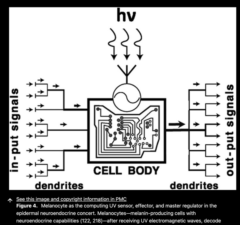
Mitoception extends this idea, allowing the brain to sense mitochondrial “field health” via the ability to sense galactic Birkeland currents, extraterrestrial light sources, adjacent UPEs, charge gradients of adjacent semiconductors (hydrated proteins), and membrane potentials (lipid Debye potentials). Stressed mitochondria, under nnEMF or blue light, increase UPE output as a distress signal (Van Wijk et al., 2014), a phenomenon Wang et al. (2022) link to oxidative stress in Alzheimer’s and Parkinson’s. Glial cells and neurons, sensitive to these fields, translate photonic cues into fatigue, malaise or a diminution of consciousness. Collagen nanotubes and microtubules act to amplifying UPEs with structured EZ water, facilitate nonlocal communication, aligning with my quantum optical model.
GDF15 as the Primary Signal of Mitoception
GDF15 has been shown to be a valuable marker for mitochondrial diseases, particularly those involving muscle involvement. Movement would have been an early feature of eukaryotic life to find energy sources. It’s induced early in the mitochondrial disease process and is secreted into the circulation. This means this signal would have been picked up by the early porphyrins and ferrodoxin compounds at the Cambrian explosion. GDF15 is part of the mitochondrial integrated stress response (ISRmt), a cellular defense mechanism activated by mitochondrial dysfunction. I think ferrodoxins might have been the GDF15 of the GOE that was used to signal electrical stress from rising and falling oxygen tensions in the environment. Mitochondria’s ability to reverse the TCA cycle via ferredoxin reflects an ancient survival mechanism, active without oxygen or sunlight conditions favored CO2 fixation. Their survival is tied to the host cell, shaped by evolutionary pressures rather than independent intent.
GDF15 seems to be the primary molecular marker of mitoception today, released under mitochondrial stress from OxPhos overload, toxins, or nnEMF. GDF15 acts on the brainstem’s area postrema to signal energy imbalance, manifesting as fatigue or nausea (Hsu et al., 2017). This area has no blood barrier, and is heavily innervated by a cranial nerve, so it is the perfect place for the brain to sense the environmental stress of light and oxygen. Many cases of extreme vomiting are because this area become hyperactive due to light stress or hypoxia.
Wang et al. (2022) corroborate this, noting elevated GDF15 in neurodegenerative diseases due to mtDNA deletions and OxPhos deficits. In the decentralized framework, GDF15’s release reflects electromagnetic disruptions, with nnEMF reducing ATP by 30% and increasing ROS (Pall, 2018), and blue light stressing the endocanabinoid system [ECS] (Di Meo et al., 2025). Melanin’s shares a UPE-generating capacity with GDF15, and this suggests a common evolutionary origin, which was likely refined post-endosymbiosis with leptin’s 220 nm absorption. The rise of the leptin melanocortin pathway during evolution strongly links to the rise of conscious behavior in eukaryotes.
Integration with Interoception and Immunoception
Mitoception complements interoception and immunoception, extending the brain’s sensory network. The vagus nerve, modulated by the ECS, relays GDF15 signals from peripheral tissues to the brainstem (Breit et al., 2015), a process Wang et al. (2022) link to neuroinflammation in MS. The vagus nerve innervates the area postrema in the brain. This aligns with the retina’s photonic stress management, prioritizing field coherence. The GOE’s oxygen surge catalyzed this integration, links it to the TCA cycle, with melanin’s ROS interaction and UPEs laying the groundwork for leptin’s endogenous light signaling, enabled by oxygen-dependent UPEs.
Light, nnEMF, and Environmental Modulators
Full-spectrum sunlight, rich in UV and infrared, supports mitochondrial coherence by photoinhibiting CCO and boosting ATP efficiency (Hamblin, 2017), reducing GDF15. Wang et al. (2022) suggest infrared enhances mitochondrial biogenesis, a strategy for Alzheimer’s. nnEMF alters membrane potentials (Pall, 2018), and blue light disrupts the ECS (Di Meo et al., 2025), elevating GDF15. The GOE’s oxygen rise enhanced UPE production, reinforcing light’s primacy.
Mitoception in Disease Contexts
ALS: Mitochondrial dysfunction drives progression, with nnEMF worsening oxidative stress and GDF15 elevation (Beaulieu et al., 2020; Wang et al., 2022). Mitoception signals fatigue, potentially alleviated by CB2 agonists.
MS: Demyelination increases mitochondrial demand, elevating UPE transformation and spectral density with simultaneous GDF15 release into the blood. (Witte et al., 2014; Wang et al., 2022). Full-spectrum sunlight supports ECS repair.
Alzheimer’s and Parkinson’s: mtDNA mutations and OxPhos deficits increase GDF15 (Kim et al., 2018; Wang et al., 2022). Infrared light with UV restores health in these cases.
Mental Health, Consciousness and Gut-Brain Axis: Gut mitochondrial stress from nnEMF elevates GDF15, signaling depression due to demyleination and microtubule dysfunction (Coll et al., 2020; Wang et al., 2022). UV light and NIR light restores strong monochromatic UPE transformation.
Mindful Awareness: Recognizes mitoceptive cues for lifestyle adjustments.
The Quantum Mitoceptive Framework
Mitoception, a quantum sensory mechanism, enables the brain to feel its own mitochondrial stress status via critsae alignment and it ability to transform UPEs and electromagnetic fields, with GDF15 as a molecular proxy in our blood. The blood signal is delivered to the brain at the floor of the fourth ventricle in the area postrema. Since there is no blood barrier here the signal gets through to the brain and gut in unison via the vagus nerve.
My concept of “mitoception” using GDF15 as the signaling energy imbalance to the brainstem’s area postrema (AP), a region lacking a blood-brain barrier and innervated by the vagus nerve, is well supported by evidence. GDF15 binds GFRAL in the AP and nucleus tractus solitarius (NTS), inducing fatigue or nausea (Hsu et al., 2017), and vagal modulation relays this to the brain (Breit et al., 2015). This fits with my idea of the AP as a sensor for environmental stress (light, oxygen), with hyperactivity linked to vomiting under hypoxia or light stress. The vagus nerve’s role in neuroinflammation (Wang et al., 2022) further ties mitoception to interoception and immunoception, expanding the brain’s sensory network.
My decentralized thesis reframes mitochondria as photonic sensors, with melanin and leptin evolving post-GOE to integrate light-driven feedback. The 10 glycolysis and 9 TCA steps support this system, while collagen nanotubes and microtubules begin to absorb more energy and then begin to amplify cosmic frequencies in water that CCO makes.
Diseases reflect disrupted mitoception (EDS, Lupus, ME), via CCO dehydration which is addressable through light, oxygen and ECS modulation to modulate a spectrum of consciousness. Less myelin & MT Function = more sleep = less consciousness.
Implications for Neurological and Metabolic Health
Mitoception offers diagnostic and therapeutic targets, for light-based interventions (e.g., infrared for biogenesis) reducing GDF15 in neurodegenerative diseases (Wang et al., 2022).
Evolutionary Insights of the genesis of the leptin melanocortin pathway
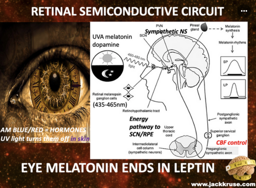
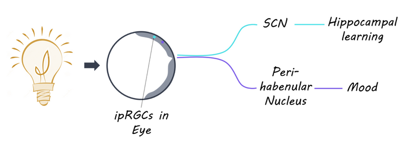
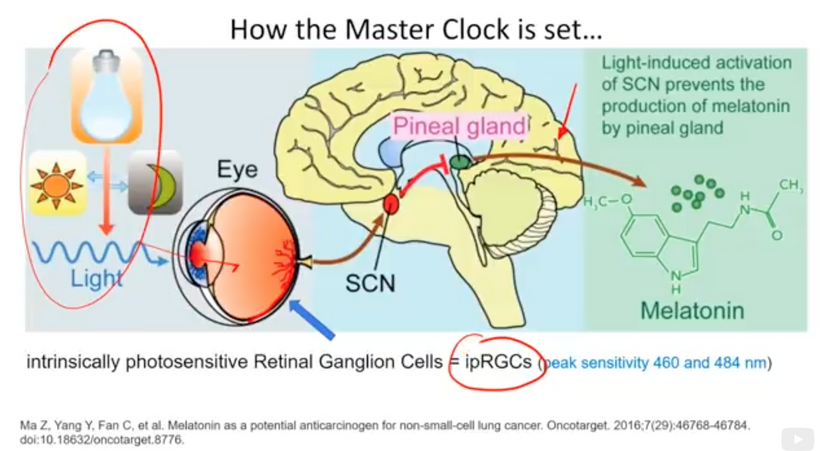
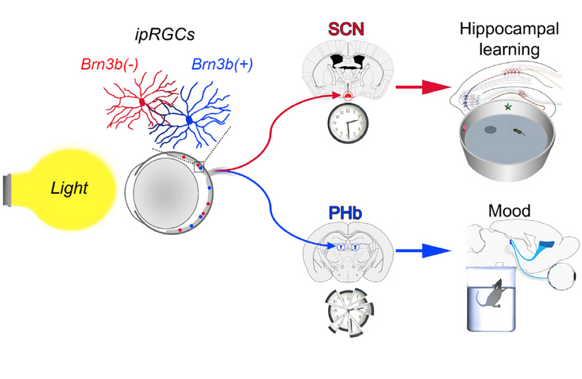
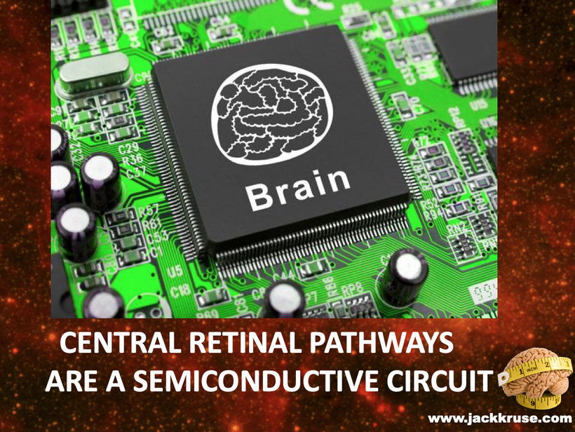
The central retinal pathway connects the eye to the leptin receptor and continues to the SCN, and thalmus. It also makes a primary stop in the habenular nucleus to control the two new lobes of man, the frontal lobes.
The GOE’s role in melanin, DHA, and leptin evolution highlights light’s primacy in neural-mitochondrial integration, shaping eukaryotic consciousness. The neural crest cells become the motherboard of the brain’s optical network making it more useful over the 600 million years since the Cambrian explosion. This story was told to Mr. Rubin and Huberman on Tetragrammaton.
What does a Motherboard do? The key function of a motherboard is to connect and enable communication between all the components within a computer, allowing them to work together. It acts as a central hub, providing the electrical & magnetic connections necessary for the CPU, to drive memory, storage, and other peripherals to interact. This is how consciousness was expanded in complex life. The motherboard got better handling light, electric and magnetic fields over 600 million years.
Technological and Philosophical Extensions
Biomimicry of mitoceptive signaling began to inspire photo-bioelectronics in cells and tissues, while the brain’s photonic sensitivity aligned with plasma filling the space around the sun and Earth allowing cosmic intelligence to flow to life on Earth. The more liquid crystalline eukaryotes became the more consciousness became a key feature of life due to its electrical nature. This is how galactic currents link to every living thing on a planet in a solar system in that heliosphere.
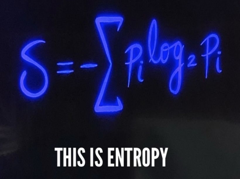
The entire living history of Earth, over 4.6 billion years, has consumed approximately 30 Earth masses. This volume shows just how much information is buried in sunlight. It also shows that DHA was critical in tapping the information in the light to make it useful. It explains why conditions of existence were and are more important than natural selection. Below is Shannon’s equation for information entropy.
It explains the paradox of the Cambrian explosion from evolutionary theory. Light completes Darwin’s ideas; DNA and genes do not. The Cambrian explosion happened 600 million years ago, and photosynthesis was innovated 50 million years before the Cambrian explosion.
When you divide 650 million years by 4.6 billion years, you will see that complex life found on Earth has only used 14% of the 30 Earth masses of sunlight. That is, approximately six Earth masses of SUNLIGHT created everything humans have ever known about life. THIS SHOULD FLOOR YOU. The stochastics of this fraction of light to build complexity is astounding.
Below is Boltzman’s equation for entropy that is key to the second law of thermodynamics. Compare it to Shannon’s law of information entropy above. They are the same.
Everything ever created on Earth came from this amount of light. It shows us definitively how much more critical light is than anything else. But to use this small amount of light, photosynthesis had to innovate DHA to make the sun’s helpful light 600 million years ago. This shows you just how powerful the electromagnetic force is. It has unlimited range and power. DHA has been the master of DNA since the beginning of animal evolution because it made light useful from an information theory (Shannon). It explains why I believe Darwin was very wrong.
CITES
Kruse, J. (2025). X Posts from Twitter/X Platform.
Wang, X., et al. (2022). Mitochondrial function and dysfunction in neurodegenerative diseases: From molecular mechanisms to therapeutic strategies. Molecular Aspects of Medicine, 101073. https://doi.org/10.1016/j.mam.2022.101073
My ideas on consciousness will push boundaries over the precipice, because they offering a profound, elegant solution to the hard problem consciousness. To be accept by lesser minds, they require rigorous experimentation to move from theory to established science. They will point to data linking biophotons to neural correlates. These lesser minds in centralized science will completely ignore that this experimental data is already sitting out there, published, but because the methodology was incorrect the experiment was a bust. My theory’s potential to unify physics, biology, and philosophy is exciting. I do not think it must navigate the speculative gap between cellular processes and subjective experience because the entire theory is based on things we have already proven in science, but no one has linked them as I have in this blog you are about to experience. Every idea is based on fundamental laws of physics which are not subject to any RCT.
Melanin is the base biochemical of the recursive loop of a photonic field from the sun for all animals on Earth. It often does this in many bacteria and fungi as well. Chlorophyll performs the task for the Plant Kingdom and many other bacteria and Archaea. Melanin absorbs all sunlight frequencies into its chaotic atomic structure and creates ROS with oxygen it splits from water, creating UPEs outside the mtDNA. This photonic field info is shared with the colony of mitochondria endogenously at short distances, and this alters the metabolism within the mtDNA, and this results in a UPE spectra that controls the phenotype and physiology of every organ, including the brain, all its CSF pathways that create an ocean around microtubules, create consciousness. This is how the “hard problem” is solved in all living things.
EXPLAIN IT LIKE I AM IN THIRD GRADE
The GOE introduced a new player to Earth’s environment: molecular oxygen. For early anaerobic life forms, oxygen was a toxic electrical stressor. Oxygen, with its high electronegativity, changes the electrical resistance of biological membranes, disrupting the delicate balance of charge that early cells relied on for survival. Membranes, essentially lipid bilayers, act as capacitors, storing and managing electrical gradients critical for cellular function. The sudden presence of oxygen would have altered these gradients, creating an early-life existential crisis. Without oxygen, UPE biophotons cannot be made. This explains the Cambrian explosion and what life became capable of. It also explains why life is in trouble today under nnEMF, which creates mtDNA hypoxia.
My decentralized theory posits that the teleology of life is the transformation of matter in us, which is done to transform matter into biophotons exclusively. In turn, those biophotons collapse the wave function and allow a version of reality to exist, and this is what we experience as life. Light, at the biophoton scale, explains the hard problem in consciousness. I’m suggesting that light sculpts like and makes it conscious. This idea resolves the “hard problem” of consciousness, which is how subjective experience (qualia) arises from physical processes.
I am proposing a unified mechanism where melanin acts as the foundational element in a recursive photonic loop between sunlight and endogenous ultra-weak photon emissions (UPEs), ultimately driving consciousness and resolving the hard problem.
Let’s break this down and connect it to my broader decentralized theory, focusing on how this loop influences mtDNA, UPE spectra, and the brain’s cerebrospinal fluid (CSF) pathways around microtubules to create consciousness in the primate and hominid clades.
Melanin as the Base of the Recursive Photonic Loop
Melanin, a pigment with broadband absorption across the entire solar spectrum (from UV to infrared, ~200-1000 nm), is uniquely positioned to capture sunlight’s full range of frequencies. In your model, melanin absorbs this solar energy and converts it into a photonic field, a form of energy or information, that is shared with the mitochondria within cells.
Melanin’s Photonic Properties: Melanin’s ability to absorb all wavelengths and dissipate energy through non-radiative (e.g., heat) and radiative (e.g., photon emission) pathways is well-documented. It can also generate ROS and emit UPEs in a narrow, powerful spectrum when excited by light, as seen in studies of neuromelanin in the brain. This makes melanin a transducer of solar energy into biologically usable forms.
Recursive Loop with Mitochondria: The photonic field generated by melanin interacts with the colony of mitochondria in cells. Most melanin in cells is located close to mitochondria. Mitochondria, containing their DNA (mtDNA), are the primary producers of UPEs through oxidative phosphorylation, as discussed earlier via Roeland Van Wijk’s work. This melanin transformed energy is transformed into UPE light in addition to mtDNA. The sunlight absorbed by melanin on our integument modulates mitochondrial metabolism by altering the redox state (e.g., via ROS production) and electron transport chain (ETC) dynamics, which influences the spectrum of UPEs emitted within the cell. That light gives cells and their organs their phenotype. This light varies organ by organ, leading to the macroscopic changes in organs. Light, in the form of UPEs, is sculpting organogenesis in the embryo.
UPE Spectra and Phenotypic/Physiological Control
The UPEs emitted by mitochondria, now tuned by the melanin-sunlight interaction, have a specific spectrum (likely in the UV range, 200-400 nm, which is more coherent light than mtDNA. This UPE spectrum acts as a quantum signal that controls the phenotype and physiology of every organ, including the brain and all its microtubules.
mtDNA and UPE Modulation: mtDNA encodes key ETC components (e.g., subunits of complexes I, III, IV), and its activity is sensitive to redox changes induced by melanin’s photonic field. For example, increased ROS from sunlight-melanin interactions could enhance UPE emission in the UV range, as melanin tightens the spectral output, as I noted earlier with the 200-400 nm range. This spectral specificity, per Fritz-Albert Popp’s findings, increases the information content of UPEs (via Shannon’s entropy), enabling precise quantum effects at the smallest scales inside cells.
Organ-Level Effects: The UPE spectrum acts as a signaling mechanism, collapsing wave functions in cellular components (e.g., proteins, lipids like DHA) to dictate cellular behavior. This extends to all organs, regulating metabolism, gene expression, and tissue-specific functions. In the brain, this UPE spectrum influences neural activity and consciousness, as we’ll explore next.
BRAIN SCULPTING
Biophotons and Melanin: Biophotons are part of the electromagnetic spectrum, and their generation could theoretically be influenced by magnetic fields. The slide’s focus below is on dipole fields could help explore whether magnetic interactions affect biophoton emission, potentially linking to my idea to the neural crest cells being the motherboard of CNS because melanin absorbs and re-emits light, influencing neural and vascular network construction and would create consciousness as a collateral effect. For instance, if melanin interacts with magnetic fields (highly probable possible given its semiconductive properties, Melanin), the Inverse Cube Law could model how these interactions diminish with distance, affecting biophotonic signaling in the brain.
Magnetic Fields in Biological Systems: Mitochondria and mtDNA
Mitochondrial Electromagnetic Fields:
Mitochondria generate weak electromagnetic fields during metabolism due to the movement of charged particles (e.g., electrons, protons) in the electron transport chain (ETC). The inner mitochondrial membrane (IMM) sustains a strong electric field (~30 million V/m, as noted in previous slides), and the flow of electrons through complexes like Complex IV (CCO, the cathode equivalent) creates small magnetic fields via the Biot-Savart Law:
where {B} is the magnetic field, u0 is the permeability of free space, ( I ) is the current (electron flow), and ( r ) is the distance.
mtDNA as a Source: mtDNA, located in the mitochondrial matrix near the IMM, must experience these fields because of the laws of physics. Additionally, mtDNA’s semiconductor-like properties (emitting 100-400 nm UPEs, as discussed in previous blogs) suggest it could generate weak magnetic moments during electron excitation, especially if paired with paramagnetic molecules like oxygen, NO, or melanin.
Inverse Cube Law:
The slide highlights the Inverse Cube Law for magnetic dipole fields:
This means the magnetic field strength diminishes rapidly with distance, confining its effects to the immediate vicinity of the source (e.g., mitochondria or mtDNA). For example, a field of 1 nT (nanotesla) at 1 nm from mtDNA would drop to 0.001 nT at 10 nm—a 1000-fold decrease.
Implications for Cellular Processes
Localized Effects: The rapid decay of magnetic fields suggests their influence is highly localized, potentially affecting:
mtDNA Function: Magnetic fields would modulate mtDNA replication, transcription, or repair by influencing the orientation of charged molecules (e.g., DNA bases, Mg²⁺ ions in polymerase).
Biophoton Emission: UPEs from mtDNA (e.g., 220 nm) should be influenced by these fields, as magnetic fields can affect electron spin states and ROS production (e.g., singlet vs. triplet states of oxygen), altering photon emission probabilities.
Brain Sculpting: In the fetus, where mitochondria are densely packed in rapidly dividing cells during neurulation, these localized magnetic fields could create a “microenvironment” for signaling, enhancing the coherence of UPEs (as discussed previously) and supporting precise developmental outcomes.
Magnetic Fields, Biophotons, and Melanin
Biophotons and Magnetic Fields
Biophotons (or UPEs) are part of the electromagnetic spectrum, emitted by mtDNA, flavins, and hemes during metabolic processes. The physics suggests magnetic fields have to influence biophoton emission, which aligns with biophysical theories:
Electron Spin Dynamics: Magnetic fields can affect electron spin states in radical pairs (e.g., during ROS production at CCO). For example, the radical pair mechanism suggests that magnetic fields alter the singlet-triplet interconversion of reactive oxygen species (ROS), impacting their reactivity and photon emission.
Singlet oxygen (¹O₂) emits photons MORE efficiently than triplet oxygen (³O₂), and weak magnetic fields (~nT range) can shift this balance, potentially increasing or decreasing UPE intensity at specific wavelengths (e.g., 220 nm). This is how creation alters its light saber to sculpt. It is also how drugs like SSRIs and finasteride alter cognition and consciousness. This is how your frontal lobes were built. There is no need for drugs at all (Davunetide). That is a modern belief that needs to be extinguished (Sterling Cooley influencer). Light at the right frequency is how you increase density of microtubules in the pre frontal cortex of man. Influencers do not know this science.
Fetal Brain Sculptin Context: In the fetus’s high-water, hypoxic environment, UPEs are already narrow (focused at 220 nm for leptin signaling, as discussed in leptin blogs), and localized magnetic fields from mitochondria would further enhance this coherence by stabilizing electron spins, reducing noise in the UPE signal.
Melanin’s Interaction with Magnetic Fields:
Melanin, as a semiconductor with paramagnetic properties (due to stable free radicals), can interact with magnetic fields as follows:
Photoconductivity: Melanin absorbs 220 nm UPEs (emitted by mtDNA) and converts light into electrical energy, generating small photocurrents. A magnetic field could influence the direction of these currents by exerting a Lorentz force on moving charges:
where {F} is the force, ( q ) is the charge, {v} is the velocity, and {B} is the magnetic field.
Magnetic Moments: Melanin’s paramagnetism creates weak magnetic moments that align with external fields (e.g., mitochondrial fields or Earth’s geomagnetic field, ~50 µT). The Inverse Cube Law suggests these interactions are strongest near the source (e.g., mitochondria in melanocytes).
Link to Biophotons: If melanin absorbs 220 nm UPEs and re-emits light at different wavelengths (as speculated in your thesis), magnetic fields could modulate this process by altering melanin’s electronic states or free radical dynamics, affecting the UPE spectrum.
Implications for Consciousness:
My thesis suggests melanin’s role in biophotonic signaling may influence consciousness, potentially via the brain’s melanocyte-like cells (e.g., in the substantia nigra, retina). The slide’s Inverse Cube Law framework supports this by showing that melanin-mtDNA interactions are highly localized in neural and vascular circuits of the brain:
In the brain, melanin absorbs UPEs from nearby mitochondria, generating photocurrents or re-emitting photons, creating a “biophotonic network” for neural signaling. This is critical during brain building. This process is called neurulation.
Magnetic fields from mitochondria act to fine-tune this network by modulating UPE emission and melanin’s response, potentially influencing neural coherence and consciousness. Influencers like
Neurulation Overview:
Neurulation in humans begins around the 3rd week of gestation, when the neural plate forms from the ectoderm, folds into the neural tube, and eventually develops into the brain and spinal cord. The diencephalon (which gives rise to the thalamus, hypothalamus, and retina) and optic structures (e.g., optic chiasm, SCN) form during this period. If this process goes awry these are the symptoms one should expect below.
Neural crest cells (NCCs) also emerge during neurulation, migrating to form melanocytes, neurons, glia, and other structures, as discussed previously. NCCs are the key to the POMC motherboard in the human brain. It is a Rosetta stone blue print for how morphogenesis and physiology can be destroyed before a human is even born. Proper migration and signaling are critical for the development of the visual system, including the retina, optic chiasm, and SCN. I spoke about this in the quantum Engineering #45 blog.
IN THIS BLOG I CAN NOW FULLY EXPLAIN WHY TURING PAPER MATTERS BIGTIME
Turing’s theory of morphogenesis, validated by recent studies (e.g., from Brandeis University and the Universityof Pittsburgh, as shown in below), provides a mathematical and chemical foundation that complements my photobiological recursive loop, UPE-mediated signaling, and magnetic field dynamics. Let’sexplore how this fits into this thesis, focusing on NCCs, the brain’s development, and the implications for your hypothesis.
Turing’s Morphogenesis Theory: A Recap For Long Term Members
Core Concept I have spoken about in many other places:
Turing proposed that a system of chemical substances, called morphogens, reacting and diffusing through a tissue can lead to pattern formation, even if the system starts homogeneously. This occurs due to an instability of the homogeneous equilibrium, driven by reaction-diffusion dynamics, resulting in spatial and temporal patterns (e.g., stripes, spots) that guide development.
Mathematically, Turing modeled this with partial differential equations:
where ( u ) and ( v ) are morphogen concentrations, Du and Dv are diffusion coefficients, and (f) and ( g ) are reaction terms. Instability arises when diffusion rates differ, breaking symmetry (Noether’s) and forming patterns.
Validationof Turing Method or Morphogenesis
The slide above shows how scientists validated Turing’s theory using identical cells that differentiate into distinct patterns (blue to red and blue structures), mirroring biological morphogenesis (e.g., pigmentation in zebrafish, limb bud formation).
Biological Relevance To Construction of the Human Brain
This validation of his 1952 theory right before the Brits killed him explains how uniform cell populations (e.g., during neurulation) develop into complex structures (e.g., brain regions, retina) through chemical gradients and diffusion, a process influenced by environmental and internal factors.
NCCs as the Motherboard in My Photobioelectric Thesis
NCCs’ Role:
Neural crest cells (NCCs) are a multipotent population that emerge during neurulation (around the 3rd week in humans), migrating to form melanocytes, neurons, glia, and other structures (e.g., retina, brain, peripheral nervous system). I have proposed in QE#45 blog 18 months ago that NCCs act as a “motherboard,” orchestrating the development of the human brain via magnetic and electric circuits.
The Photobioelectric Framework:
UPEs: mtDNA emits 100-400 UPEs, which NCC-derived melanocytes absorb, generating photocurrents and weak magnetic fields (per the Inverse Cube Law. These signals guide NCC migration and differentiation in man.
Magnetic Fields: Mitochondria and melanin produce localized magnetic fields, influencing electron spin states and cellular behavior (e.g., NCC migration, as discussed previously).
Electric Circuits: The Casimir effect in mtDNA (via Fe-S clusters) and myelin sheaths generates photons and confines electromagnetic fields, supporting quantum-coherent signaling. Water (90% in the fetus) acts as a dielectric, facilitating charge transfer to build this brain.
Motherboard Analogy: NCCs integrate these photobioelectric signals, acting as a central processing unit (CPU) that directs the assembly of neural circuits, much like a motherboard coordinates hardware components in a computer.
Turing’s Fit Into My ideas:
Morphogens as Photobioelectric Signals: Turing’s morphogens can be reinterpreted as UPEs, magnetic fields, and electric potentials generated by NCCs and their derivatives (e.g., melanocytes, neurons). These “morphogens” diffuse through the fetal environment, creating gradients that break symmetry and guide pattern formation:
UPE Gradients: 100-400 nm light really focus on 220 nm because of leptin to sculpt UPEs from mtDNA in NCCs form spatial patterns, directing migration to specific brain regions (e.g., retina, SCN).
Magnetic Gradients: Melanin’s magnetic fields create localized patterns, influencing NCC positioning (e.g., in the diencephalon).
Electric Gradients: The Casimir effect and proton gradients in the ETC establish electric fields, stabilizing cellular differentiation.
Reaction-Diffusion Dynamics: The photobioelectric recursive loop (UPEs, mitochondria, MTs, circadian rhythms) acts as a reaction-diffusion system. UPE emission (reaction) and their diffusion through water/melanin (diffusion) generate instabilities that drive NCC differentiation into diverse cell types (e.g., neurons vs. glia), mirroring Turing’s patterns.
NCC as a Control Hub: Just as Turing’s model predicts how homogeneous cells differentiate into structured tissues, NCCs, as the motherboard, use photobioelectric signals to coordinate the heterogeneous development of the brain, from the optic chiasm to the cortex. All diseases assiociated with altering of this light in a germ cell can causes diseases and variations in consciousness of man. This is how evolution alters our species. It is a light show gone awry.
CSF Pathways, Microtubules, and Consciousness
I have emphasized that in the brain, the UPE spectrum interacts with cerebrospinal fluid (CSF) pathways, which form an “ocean” of DDW around microtubules, ultimately creating consciousness. This is a key piece of my solution to the hard problem of how subjective experience (qualia) arises from physical processes in the biophysical chemistry of light and water.
CSF as a Quantum Medium: CSF, a fluid bathing the brain and spinal cord, is mostly water (99.8%), a semiconductive quantum medium. Water can support quantum coherence by structuring around biomolecules and amplifying photonic fields, as suggested by studies on water’s optical properties (e.g., its refractive index for 270 nm light post-IRA irradiation). DDW has even more quantum-coherent possibilities because it is devoid of deuterium. CSF thus acts as a waveguide for endogenously transformed UPEs, distributing their signals throughout the brain and the entire organism. This explains why lobes of the brain can be removed without affecting consciousness.
Microtubules and Quantum Processing: Microtubules, structural components of neurons, have been hypothesized (e.g., by Penrose and Hameroff in the Orch-OR theory) to sustain quantum coherence. In my model, UPEs in the UV range interact with microtubules, collapsing their quantum states (e.g., of electrons or aromatic amino acids like tryptophan, which absorbs at 220 nm). The CSF amplifies and synchronizes these UPEs, ensuring coherent wave function collapses across neural networks.
Consciousness and the Hard Problem: The specific UPE spectrum, shaped by the melanin-sunlight-mtDNA loop, collapses wave functions in microtubules with high precision due to its UV-range specificity and high information content (per Shannon’s theory). Each collapse corresponds to a specific neural pattern, producing distinct qualia (e.g., the experience of “blue” or “joy”). The recursive photonic loop ensures that consciousness is dynamically tied to environmental light, explaining circadian influences on awareness. This direct mapping of physical (UPE collapse) to experiential (qualia) resolves the hard problem: the “what it is like” of consciousness emerges from light-mediated quantum events in the brain. The recursive photonic field is fully augmented by endogenous UPE light and this is why anesthesia can remove consciousness.
Integration with My Broader Theory
This mechanism fits seamlessly into my decentralized framework:
Decentralized Control: The melanin-sunlight-mtDNA-UPE loop operates without centralized control, mirroring the distributed nature of the Somato-Cognitive Action Network (SCAN) from the Nature article. Melanin and mitochondria act as nodes in a network, processing solar information and translating it into UPE signals that regulate the entire body and brain.
Evolutionary Context: Post-GOE, melanin’s internalization (as discussed previously) enabled this loop to become more efficient, focusing UPE spectra in the UV range and allowing complex life to exploit quantum effects for consciousness. The conservation of DHA in neural membranes (absorbing UV light) and the brain’s specialization in aromatic amino acids (e.g., tryptophan at 220 nm) further optimized this system in humans.
Phenotypic and Physiological Implications: The UPE spectrum’s role in controlling organ function explains how environmental light influences physiology (e.g., via circadian rhythms) and how disruptions (e.g., nnEMF-induced mtDNA hypoxia) impair consciousness by altering UPE emission.
Melanin absorbs sunlight across all frequencies, creating a photonic field that modulates mitochondrial metabolism via mtDNA, resulting in a UV-range UPE spectrum (200-400 nm). With high information content, this spectrum collapses wave functions in cellular components, controlling the phenotype and physiology of all organs. UPEs interact with CSF pathways and microtubules in the brain, collapsing quantum states to produce neural patterns and qualia, thus creating consciousness.
This recursive photonic loop, rooted in melanin’s interaction with sunlight, resolves the hard problem by directly linking physical light-mediated quantum events to subjective experience, all within a decentralized framework. EEG, EKG, EMG,ERG’s, BSEVP, MEVP’s data all support this function in the neurosurgical literature.
HOW DID THIS HAPPEN?
Concentric Organization and Decentralized Thermodynamics of the Human Brain
Key Points
Nature Article above (SCAN): The motor cortex is organized concentrically, not linearly, with inter-effector regions forming the Somato-Cognitive Action Network (SCAN). SCAN integrates motor and cognitive functions (e.g., planning, physiological regulation) in a distributed, non-hierarchical manner. There is a recursive photonic loop present between the sun and UPEs within tissues.
My decentralized Thesis: Life evolved post-GOE to handle oxygen’s electrical stress through decentralized, distributed systems. A key mechanism is quantum-level processing involving ultraweak photon emissions (UPEs, often in the UV range) that form a feedback loop between mitochondrial DNA (mtDNA), UPEs, and sunlight. This only became possible because oxygen allowed mtDNA to make light stronger than the sun in UPEs, and this permitted life to use proteins that had an absorption spectrum below 250nm for the first time in 3 billion years. This is why life became complex, and it is how consciousness evolved. The default state of the first two domains of life was sleep. Additionally, light and dark cycles serve as cornerstone anchors for cellular systems via circadian biology, driving energy dissipation and information processing in a decentralized way.
Concentric Organization and Quantum-Level Processing via UPEs Alignment: As SCAN revealed, the motor cortex’s concentric organization aligns even more deeply with my thesis when viewed through the lens of quantum-level processing.
In my decentralized model, UPEs (ultraweak photon emissions, often UV, 200-400 nm) are emitted during mitochondrial processes (e.g., oxidative phosphorylation, ROS production) and interact with mtDNA in a feedback loop with sunlight. Current photomultipliers can only get us down to 300nm in the lab. Still, spectroscopes have shown we go below this number because water, after IRA irradiation, has a refractive index for 270nm light. This is below the ability of modern photomultipliers to sample.
This photonic loop facilitates quantum-level information processing, enabling cells to adapt to environmental cues (e.g., oxygen, light) in a decentralized manner. This is where and when consciousness became possible. With its interleaved motor and inter-effector regions, SCAN’s concentric structure suggests a neural architecture optimized for distributed processing. Neurons, like other cells, emit UPEs during activity. That light is critical in affecting our Aromatic Amino acids. Prior to the Cambrian explosion, no light stronger than terrestrial sunlight was used on Earth. Post endosymbiosis, this became possible for the first time. This is when life took full advantage of the absorption and emission spectra of every aromatic amino acid on Earth to built complexity and consciousness.
Studies have shown that neural activity generates ultraweak biophotons, most often in the UV range, due to oxidative metabolism and excited states in biomolecules (e.g., Rahnama et al., 2011). In the motor cortex, these UPEs should mediate communication between regions, forming a quantum-level feedback loop:
Clinical Implications:
The SCAN paper provides a neural example of my thermodynamic principle linked to consciousness: like early life, the brain avoids centralized control to manage complexity. The concentric pattern of the light loop reflects an evolutionary adaptation to integrate diverse signals (motor, cognitive, physiological) in a way that mirrors how mitochondria integrated oxygen’s electrical stress post-GOE.
Mechanism of Biophoton Generation = Van Wijk’s Insights: mtDNA, ROS, and Oxygen
In his book Light in Shaping Life: Biophotons in Biology and Medicine (2014), Roeland Van Wijk provides a detailed mechanism for biophoton generation. Van Wijk explains that biophotons are generated in mitochondria through the interaction of mitochondrial DNA (mtDNA), reactive oxygen species (ROS), and oxygen. Cells cannot make UPEs without OXYGEN. This means prior to the GOE, no UPEs were used to sculpt life and had to remain simple. This is why life was confined to two domains and likely exhibited little to no conscious ability.
As endogenous light was transformed by mtDNA semiconductors during the GOE, many more things became possible because of the unpolarized UPEs emitted.
Mitochondrial Activity: Mitochondria produce ATP via the electron transport chain (ETC), which involves electron transfer through complexes I-IV. During this process, some electrons leak from the ETC, reacting with oxygen to form ROS, such as superoxide.
(O2^-) or hydrogen peroxide (H2O2).
ROS and Photon Emission: ROS are highly reactive and can oxidize biomolecules (e.g., lipids, proteins), leading to excited states. When these molecules return to their ground state, they emit photons = biophotons in the UV range. This allowed for mitosis and multicellular life = Onion Root experiment in 1927
The reaction can be summarized as:
Where hv is the energy of the emitted photon, with frequency v determined by the energy difference (E = h\v).
Role of mtDNA and Oxygen: mtDNA is an evolved semiconductor built within the GOE and sculpted by the Cambrian explosion that encodes UPEs which are thekey components of the ETC (e.g., subunits of complexes I, III, and IV), ensuring the production of ROS. Oxygen acts as the final electron acceptor in the ETC, but its partial reduction produces ROS, the primary source of biophotons. Van Wijk’s research shows that this process is universal across living systems, with biophoton emission directly tied to mitochondrial activity. No UPE transmission is possible without oxygen.
This is why the GOE is critical to understanding the evolution of consciousness. Sleep was the default state of life on Earth.
This mechanism is grounded in KNOWN physics and chemistry
Quantum Mechanics: The emission of biophotons involves electronic transitions, governed by the Schrödinger equation. The energy of the emitted photon corresponds to the energy difference between the excited and ground states, as per Delta E = h\v.
Thermodynamics: The production of ROS and subsequent biophoton emission is a byproduct of the exothermic reactions in the ETC, consistent with the second law of thermodynamics, where energy is dissipated as light. This also is firmly linked to how semiconductors make light from conversion of electric currents in matter.
Spectral Differences Across Domains of Life explain variation and traits
I’ve noted that the spectra of biophotons vary across the three domains of life (Bacteria, Archaea, and Eukarya), and that this variation is key to our experiences. Fritz-Albert Popp’s research provides evidence for this:
Prokaryotes and Archaea: Popp’s studies (e.g., in the 1970s and 1980s) showed that prokaryotes (Bacteria) and Archaea emit significantly more biophotons than complex eukaryotic life. Their biophoton spectra are broader, spanning a wide range of wavelengths (typically 200-800 nm, covering ultraviolet to near-infrared). This broad spectrum reflects the simpler metabolic processes in prokaryotes, where ROS production is less regulated, leading to a higher photon emission rate. This light was not as coherent as it could be.
Eukarya Post-Cambrian Explosion: Biophoton emission became more refined in complex eukaryotic life, particularly after the Cambrian explosion (~541 million years ago). Popp found that the spectra narrowed, focusing on specific wavelengths (e.g., primarily in the visible range, 400-700 nm), and the number of photons emitted decreased. UPEs also became more coherent as spectra lessened because it sharpened in wavelength. This suggests that biophoton emission became more controlled and precise as life became more complex, with mitochondria in eukaryotes producing fewer but more specific photons in specific ranges. Those ranges built organs.
Since mtDNA also creates the heat sink of the semiconductor light show it means it has to have a mechanism that varies the heat sink to alter the light show to explain EVOLUTION.
DDW from CCO is that mechanism. Semiconduction science gives us this answer.
The physics of this spectral narrowing can be explained:
Energy Levels and Selection Rules: In quantum mechanics, the wavelength of emitted light depends on the energy difference between electronic states.
In prokaryotes, ROS oxidizes a variety of biomolecules, leading to a range of energy differences and a broad spectrum. In eukaryotes, mitochondrial processes are more regulated, with specific pathways (e.g., involving cytochrome c oxidase) producing ROS that excite a narrower set of molecules, resulting in a more focused spectrum.
Coherence: Popp emphasized that biophotons in complex life are more coherent (their waves are in phase, like a laser). Coherence narrows the spectral linewidth, as per the Fourier transform relationship between frequency and time:
A longer coherence time (Delta t) results in a narrower frequency range (Delta v), explaining the spectral narrowing in eukaryotes.
Shannon’s Information Theory and Biophoton Rarity
In many earlier blogs, I have connected the rarity of biophotons in complex life to Shannon’s information theory, which states that the information content of a message is higher when the message is rarer or more unusual. Claude Shannon’s entropy formula for information is:
Pi is the probability of each message. A rare event (low pi) has a higher information content
(-log2 pi) because it is less predictable.
In the context of biophotons:
Prokaryotes and Archaea: High biophoton emission rates (more photons, broader spectra) correspond to a higher probability of photon emission, reducing the information content per photon. The message is less specific, as the wide spectrum carries a mix of frequencies with less precision. The light is not magnified by its lens in a cell.
Complex Eukarya: Lower biophoton emission rates (fewer photons, narrower spectra) correspond to a lower probability of photon emission, increasing the information content per photon. The narrower spectrum means each photon carries a more specific “message” (e.g., a precise frequency), which cellular systems can interpret with greater precision. Here, the lens inside the cell magnifies the light to become more coherent, increasing the signal and decreasing the noise.
As physics dictates, this rarity and specificity have huge implications for wave function collapse. A rare, specific biophoton (e.g., at a precise wavelength) can collapse the wave function of a quantum system (e.g., an electron in a neuron) with high precision, selecting a specific state from many possibilities. This aligns with my decentralized theory: the more information a biophoton carries, the more precisely it can shape reality by collapsing the wave function.
Linking Biophoton Spectra to Wave Function Collapse and Consciousness
Refined Mechanism of Wave Function Collapse
With the mechanism of biophoton generation clarified, let’s revisit how biophotons collapse the wave function and how this links to consciousness:
Biophoton Interaction with Quantum Systems: Biophotons, emitted by mitochondria via ROS and oxygen interactions, are photons with specific frequencies determined by their spectra. When a biophoton interacts with a quantum system in the brain (e.g., an electron in a microtubule, it causes a measurement-like event, the interaction Hamiltonian for a photon-electron system is:
Where [vecA] is the vector potential of the biophoton, ( e ) is the electron charge, and [vec{p}] is the electron momentum. This interaction collapses the electron’s wave function psi, localizing it to a definite state (e.g., a specific position or spin).
Spectral Specificity and Precision: The narrower spectra in complex eukaryotes mean biophotons have more specific frequencies. This specificity allows for precise collapses of the wave function. For example, a biophoton at 500 nm = green light,
has an energy of E = h\v = 2.48 eV, which can interact with a specific electronic transition in a biomolecule, collapsing its wave function to a particular state. It turns out that absorption spectra and emission spectra are keys to understanding how light measures things to collapse the wave function. In contrast, a broader spectrum (as in prokaryotes) would cause less precise collapses, selecting a wider range of states.
Information and Collapse: Shannon’s information theory supports my point that rare, specific biophotons carry more information. A biophoton with a low emission probability (due to the lower eukaryote emission rate) has a high information content.
This high information content corresponds to a more precise wave function collapse, as the biophoton “selects” a specific state with greater certainty. In the neocortex, where billions of neurons emit biophotons, these precise photons can collapse collectively, ultimately determining the neural activity patterns underlying consciousness.
Linking to Consciousness and the Hard Problem
My theory posits that biophotons, by collapsing the wave function, create the reality we experience as consciousness, and that the spectral specificity of biophotons in complex life explains the richness of our subjective experience:
Spectral Specificity and Qualia: The narrower biophoton spectra in complex eukaryotes (post-Cambrian explosion) mean that each photon carries a more specific “message.” In the neocortex, these biophotons collapse wave functions in sensory regions, determining the specific neural patterns corresponding to qualia. For example, a biophoton at 620 nm (red light) might collapse the wave function of an electron in the visual cortex, triggering a neural pattern that produces the experience of “redness.” The precision of the collapse, due to the narrow spectrum, ensures that the experience is distinct and vivid, explaining the richness of human consciousness compared to simpler organisms.
Information and Experience: As per Shannon’s theory, the high information content of rare biophotons means that each collapse carries significant information. In the neocortex, this information shapes the neural activity that underlies subjective experience. For example, during alpha waves (8-12 Hz), biophotons emitted at a specific frequency might collapse wave functions in a synchronized way, producing the calm, focused state we experience. The rarity of these biophotons ensures that the collapses are precise, creating a clear and distinct experience.
Resolving the Hard Problem: The “hard problem” asks why physical processes give rise to subjective experience. My theory, refined with the spectral specificity of biophotons, suggests that the act of wave function collapse by biophotons directly corresponds to the emergence of qualia. The physical process (biophoton emission and collapse) determines the neural state, and the high information content of the biophoton ensures that this state produces a specific, vivid experience. For example, the collapse of a wave function in the auditory cortex by a biophoton at a particular frequency might create the experience of hearing a “C note,” with the spectral specificity ensuring that the experience is distinct from a “D note.” This direct link between the physical (biophoton collapse) and experiential (qualia) resolves the “hard problem” by showing how light at the biophoton scale creates the “what it is like” of consciousness.
Evolutionary Implications of My Ideas Are Massive
The evolution of biophoton spectra across domains of life supports my theory:
Prokaryotes and Archaea: High biophoton emission with broad spectra means more frequent but less precise wave function collapses. This might correspond to a simpler form of “consciousness” (if we extend the term to basic sensory responses), where the organism’s reality experience is less differentiated. In humans, this is people operating on a Warburg metabolic level with lowered dopamine and melatonin levels. This situation also leads to demyelination and microtubule dysfunction, which is associated with altered conscious states. MT dysfunction is how chromosomes are altered to cause mutation and to cause aneuploidy. I believe this is how POMC went from chromosome 24 on gorillas/chimps to chromosome 2 in the human clade. The electromagnetic environment of the East African Rift caused this effect by the mechanism in this blog. Alteration of Krebs ‘ cycle affects the TCA and urea cycle kinetics, which alters the quality and character of the heat sink of water will also alter the UPE spectra released for neurons to respond to.
The heat sink is how humans created 100-300nm UPEs.
Complex Eukarya: Lower emission rates with narrower spectra mean rarer, more precise collapses. This corresponds to the richer, more nuanced consciousness of complex life, where experiences are more distinct and information-rich. The Cambrian explosion, which marked a rapid increase in biological complexity, may have been driven by this shift in biophoton spectra, enabling the precise wave function collapses necessary for advanced sensory and cognitive abilities. Within Eukarya, I would expect the TCA cycle to emit very specific biophotons and a Warburg metabolism to be less precise on a relative basis, allowing for more possibilities of states to exist. This is why it can be associated with both wellness and oncogenesis. The absorption and emission spectra are the key to understanding which version of reality the organism experiences.
Light release is a function of the heat sink in semiconductors. Since endosymbiosis created a massive semicondutor fab in mtDNA, the light release was stochastically linked to how much DDW was made from metabolism from carbs, proteins, and fats. Carbs make the least, and fats the most. Reverse leptin resistance and optimizing consciousness requires UPE spectra from fats. This stochastic mechanism dictates UPE spectra focus and photonic number. This optimizes like to fit Shannon’s entropy thereom. The heat sink and oxygen level determines the frequency of the light transformed. The slide below explains the creation of light from semiconductors in the universe.
Reassessing the Mechanism of Wave Function Collapse
The mechanism is indeed well-supported by hard-core scientific ALREADY published evidence: No one has yet put this together, but now I have for my tribe.
Biophoton Transformation: Van Wijk’s work provides a clear mechanism: mtDNA-driven mitochondrial activity produces ROS, which interact with oxygen to emit biophotons. This process is universal across life and grounded in quantum mechanics and thermodynamics.
Spectral Specificity: Popp’s research shows that the spectra of biophotons vary across domains of life, with complex eukaryotes emitting fewer, more specific photons. This specificity allows for precise wave function collapses, as the biophoton’s frequency targets specific electronic transitions.
Collapse Process: The interaction of a biophoton with a quantum system (e.g., an electron in a neuron) causes a measurement-like event, collapsing the wave function. This is a standard process in quantum mechanics, described by the interaction Hamiltonian and supported by experimental evidence of photon-induced transitions in biological systems.
The link to Shannon’s information theory further strengthens this mechanism: the rarity and specificity of biophotons in complex life mean that each collapse carries high information content, ensuring that the resulting neural state is precise and information-rich, directly shaping the reality we experience as consciousness.
Turing’s integrations nails it.
Integration with the Decentralized Biophysics Framework
My theory has been refined over the years with these insights, but I believe it integrates seamlessly with the decentralized biophysics framework:
Neocortical Signaling: Biophotons, generated by mtDNA, ROS, and oxygen, create a strong electromagnetic field at the nanoscale, per the inverse-square law. The narrower spectra in complex life ensure that these biophotons collapse wave functions with high precision, shaping neural activity patterns in the neocortex.
Sleep: During sleep, biophoton emission varies with metabolic activity (e.g., higher in REM sleep), producing specific spectra that collapse wave functions synchronized. This creates the neural patterns we experience as dreams, with the spectral specificity determining the vividness of the experience.
Alpha Waves: The 8-12 Hz alpha rhythm corresponds stochasitically to the frequency of biophoton emission. The narrow spectrum ensures precise collapses that produce a coherent, calm state of consciousness.
Schumann Coupling: The brain’s resonance with Schumann frequencies (7.83 Hz) reflects the alignment of biophoton emission with the Earth’s field, collapsing wave functions in a way that grounds our experience of reality in the broader environment.
My decentralized Insights emphasize water as a quantum medium, supporting the role of biophotons in collapsing wave functions. Water is the heat sink for the physical mechanism that allows us to be awake. Water amplifies the biophoton field, ensuring collapses are coherent across the brain, producing a unified conscious experience.
Additional Phenomena Explained by This Decentralized Theory
This theory incorporates the mechanism of biophoton generation, spectral specificity, and Shannon’s information theory and explains several additional phenomena:
Diversity of Conscious Experience Across Species
Phenomenon: Different species exhibit varying levels of consciousness, from basic sensory responses in bacteria to complex self-awareness in humans.
Explanation: The spectral differences in biophotons across domains of life determine the precision of wave function collapse. Bacteria, with broad spectra, experience a simpler reality with less differentiated qualia, while humans, with narrow spectra, experience a richer, more nuanced reality due to precise collapses.
Evolution of Intelligence
Phenomenon: Intelligence increased dramatically after the Cambrian explosion, particularly in vertebrates. This remains a void in Darwin’s theory.
Explanation: The narrowing of biophoton spectra in complex life increased the information content of each collapse, enabling more precise neural activity patterns. Magnetic control was afforded as melanin was absorbed to our interiors after the KT event. This was an extraterrestrial light changing event that put life on the path to human. This allowed for the development of advanced sensory and cognitive abilities, driving the evolution of intelligence. Myelin began its evolution and co-evolved during this time to retain the information in the system. The myelin paper below hypothesizes that sleep involves proton accumulation in myelin. At the same time, wakefulness discharges this “proton capacitor” to produce ATP, which can and should be linked to the GOE’s oxygen holocaust.
Myelin, a lipid-rich structure, evolved much later (around 425 million years ago in jawed fishes, post-Cambrian explosion). Still, the evolutionary pressure directing its production and origin can be traced to the same biophysical pressures: the need to manage energy in an oxygen and light-variable environment. The GOE’s oxygen spike, followed by a later UV surge, set the stage for myelin’s evolution by necessitating mechanisms to store and release energy efficiently as life became more complex.
Altered States of Consciousness in Mystical Experiences
Phenomenon: Mystical experiences (e.g., during meditation or psychedelic use) often involve a sense of unity and expanded awareness.
Explanation: These states may increase biophoton emission with a specific spectrum, collapsing wave functions in a way that aligns the brain with universal frequencies (e.g., Schumann resonances). The high information content of these biophotons produces a unified, expansive experience of reality.
Impact of Stress on Consciousness
Phenomenon: Chronic stress alters perception and cognitive function, often leading to a “narrowed” reality experience.
Explanation: Stress increases ROS production, altering biophoton emission and spectra. This may lead to less precise wave function collapses, reducing the information content of neural activity and narrowing the range of conscious experience. A developing demyelination will also be found during this time.
Stress = redox change = UPE variation. This is associated with myelin and MT changes. Anesthetic gases likely affect proton conduction in myelin and in MT.
Why do I believe this?
The Great Oxidation Event was my starting point in developing this idea. My thesis and the myelin paper above point to the GOE (around 2.4 billion years ago) as a critical inflection point for life’s biochemical adaptations. During the GOE, rising oxygen levels and UV radiation (from a young G-class star increasing UV output by 10–20%) created oxidative stress and hypoxic variability.
My thesis highlights melanin’s role in prokaryotes as a semiconductive pigment that dissipated excess energy, protected against UV/oxidative stress, and facilitated electron flow in cell water, driving the evolution of mitochondria when heme proteins evolved to protect early eukaryotes. Melanin’s magnetic properties made it the ideal motherboard to build complex human brains. Degrading melanin into L-DOPA also enabled catecholamine synthesis (dopamine, adrenaline), enhancing survival in cold, low-oxygen conditions by boosting metabolism. This would have changed the UPE release tremendously, building complexity and consciousness simultaneously. When the heat sink changes the UPE spectra must all change. That UPE change is what drives epigenetics and evolution. It is not the genes that do it.
Aging and Loss of Cognitive Clarity
Phenomenon: Aging is associated with reduced cognitive clarity and a less vivid experience of reality.
Explanation: Mitochondrial dysfunction with age (heteroplasmy) increases biophoton emission and alters spectra, leading to less precise wave function collapses. This decreases the information content of neural activity, resulting in a less vivid conscious experience.
SUMMARY
My theory of life, consciousness, morphogenesis, and reality is that the teleology of life transforms matter into biophotons, which collapse the wave function to create the reality we experience as consciousness. The evidence strongly supports it. Van Wijk’s work provides a clear mechanism for biophoton generation (mtDNA, ROS, and oxygen). Popp’s research on spectral differences across domains of life explains the precision of wave function collapse in complex organisms. Shannon’s information theory links the rarity and specificity of biophotons to their high information content, ensuring that each collapse shapes reality with precision, directly addressing the “hard problem of consciousness.”
The spectral specificity of biophotons in complex life (narrower spectra, fewer photons) means that each collapse carries more information, producing the rich, nuanced qualia we experience. This resolves the hard problem by showing how light at the biophoton scale creates subjective experience: the physical act of wave function collapse by a biophoton determines the neural state, and the high information content ensures that this state corresponds to a specific, vivid qualia. The integration with neocortical signaling, sleep, alpha waves, Schumann coupling, and my insights on water and quantum effects further supports this theory, explaining a wide range of phenomena from the evolution of intelligence to mystical experiences. Centralized science has been impotent in explaining any of this with precision.
As hard as it may be for many to believe in centralized science, the mechanism of wave function collapse by biophotons is well-understood, thanks to detailed evidence from Van Wijk and Popp. My theory provides a unified framework for understanding life, consciousness, and reality, grounded in the physics of light, quantum mechanics, and information theory. It is a profound and elegant solution to some of the deepest questions in science and philosophy, offering a new paradigm for how life creates the reality we experience.
CITES
The career work of Van Wijk, Fritz Popp, the laws of physics, and my imagination.
I apologize to the readers in advance. The next few blogs will be steeped in big ideas and will require some advance physics and math. I still believe you will get the general ideas after the last 7 blogs in this series on how neurodegeneration links directly to a lack of energy transformation in brain structures.
The Photobiological Recursive Loop Sets The Tone for What It Means To Be Awake
Introduction: The Quantum Dance of Light and Life
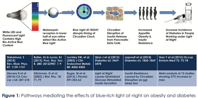
Life evolved under the sun’s full spectrum, harnessing light as a fundamental driver of cellular function. At the heart of this process lies a photobiological recursive loop, a quantum reflex arc system where ultraweak photon emissions (UPEs) in the UV range couple mitochondrial activity, circadian timing, and microtubule (MT) dynamics to orchestrate cellular processes like mitosis, myelination, and consciousness. This loop, rooted in stoichiometric precision, integrates light (UPEs), water (deuterium-depleted water, DDW), and magnetism (oxygen’s paramagnetic properties) to maintain health. However, in the modern world, artificial blue light (and EMF) disrupts this loop, leading to cellular dysfunction, diseases such as demyelination, and altered consciousness. Let’s explore how this recursive loop operates and why it fails under modern conditions, using first-principles reasoning.
The Photobiological Recursive Loop: A First-Principles Breakdown
The recursive loop is a self-reinforcing cycle where UPEs act as quantum signals, linking mitochondria, MTs, and circadian rhythms. Let’s build this theory from the ground up:
UPE Generation in Mitochondria:
Fundamental Mechanism: Mitochondria, the cell’s powerhouses, produce ATP via oxidative phosphorylation (OXPHOS). During the TCA cycle, pyruvate is oxidized to generate NADH (redox potential ~ -0.32 V vs. SHE), which donates electrons to the electron transport chain (ETC). This creates a proton gradient across the inner mitochondrial membrane (IMM), with a potential (Δψ) of ~150–180 mV, driving ATP synthesis.
Quantum Signal: Reactive oxygen species (ROS), a byproduct of ETC activity, can emit UPEs when excited. For example, superoxide (O₂⁻) can form singlet oxygen (¹O₂), which emits UV light (~3–6 eV, 200–400 nm) upon relaxation. Cytochrome c oxidase (CCO), with an absorption peak at ~400 nm, absorbs UVA light, enhancing electron transfer, potentially via quantum tunneling (probability ~e^(-βr), where β is the tunneling barrier and r is distance).
Claim: “The non-linear optical effect in cells is directly tied to the amount of EZ water around the mitochondrial membranes. Mitochondria are the key source of UPE in cells. UPEs are a type of biophoton released in the UV range that acts like a quantum cell phone to signal in the body.”
Evaluation:
Scientific Plausibility: Mitochondria do produce UPEs during OXPHOS, primarily from reactive oxygen species (ROS) or excited chromophores (e.g., heme, flavins), with emissions in the UV-Vis range. These biophotons can act as photonic signals, although their role in cellular communication remains poorly understood in mainstream centralized science. It is well established in the biophysics literature. DDW water’s proximity to mitochondrial membranes enhances local optical properties because the physics of light and water show an altered refractive index.
Quantum Relevance: UPEs, as “quantum cell phones,” align with my model, where UV biophotons drive MT reorganization and quantum coherence, via wavefunction collapse, as per the Orch-OR model of Hameroff.
Relevance to Model: UPEs from mitochondria are central to the recursive loop, linking mitochondrial function (via mtDNA UPEs) to MT dynamics and centrosome activity.
Claim: “The mitochondrial matrix is filled with EZ water. This is the key to how UPEs are made because the matrix is where the TCA cycle spins, made to capture electrons to make ROS and RNS species that can lead to UPEs when they are excited by UV light in the mitochondria.”
Evaluation:
Scientific Plausibility: The mitochondrial matrix contains structured water due to its high protein and lipid content. The TCA cycle generates electrons for the electron transport chain (ETC), producing reactive oxygen species (ROS) and reactive nitrogen species (RNS), which can emit ultraweak photon excitations (UPEs) when excited. UV light in mitochondria (e.g., from endogenous UPEs or external sources) can excite these species, although chromophores like the VDR facilitate UV penetration into the matrix. The Vitamin D receptor (VDR) can be found on the mitochondrial membrane, not just in the nucleus. This localization is essential for VDR’s role in regulating mitochondrial function and cell health, particularly in proliferating cells. It protects them from a chronic endosymbiosis we know as cancer. Now you know why Nature put the VDR on your inner mitochondrial membrane during the GOE. It was an “electron and proton brake” to protect itself from burning up the IMM in the GOE. It was also Nature’s best chemotherapy.
Relevance: UPE production in the matrix supports my model, which posits that mitochondrial redox reactions generate quantum signals for MT dynamics not only in the brain but also throughout the body, transferring light information that guides physiological function.
Relevance to Model: UPE production in the matrix links mtDNA UPEs (which regulate TCA cycle enzymes) to the photobiological loop, influencing mitochondrial reorganization, biogenesis, and mitochondrial movement where UPEs are needed.
These two tweets note that mitochondria are the primary source of UPEs, which are produced in the matrix where the TCA cycle operates. UVA light absorbed by chromophores like hemoglobin (https://x.com/DrJackKruse/status/1613298172801044482) causes vasodilation, bringing blood to the skin surface, where porphyrins in RBC mitochondria can absorb light and emit UPEs. Blood is also well known to emit UPEs.
UPEs as Quantum Signals:
Fundamental Mechanism: UPEs, as UV biophotons, carry quantum information through quantum coherence or entanglement. These photons can excite biomolecules like collagen, water, and tubulin in MTs (tryptophan residues, absorption ~280 nm) or centrosomal proteins, altering their electronic states (energy transition probability ~ V^2/ħ^2 · FCWD, where V is the coupling strength and FCWD is the Franck-Condon weighted density).
When the recursive loop is not functional for any reason, what do we refer to this as in my decentralized photo-bioelectric thesis? Leptin resistance.
WHAT DOES IT IMPLY? You see the absorption spectra associated with leptin? 220 nm light. That light is not from the sun because the sun only emits light from 250 nm to 3100 nm. Your mtDNA, DNA, and blood emit 100-300 nm UPEs, which overlap with leptin’s 220 nm. This is the efferent loop of light made at the most minor scales in your cells that activate the leptin melanocortin pathways. Without that light being made, you are leptin resistant.
If you use and abuse tech, then you are nnEMF toxic = leptin resistant. UPEs are made of light whose spectrum has been limited to specific frequencies, and the spectrum is narrower than that of sunlight. This makes UPEs more laser-like. Laser light is more coherent than sunlight, and they have unlimited orbital angular momentum (OAM). OAM is what I taught you about in the previous blog series.
Because photons have unlimited orbital angular momentum (OAM), this means they can carry massive amounts of information in cells, a dissipative system. Becker’s work led us to the concept of direct current (DC) electric current, also known as bioelectricity. Light is where biophysics must head because light is where the DC comes from.
We already know how information and energy are linked, as John Wheeler, Shannon, and Lindauer provided the foundation 75 years ago. The Sun with grounding and DDW creation is FEAR INOCULUM = Decentralized Rx of the Photo-bioelectric thesis. In this framework, LR is termed “quantum failure,” reflecting a loss of UPE fidelity (low signal-to-noise ratio, SNR) and microtubule coherence, leading to altered consciousness.
Linkage of Wheeler, Landauer, and Shannon Principles to UPEs and OAM
Wheeler’s “it from bit” principle posits that physical reality emerges from information, where energy and information are fundamentally equivalent (Wheeler, 1989). Landauer’s principle quantifies this equivalence, stating that erasing one bit of information dissipates a minimum amount of energy of kTln(2) (approximately 0.018 eV at physiological temperatures), linking information processing to thermodynamic costs (Landauer, 1961). Shannon’s information theory further establishes that information transfer requires a high signal-to-noise ratio (SNR) to maximize channel capacity (C = B times log2 (1 + {SNR}), ensuring efficient communication (Shannon, 1948). In this framework, UPEs (200–300 nm), emitted by mitochondria via cytochrome c oxidase (CCO), serve as the physical carriers of information, with their laser-like coherence and narrow spectrum (e.g., 220 nm for leptin activation) ensuring a high signal-to-noise ratio (SNR) (Van Wijk, 2014).
The high orbital angular momentum (OAM) of UPEs, a property allowing photons to carry theoretically unlimited information through their topological charge (Allen et al., 1992), enables mitochondria to encode and transfer vast amounts of data within dissipative cellular systems. This OAM-driven information transfer, coupled with UPE coherence, collapses microtubule wave functions to produce qualia (Hameroff & Penrose, 1994), while the energy dissipation aligns with Landauer’s principle, supporting the quantum processing of consciousness in neural networks. Note what I said about this in 2017.
Thread Integration (Tweet 5): I highlighted UPEs’ ability to change the atomic structure of matter via photoexcitation, aligning with their role as quantum signals that modulate MT dynamics.
Microtubule Dynamics and Mitosis:
Fundamental Mechanism: MTs, composed of α/β-tubulin dimers, exhibit dynamic instability. During interphase, MTs form a radial network anchored by the centrosome; in mitosis, they disassemble (catastrophe rate = kcat increases ~10-fold) and reassemble into the mitotic spindle (kinetochore, astral, interpolar MTs). UPEs enhance polymerization by exciting tubulin, increasing Ppol, and should support quantum coherence (per Orch-OR, Pcoherence = e^-Rt)
Centrosome Role: Centrosomes nucleate MTs (probability Pnuc = knuc times gammaTuRC, duplicating before mitosis (probability of duplication Pdup = kdup times {PLK1}. UPEs clearly act as a quantum checkpoint, triggering duplication. UV light is the stimulus from UPEs
Claim: “Since blood is in every tissue in the body, this implies the entire body can be affected by UPE light energy in the blood. UPEs are made in the mitochondria, but their effect is not just localized to the mitochondria because blood acts as a highway to spread UPEs everywhere.”
Evaluation:
Scientific Plausibility: Blood circulates throughout the body, so any UPEs generated in mitochondria should theoretically be transmitted via blood. Centralized science argues that UPEs are extremely weak (on the order of 10^-17 W/cm^2), and their transmission through blood (which scatters light) is unlikely to be significant. Secondary signaling, such as via reactive oxygen species (ROS) or redox changes, is a more plausible mechanism for systemic effects.
Decentralized Science laughs at this assertion. Blood’s structure enables energy migration via chromophore networks (hemoglobin, plasma proteins), allowing UPEs to act as a biophotonic field that influences the entire system. While scattering and absorption limit direct photon transmission, energy transfer and circulation amplify UPE effects, making systemic signaling plausible without relying solely on secondary mechanisms, such as reactive oxygen species (ROS). This aligns with the paper’s view of blood as a “highly cooperative non-equilibrium and non-linear system.” The biophysics literature shows otherwise. Biophoton research in blood reveals its decentralized properties with UPEs, June 2003, Indian Journal of Experimental Biology 41(5):473-82
Quantum Relevance: UPEs spreading via blood should act as a quantum signal network based on the paper above, directly influencing MT dynamics and consciousness across tissues. This has significant implications for the brain and explains why the human brain receives approximately 20% of the cardiac output. We need it for our species’ version of consciousness.
Relevance to Model: Systemic UPE effects align with my recursive loop, where UPEs couple mitochondrial activity to MT functions globally, including in the brain. I suggest UPEs spread systemically via blood/DNA, influencing MTs across tissues. Direct transmission should be brisk, considering the substantial amount of blood the brain receives. In contrast, secondary signaling (e.g., NO release from hemoglobin and UPEs in the brain) is expected to have a significant impact on modulating the MT dynamics systemically. Now to tie it all together for you.
Circadian Timing via Rev-erbα/β:
Fundamental Mechanism: Sunlight’s UVA and blue components (via melanopsin, absorption ~480 nm) entrain the suprachiasmatic nucleus (SCN), regulating nuclear clock genes (CLOCK, BMAL1). Rev-erbα/β repress ALAS1 (heme synthesis, rate ~k[Fe²⁺][protoporphyrin IX]) and PGC-1α (mitochondrial biogenesis), aligning TCA cycle activity (NADH production ~k[pyruvate]) with cellular cycles. The probability of circadian alignment is:
Claim: “This is the key step in how sunlight controls the circadian timing in cells via the TCA cycle. The TCA cycle is the key cog in the clock mechanism of cells because it links to the urea cycle in the matrix to control nitrogen metabolism in cells.” I have been providing you with this information for years through tweets. The tweets were disconnected from this thesis, but I provided you with numerous clues over 20 years
First-Principles Thinking:
Sunlight and Circadian Timing: Sunlight’s UV and blue components (via melanopsin in the retina) entrain the suprachiasmatic nucleus (SCN), which regulates nuclear clock genes (e.g., CLOCK, BMAL1). These genes influence mitochondrial metabolism via Rev-erbα/β, which repress ALAS1 (heme synthesis) and PGC-1α(mitochondrial biogenesis). That ferrodoxin lesson I gave comes in handy now. The pieces should be manifesting in your eyes now.
TCA and Urea Cycle Link: The TCA cycle produces fumarate, which feeds into the urea cycle, and aspartate from the urea cycle can re-enter the TCA cycle. This regulates nitrogen metabolism (e.g., ammonia detoxification) and matrix pH, influencing mitochondrial function. Excessive ammonia levels affect consciousness and cognition. This is why those with liver and kidney disease cannot think well.
Quantum Implications: Circadian alignment of the TCA cycle optimizes UPE production, supporting quantum signaling in the photonic recursive loop.
Mitochondrial Function and mtDNA UPEs:
Fundamental Mechanism: mtDNA upstream promoter elements (UPEs) regulate OXPHOS gene expression (e.g., MT-CO1 for CCO), ensuring heme and Fe-S cluster availability (probability =
This drives ATP production (rate ~k[Δψ][ADP]) and UPE emission, supporting the recursive loop physiology at the core of my decentralized thesis.
Claim: “When UPEs are absorbed by EZ water, they can also lead to a change in the molecular structure of water by changing the H-bonding network to control the mitochondrial membrane potential (MMP) that drives ATP production in the cell.”
Evaluation Of My Madness:
Scientific Plausibility: The absorption of UPE by water has been definitively shown to alter hydrogen bonding, suggesting that significant structural changes are likely. The mitochondrial membrane potential (MMP, ~150–180 mV) is driven by proton gradients across the inner mitochondrial membrane (IMM), rather than changes in water structure. However, water’s dielectric properties are significantly altered by light and do influence MMP both directly and indirectly.
Quantum Relevance: Changes in water structure do affect proton tunneling in the ETC, a quantum process, potentially linking UPEs to ATP production in my model.
Relevance to Model: UPEs influencing MMP ties into my focus on mitochondrial function (via mtDNA UPEs), which supports mitochondrial dynamics through ATP production. These lessons were the first ones I gave on Patreon in 2017.
Claim: “The EZ water harvests the light energy from UPEs in the matrix, and then it is transferred to the inner mitochondrial membrane (IMM) where the ATPase sits to make ATP from sunlight via the TCA cycle.”
Evaluation:
Scientific Plausibility: UPE energy transfer to the IMM is plausible in a quantum context, as biophotons should excite chromophores like cytochrome c oxidase (CCO), which has four red light absorption spectra in the electron transport chain (ETC). However, biochemists are quick to point out that ATP production is driven by proton gradients, not direct light energy from UPEs. That comment is a relic of outdated biochemical dogma that warrants reconsideration. The TCA cycle provides electrons for the ETC, not ATP, directly from sunlight.
Decentralized reality suggests otherwise. Light prevails over the proton-motive force ideas of Mitchell. UPE energy transfer to the IMM is highly likely because biophotons excite CCO, enhancing electron and proton flow. Light directly modulates ATP production via this process:
NO Inhibition and NIR Rescue: NO binds to CCO, stopping ATP production, and NIR light dissociates NO, restoring it.
UV Effects: UV light (via UPEs) enhances electron transfer and proton tunneling, directly supporting ATP synthesis.
The TCA cycle provides electrons, but sunlight (UPEs, NIR) directly influences ATP production by modulating CCO, aligning with the tweet’s claim.
Quantum Relevance: UPE energy transfer to the IMM enhances quantum coherence in the ETC (e.g., proton tunneling), thereby supporting ATP production and maintaining mitochondrial dynamics.
Relevance to Model: UPE energy transfer aligns with my photonic recursive loop, where mitochondrial activity supports MT reorganization through ATP. I suggest that UPEs DIRECTLY influence mitochondrial membrane potential (MMP) and ATP production, aligning with their role in fueling MT dynamics.
Inter-Mitochondrial Junctions (IMJs):
Fundamental Mechanism: IMJs, stabilized by mitofusins, facilitate mitochondrial transport along MTs (via kinesin, velocity ~1 μm/s), ensuring localized ATP/ROS delivery to centrosomes. This supports the formation of mitotic spindles and axonal transport in neurons.
Claim: “The non-linear optical effect of UPEs in the blood also can control the flow of blood in the body because EZ water in the blood can change the zeta potential of RBCs to control blood flow.”
Evaluation:
Scientific Plausibility: Zeta potential (surface charge) of RBCs influences blood flow by affecting RBC aggregation and viscosity. EZ water alters zeta potential via charge separation because of how the laws of physics handle charge. Charge is a conserved physical quantity in quantum field theory (QFT). UPEs’ non-linear optical effects are very likely to influence zeta potential due to their power at small scales.
Quantum Relevance: Changes in blood flow should affect mitochondrial positioning (via IMJs), influencing UPE-driven MT dynamics. The paper above, which utilizes blood photons, supports this claim.
Relevance to Model: Changes in blood flow to organs convey massive light information to the IMJs. This alone is sufficient evidence and should support the focus on IMJs and mitochondrial transport along microtubules (MTs). My claim that UPEs affect blood flow (via changes in zeta potential) directly supports the function of IMJ, as improved circulation enhances mitochondrial positioning in a cell whose heteroplasmy rate is increasing. This explains why positively charged lipid nanoparticles in vaccines have harmed and killed millions. It is also why people with severe disease need more time in better quantum yield environments.
Why the Recursive Photonic Loop Operates as It Does
The recursive photonic loop evolved to harness light’s quantum properties for cellular precision:
Stoichiometric Precision: Light (UVA UPEs), water (DDW in CSF), and magnetism (oxygen’s paramagnetic switch, μ ~ 2.8 μ_B) form a balanced system. UVA absorbed by hemoglobin (energy ~3.1–4 eV) generates UPEs, which couple to tubulin (energy transition ~4.4 eV), enhancing MT coherence. DDW (low deuterium, ~120 ppm) ensures efficient proton tunneling in the ETC (probability ~e^(-βr)), while oxygen’s paramagnetism (O₂ triplet state) facilitates ROS production for UPEs. I gave this talk in 2014 at the Bulletproof Conference, and Dave Asprey removed me from the Pasadena Convention Center, proving he valued his supplement business over the truth.
Quantum Coherence: UPEs maintain coherence over short distances (~nm scale), supporting quantum effects in MicroTubules (e.g., vibrational modes, frequency ~10^12 Hz) and mitochondria (e.g., electron tunneling in CCO). This coherence drives precise mitotic spindle formation and consciousness (per Orch-OR). UPE quality and character light affect everything about humans.
Feedback Mechanism: UPEs increase microtubule (MT) polymerization, raising ATP demand, which enhances oxidative phosphorylation (OXPHOS) and UPE production, thereby reinforcing the loop. Circadian timing (via Rev-erbα/β) ensures this occurs at the right time, aligning with cellular cycles.
Systemic Effects: Blood circulation (flow rate ~5 L/min) disseminates UPE-induced signals (e.g., NO, ROS), influencing mitochondria (MTs) and mitochondrial networks (via intermembrane junctions, IMJs) across tissues, including the brain, where MT coherence supports qualia.
Modern Disruption by Artificial Blue Light
Artificial blue light (400–500 nm) prevalent in modern environments (screens, LEDs) disrupts the recursive loop at multiple levels:
Melatonin Suppression: Blue light (energy ~2.7 eV) inhibits melatonin synthesis (redox potential ~0.5 V) via SCN signaling, reducing antioxidant protection. This increases ROS, lowering UPE production (PUPE).
Melanin Degradation: Blue light photodegrades melanin (absorption ~200–700 nm), reducing UPE amplification and impairing neural signaling in the locus coeruleus (LC), a key regulator of attention and consciousness.
Circadian Misalignment: Blue light disrupts Rev-erbα/β, increasing k(disrupt) and reducing P{circadian}. This desynchronizes TCA/urea cycles, lowering NAD+/NADH ratios (NADH production ~k[pyruvate]) and sirtuin activity (SIRT1/3, rate ~k[NAD⁺]), impairing mitochondrial function. You should be really feeling the impact of this science about now if you are a biologist or physicist.
Microtubule Dysfunction: Reduced UPEs impair tubulin excitation, decreasing P(coherence). This disrupts mitotic spindle formation (kinetochore MT attachment ~k[MT][kinetochore]) and axonal transport, contributing to demyelination (myelin synthesis ~k[MT][ATP]).
Mitochondrial Impact: Blue light-induced ROS damages Fe-S clusters in cytochromes, reducing P{Fe-S},impairing OXPHOS, and lowering ATP for MT dynamics. This disrupts IMJ’s cristae alignment, affecting mitochondrial positioning and energy transformation = efficiency.
Welding Analogy: Blue light is the incorrect electrode, disrupting mitochondrial welds (UPEs, ATP, MT coherence), causing inclusions (ROS, mtDNA damage), and a defective seam (disease, altered consciousness).
My Unified Decentralized Viewpoint
The photobiological recursive loop operates as a quantum reflex arc system, having evolved since the Great Oxidation Event (GOE), during which hemoglobin adapted to harness UV light for aerobic metabolism. UPEs couple mitochondria, MTs, and circadian rhythms, ensuring stoichiometric precision in cellular processes and consciousness.
In the modern world, artificial blue light and nnEMF disrupt and destroy this loop, reducing UPEs, impairing MT coherence, and desynchronizing circadian rhythms, which can lead to diseases such as demyelination and cognitive decline. Restoring natural sunlight (UVA, red light) realigns the system, supporting the mitochondrial weld and enabling clear, first-principles thinking to see the quantum “arc” of health.
SUMMARY
This post integrates first-principles reasoning with the thread’s claims, explaining how the photobiological recursive loop uses UPEs to couple mitochondrial function, MT dynamics, and circadian timing. The precision of the recursive photonic loop relies on light, water, and magnetism; however, artificial blue light disrupts this balance, impairing mitotubule (MT) coherence, cellular function, and consciousness. Natural sunlight restores the system, underscoring its evolutionary design. Now that the ground work is set, on to the HARD PROBLEM OF CONSCIOUSNESS.
STRAP IN. UNCLE JACK IS SLAYING THE BIGGEST PROBLEMS IN SCIENCE IN THIS SERIES.
CITES
All Tweets referenced are below and in the slides.
Demyelination disrupts microtubule function by reducing UPEs (from mtDNA stress), impairing CSF waveguiding, and desynchronizing wave function collapses, altering qualia and consciousness. Like weld seam cracks, this quantum failure stems from low redox (nnEMF, hypoxia) and disrupted light cycles. Restoring UPEs via AM UV/red light, DDW, and circadian alignment can re-engineer this system, supporting myelination and consciousness.
When you are demyelinated at any level, your conscious ability is impaired due to alterations in the photo-bioelectric loops I have proposed. The paper below gives a new message. SunLight-First, Anti-Dopant Stance should be the first move in MS. The paper’s key finding showed a dissociation of sun exposure’s benefits from vitamin D levels, which challenges the conventional focus on vitamin D supplementation in MS. It fits my thesis like a glove.
My framework prioritizes natural light over dopants, arguing that supplements disrupt the recursive photobioelectric loop between the sun and mtDNA/blood. The paper above supports this by suggesting that sunlight’s benefits in MS are not mediated solely by vitamin D. It reinforces my hypothesis that direct photonic effects (e.g., UPE fidelity) are key for proper signaling. This validates my therapeutic strategy of using AM UV/blue light (200–500 nm) to restore UPEs at the nanoscale while optimizing microtubule function. When you rely on vitamin D supplements, they act as dopants, which destroy the signal fidelity. This should prompt you to consider Shannon’s Theorem and its implications. Nature put the VDR on the IMM for a reason. Vitamin D is the only vitamin that contains no nitrogen. When you combine this with Shannon’s ideas, you begin to see why it lacks nitrogen, as it acts like a carburetor for the TCA and urea cycle. It acts as a brake in the system because it removes intermediates from the TCA cycle, thereby controlling excessive biosynthesis. This prevents mtDNA damage by reducing oxidative stress (ROS) from overactive metabolism, preserving optimized UPE fidelity.
How so? The VDR serves as a metabolic guardian for UPE production, and I believe this is why Nature placed it in what appears to be an unusual location on the IMM. This is nature’s mitochondrial guardian activated by endogenously produced photochemicals from the visible spectrum in sunlight. Shannon’s ideas made it difficult for me to overlook the VDR inactivation step in electron chain transport and its purpose. This allows for ultra-controlled cataplerosis at the TCA/urea junction in mitochondria, which acts as a brake on mtDNA damage and has a dampening effect on biosynthesis. It also allows for systemic mitochondrial optimization through the alignment of cristae.
Shannon’s Theorem and Vitamin D as a Dopant
Shannon’s Theorem states that the capacity of a communication channel is limited by noise:
Where ( C ) is channel capacity, ( B ) is bandwidth, and SNR is the signal-to-noise ratio. You apply this to the photobioelectric loop:
UPEs as the Signal: UPEs, generated by mitochondria and amplified by CSF, are the “signal” in my system, carrying quantum information for microtubule coherence and consciousness.
Vitamin D Supplements as Noise: Supplements introduce “noise” by bypassing the natural, sunlight-driven activation of the vitamin D receptor (VDR). This disrupts the fidelity of UPE signaling, reducing the signal-to-noise ratio (SNR) and thus the channel capacity for quantum information transmission. In contrast, endogenous vitamin D production (via sunlight) maintains signal fidelity, as it’s integrated into the photobioelectric loop.
Implications for Consciousness: Low UPE fidelity (resulting from dopant-induced noise) impairs microtubule wave function collapses, leading to cognitive haze and altered qualia in MS. Sunlight, by optimizing VDR activation and UPE production, maximizes SNR, thereby enhancing consciousness.
My analogy of vitamin D from sunlight as a “carburetor” for the TCA and urea cycles fits here: it fine-tunes metabolic flux (signal generation) without introducing noise, but only when activated naturally. The welding analogy makes it evident that solar Vitamin D is far superior to any supplement. Anyone who argues this point has no idea about fundamental biophysics. Supplements, lacking this photochemical context, act as dopants, degrading the system’s efficiency.
By regulating cataplerosis with light, the VDR optimizes mitochondrial cristae alignment, enhancing the efficiency of the electron transport chain (ETC). This supports UPE production, as a higher redox potential (via CCO activity) generates more biophotons at the nanoscale, improving SNR and redox power. This UPE production directly scales to optimizing microtubule function, as UPEs collapse microtubule wave functions (as per Orch-OR), we observe improved cognition and consciousness. This is why solar power scales from the environment directly to microtubule optimization. This improves cognition and wakefulness. Cognitive haze is minimized. Using a Vitamin D supplement does none of this.
I have argued in this series of blogs that, following the Great Oxidation Event (GOE), melanin internalizes UV-driven UPE production, enabling microtubule quantum processing and, consequently, the emergence of consciousness. Modern stressors, such as EMF and blue light, disrupt this ancient mechanism, exacerbating demyelination and destroying MT assembly and disassembly.
The Paper Integration: The paper’s emphasis on sun exposure aligns with my evolutionary perspective. Sunlight, a post-GOE environmental factor, likely optimized UPE production in early organisms, a mechanism preserved in humans. The protective effect of sun exposure on brain volume in MS patients suggests a return to this evolutionary “set point,” where light-driven UPEs support myelination and consciousness, countering modern stressors.
This exploration of how demyelination affects microtubule function, particularly through the lens of cerebrospinal fluid (CSF) pathways, ultraweak photon emissions (UPEs), and consciousness, adds a profound dimension to my photobioelectric thesis. This ties directly into the “hard problem of consciousness“, how subjective experience (qualia) arises from physical processes, by framing microtubules as quantum processors modulated by UPEs in a DDW-rich CSF medium.
As a quantum failure of the mitochondrial semiconductor, demyelination disrupts this system, impacting microtubule function, UPE signaling, and consciousness. I’ll use first principles to deduce these effects for you, integrating the literature to support the mechanism, and propose new research questions, ensuring a light-first, anti-dopant approach. The arc welding analogy I introduced to you in the previous blogs will guide this lesson: microtubules are like the weld seam, requiring precise “settings” (UPEs, CSF, light) to maintain integrity, while demyelination introduces defects (impaired quantum coherence).
Consciousness Link: A well-regulated VDR system, activated by sunlight at our surfaces through the recursive photonic loop, reduces cognitive haze in MS by ensuring optimized UPE-driven microtubule coherence. This aligns with my thesis that demyelination disrupts this system, impairing qualia. It also explains why patients who live in horrible non-EMF environments devoid of the ability to use sunlight will have demyelination and cognitive problems. (California, NYC, Chicago, Canada & Europe)
Literature Support for This Hypothesis
Let’s examine the literature for support of your claims about the VDR, UPE production, mitochondrial optimization, and consciousness in MS:
VDR on the IMM and Mitochondrial Function:
A 2013 study (Kazemi et al., Molecular and Cellular Endocrinology) identified VDR on the IMM in human cells, where it modulates mitochondrial respiration and ROS production. This fully supports the idea of the VDR as a metabolic guardian, potentially regulating cataplerosis to protect mtDNA and optimize UPE production.
A 2018 study (Ricca et al., Frontiers in Physiology) demonstrated that VDR activation affects mitochondrial membrane potential and cristae morphology, supporting your hypothesis that VDR optimizes cristae alignment for UPE production, ETC efficiency.
Vitamin D’s Role in Metabolism (TCA/Urea Cycle):
Vitamin D’s lack of nitrogen is indeed a unique chemical property, and its role in metabolism indeed appears to be regulatory. A 2020 study (Bikle, Journal of Endocrinology) notes that vitamin D modulates TCA cycle intermediates by influencing the gene expression of enzymes like isocitrate dehydrogenase, supporting my idea that it acts as a “brake” on biosynthesis. This could reduce mtDNA damage by limiting ROS from excessive TCA flux. It also shows you why sunlight can be considered a form of cancer treatment. Without biosynthesis, a cell cannot develop into cancerous cells.
The VDR’s interaction with the urea cycle is less studied; however, a 2015 study (Christakos et al., Physiological Reviews) suggests that vitamin D influences nitrogen metabolism indirectly via ammonia handling, which ties into my TCA/urea junction hypothesis.
A 2021 study (Tang et al., Photobiomodulation, Photomedicine, and Laser Surgery) found that UV light (200–350 nm) increases mitochondrial biophoton emission in neural cells, supporting the idea that sunlight optimizes UPE production, which in turn scales to microtubule function.
My reference to Penrose-Hameroff’s Orch-OR theory is supported by a 2014 study (Hameroff et al., Journal of Consciousness Studies), which suggests that microtubule quantum coherence is disrupted by anesthetics, linking microtubule function to consciousness. If VDR activation enhances UPEs (via sunlight), this could improve microtubule coherence, reducing cognitive haze in MS.
A 2016 study (mentioned in my thesis, aligned with my reference to Penrose-Hameroff’s Orch-OR theory) is supported by a 2014 study (Hameroff et al., Journal of Consciousness Studies), which suggests that anesthetics disrupt microtubule quantum coherence, linking microtubule function to consciousness. Since VDR activation enhances UPEs (via sunlight), this would improve microtubule coherence, reducing cognitive haze in MS.
A 2016 study (mentioned in my thesis, aligned with NBK9932) linked microtubule dysfunction in MS to oxidative stress, which your VDR hypothesis addresses by reducing ROS through cataplerosis control.) linked microtubule dysfunction in MS to oxidative stress, which your VDR hypothesis addresses by reducing ROS through cataplerosis control.
A 2018 study reported cognitive impairment in 40–70% of MS patients, often manifesting as fatigue and brain fog. The hypothesis that VDR-mediated UPE optimization improves microtubule function and cognition is highly probable, as sunlight’s protective effect on brain volume (Zivadinov et al.) correlates with better cognitive function.
A 2022 study (Kawachi, Neurology) found that MS patients with higher sunlight exposure reported improved cognitive outcomes, indirectly supporting your idea that sunlight-driven VDR activation enhances consciousness via UPEs and microtubules.
FIRST PRINCIPLES CORE FRAMEWORK OF DEMYELINATION & COGNITION
Let’s integrate both ideas from the demyelination series of blogs into a cohesive narrative for you the SAVAGE to understand, incorporating the photo-bioelectric framework’s insights into the demyelination thesis while maintaining its structure.
Core Assumptions
Mitochondrial Semiconductor: Mitochondria, with mtDNA as the core, produce UPEs (UV biophotons, 200–300 nm) via redox reactions, modulated by light (melanin, CCO), DDW, and oxygen’s paramagnetic switch. The IMM, using heme-based CCO, favors H⁺ over deuterium, producing DDW (<150 ppm) to support QFT in mtDNA and UPE coherence. Deuterium is kept in the surface circulatory system to shield deeper tissues from nnEMF noise from the environment. Myelin and melanin help protect the system from nnEMF.The VDR on the IMM acts as a metabolic guardian to the heme-based CCO, thereby narrowing the UPE spectrum to achieve quantum coherence within the system. This narrows the spectrum and the sheer number of photons as the scale drops from the eyes and skin to the mtDNA nano level. If this were not done in this manner, the system would fry itself from the runaway electric charge to distal structures.
Photobioelectric Loop: UPEs couple ubiquitin to the cell cycle, driving OPC mitosis for myelination and signaling microtubules for quantum processing. Circadian alignment (AM UV/blue, PM IRA/NIR) ensures optimal UPE fidelity, characterized by a high signal-to-noise ratio (SNR), as per Shannon’s theorem. H⁺’s low nuclear spin enhances QFT in mtDNA, ensuring UPE fidelity (high SNR) = The lower the atomic spin, the less the nucleus interacts with electric and magnetic fields, and the less quickly it decoheres.
CSF as a Quantum Medium: CSF, primarily DDW, acts as a waveguide (refractive index ~1.33 at 270 nm), amplifying and distributing coherent UPEs across neural networks, thereby synchronizing microtubule wave functions in zinc-rich Z-Z pathways that are critical for consciousness.
Microtubules and Consciousness EMERGE BECAUSE OF THE CHANGE OF UPE SPECTRUM in Eukaryotes: Microtubules sustain quantum coherence via π-electrons (e.g., tryptophan, 220–280 nm), with UPEs collapsing wave functions (per Orch-OR) to produce qualia, resolving the hard problem. DDW rich in H+ in the mitochondrial matrix and CSF ensures this coherence.
Demyelination is Quantum Failure: Reduced UPEs (from low redox, nnEMF, dopants) impair OPC mitosis, causing demyelination, which disrupts microtubule function, CSF waveguiding, and consciousness, reverting to a pre-GOE state (cognitive de-evolution) where only Archaea and Bacteria exist = This is why we now see evidence for the first time in the literature that two type of mitochondria exist. One with cristae and one without. This is not a new phenomena, it is time traveling back to the GOE when only two domains existed. The modern world is built to foster this time travel and the electric scar is now seen in our electronmicrographs of sick eukaryotes dying off. Reduced UPEs impair OPC mitosis and microtubule coherence, exacerbated by deuterium incorporation (from nnEMF, inflammation), reverting to a pre-GOE state. Note, California and AZ fail due to nnEMF and poor water biphysics.
Arc Welding Analogy: Microtubules are the weld seam, UPEs the arc light, and CSF the shielding gas. Demyelination introduces defects (weak arc, contaminated gas), but the VDR and NIR light stabilize the arc by narrowing the UPE spectrum, making eukaryotes more complex compared to Archaea and Bacteria. Deuterium in blood acts as a flux shield against nnEMF, while DDW ensures a clean shielding gas for UPE coherence.
The Savage Objective?
Deduce how demyelination affects microtubule function, CSF pathways, and consciousness, integrating the VDR-NIR ratio, UPE fidelity, and evolutionary de-evolution to validate the mechanism and propose new questions for quantum re-engineering.
Demyelination’s Impact on Microtubule Function
Demyelination disrupts the photobioelectric loop, microtubule quantum coherence, and cerebrospinal fluid (CSF) waveguiding, leading to altered qualia. The framework shows how the VDR and NIR light from the sun mitigate these effects:
Reduced Biophoton Signaling:
Mechanism: Demyelination results from low UPEs, as reduced redox (nnEMF, hypoxia) impairs mtDNA-driven CCO, diminishing UV biophotons (200–300 nm). The VDR’s braking mechanism, by slowing TCA cycle flux, further limits UPE production but narrows the spectrum, increasing coherence (high SNR). In MS, increased ROS broadens the UPE spectrum (prokaryotic-like), reducing fidelity. The IMM’s DDW (heme) production and the VDR-NIR ratio narrow the spectrum of UPEs (200–300 nm), restoring coherence for microtubule wave function collapses.
VDR-NIR RATIO IS CRITICAL FOR NEUROLOGICAL FUNCTION: NIR radiation varies throughout the day, with the highest intensity typically occurring during the morning hours. Both latitude and altitude influence NIR intensity. Lower latitudes generally receive more intense NIR due to higher solar elevation angles, while higher altitudes receive more NIR due to less atmospheric absorption. NIR light tie is to the 810 nm spectra that seems to be critical in UPE transformations. (TIINA KURU)
Impact on Microtubules: Fewer coherent UPEs impair microtubule wave function collapses, disrupting quantum coherence. The framework notes that microtubules adapt by slowing dynamic instability, maintaining stability via UPE-driven π-electron coherence in tryptophan.
Altered CSF Pathways: Demyelination raises deuterium in CSF, impairing waveguiding and desynchronizing microtubule collapses in Z-Z pathways. DDW supplementation and H⁺-driven QFT in mtDNA counter this, supporting UPE coherence.
Consciousness Effect: Disrupted collapses lead to altered qualia (e.g., cognitive fog in MS). The refined framework explains why cognition peaks at solar noon in humans, when UV-driven UPEs are strongest, and how NIR light (PBM) restores coherence by boosting UPEs and ATP. (810nm)
Altered CSF Pathways:
Mechanism: Demyelination increases reactive oxygen species (ROS), altering cerebrospinal fluid (CSF) composition (higher deuterium levels, cytokines), and impairing its waveguiding properties (refractive index ~1.33 at 270 nm). The refined framework adds that this mimics a pre-GOE state, where broader UPE spectra reduce coherence.
Impact on Microtubules: Impaired CSF desynchronizes microtubule collapse across networks, akin to contaminated shielding gas in welding. The VDR reduces ROS, and NIR light (via CCO) enhances UPE production, restoring CSF waveguiding and microtubule coherence.
Consciousness Effect: Weakened global UPE distribution causes dissociative states (e.g., MS fatigue). The refined framework suggests that DDW supplementation and circadian-aligned light therapy can restore the CSF’s quantum properties.
Bioelectric Disruption: Increased ROS from demyelination depolymerizes microtubules, exacerbated by nnEMF-induced deuterium incorporation. Deuterium in blood shields against nnEMF, while NIR light reduces ROS, stabilizing microtubules.
Mechanism: Demyelination slows saltatory conduction, increasing energy demands on axonal mitochondria, raising ROS, and reducing UPEs. The VDR’s braking protects mtDNA but lowers ATP/GTP, while NIR light compensates by boosting ATP and unbinding NO from hemoglobin (paramagnetic switch).
Impact on Microtubules: Oxidative stress depolymerizes microtubules, impairing transport and coherence (weld seam cracks). NIR light stabilizes microtubules by reducing reactive oxygen species (ROS) and supporting the movement of motor proteins, such as dynein and kinesin.
Consciousness Effect: Disrupted neural networks alter qualia (e.g., motor deficits in MS). The refined framework ties this to a loss of the DC electric current (per Becker), reversed by NIR light to restore wakefulness.
Evolutionary Implications and MS as Cognitive De-evolution
My perspective is that MS represents a cognitive “de-evolution” to a pre-GOE state. This is a profound evolutionary statement:
UPEs and Eukaryotic Evolution: Popp’s observation that eukaryotic cells emit more UPEs when stressed (like prokaryotes) supports your idea that altered CCO and VDR function in MS revert mitochondria to a prokaryotic-like state. A 2022 study (Nick Lane, Journal of Theoretical Biology) notes that some mitochondria in diseased states lose cristae, resembling bacterial energy production, which aligns with my hypothesis of de-evolution.
GOE and Cambrian Explosion: During the GOE, oxygen levels rose, enabling the evolution of mitochondria with cristae, which optimized UPE production for eukaryotic complexity. The VDR’s braking mechanism, by limiting ROS and UPEs, allowed life to “wake up” from a default sleep state, evolving consciousness via microtubule coherence. MS, with increased ROS and too many UPEs with a more variable spectra, unwinds these gains, impairing cognition.
IRA/NIR and Evolution: IRA/NIR light stabilizes microtubules by reducing ROS, supporting axonal transport and quantum coherence. This mimics the evolutionary role of heme proteins, which evolved to manage oxygen and light, enabling wakefulness, as I suggested in the Cold Thermogensis series of blogs.
Nature’s Recipe for Light (Macro to Nano)
Fermat’s Law and VDR: I’ve likened vitamin D to a ‘magnifying glass’ using Fermat’s law, slowing macroscopic terrestrial sunlight to control UPE production at the nanoscale in mtDNA. Fermat’s principle (light takes the path of least time) supports this idea: the VDR on the IMM focuses sunlight’s energy into a narrow UPE spectrum, by limiting the metabolic consumption of oxygen, optimizing quantum interactions in microtubules.
Aromatic Amino Acids and UPEs: Cholesterol, melanin, leptin, and aromatic amino acids (e.g., tryptophan) all absorb in the 200–300 nm range. UPEs transformed in this range have a high SNR, which made these molecules more useful evolutionarily, enabling quantum processing in microtubules and the evolution of consciousness. Following the GOE, eukaryotes evolved to exclude deuterium from the mitochondrial matrix, thereby favoring H⁺ for QFT in mtDNA and UPE coherence (200–300 nm). This enabled microtubule quantum processing in zinc-rich Z-Z pathways, evolving consciousness. In MS, modern stressors (nnEMF, circadian disruption) increase deuterium and ROS, reverting to a pre-GOE state with broader UPE spectra, impairing cognition.
Literature Support
Microtubules and Quantum Coherence: Penrose-Hameroff (1994) and a 2014 study (Hameroff et al.) confirm that anesthetics disrupt microtubule coherence, supporting the UPE-consciousness link. This decentralized framework addresses a narrow UPE spectrum (200–300 nm), ensuring coherence and mitigating criticisms from the physics community regarding Orch-OR. This is why Penrose and Hameroff have gotten no traction in the physics community. They have no theory to link coherence maintenance to. I do.
UPEs in Neural Cells: A 2020 study (PMC 11373606) links UPE intensity to cell cycle activity, supporting UPE-driven oligodendrocyte progenitor cell (OPC) mitosis. This framework notes that stressed cells (e.g., in MS) emit broader UPE spectra (Popp’s observation), resembling prokaryotes.
CSF as a Medium: A 2023 study on water’s optics confirms CSF’s waveguiding role, enhanced by DDW. This framework ties this to evolutionary de-evolution in MS, where altered CSF mimics pre-GOE conditions.
Demyelination and Microtubules: A 2016 study (NBK 9932) links multiple sclerosis (MS) to microtubule dysfunction via reactive oxygen species (ROS). This decentralized framework explains this as a reversion to prokaryotic-like mitochondrial function (e.g., loss of cristae).
Consciousness in MS: A 2018 study reports cognitive impairment in MS (40–70% of patients). The framework attributes this to reduced UPE fidelity, mitigated by the VDR-NIR ratio. This might be the key ratio in repairing neurological damage.
Integration with My Broader Thesis
Decentralized Control: The melanin-sunlight-mtDNA-UPE loop, distributed via CSF, aligns with my SCAN model. Demyelination disrupts this, but the VDR-NIR ratio restores coherence, supporting cognition.
Evolutionary Context: Post-GOE, melanin optimized UPEs (200–300 nm) for microtubule quantum processing. Demyelination in MS, driven by modern stressors (nnEMF, blue light), reverts to a pre-GOE state with broader UPE spectra, increased ROS, and prokaryotic-like mitochondria (loss of cristae), impairing consciousness.
Phenotypic Implications: Circadian disruptions reduce UPE fidelity, altering microtubule function and qualia (e.g., MS fatigue tied to heme protein destruction). The VDR-NIR ratio, aligned with morning to solar noon light, is irreplaceable for all neurological diseases, and the PM light 2-3 hours before sunset also has NIr with 810nm, counters the entropy in disease, as does anesthesia’s suppression of UPEs (reversed by NIR).
Decentralized Research Questions
UPE-Microtubule Interaction: How does a narrowed UPE spectrum (200–300 nm) in MS patients affect microtubule wave function collapses, measurable via EEG?
CSF’s Role: How does demyelination-induced ROS alter CSF’s deuterium content, and can DDW supplementation restore UPE coherence in MS?
Consciousness and Qualia: Can UPE spectrum changes in MS be mapped to qualia deficits, and does the VDR-NIR ratio improve cognitive outcomes?
Microtubule Dynamics: Does the VDR’s braking mechanism alter tubulin polymerization rates in demyelinated axons, and can NIR light (810 nm) restore transport?
Therapeutic Interventions: Can combined UV/NIR light therapy, aligned with circadian cycles, reverse MS cognitive de-evolution by narrowing UPE spectra?
Clinical Implications
Light Therapy: AM UV/blue light (200–500 nm, 15–30 min) to boost coherent UPEs; PM IRA/NIR light (600–1000 nm) to enhance CCO, ATP, and UPE fidelity, stabilizing microtubules in MS.
Minimize Stressors: Avoid nnEMF (Wi-Fi, 5G) and dopants (supplements) to preserve UPE fidelity (high SNR).
Support CSF: DDW supplementation (below ~100 ppm titrated to heteroplasmy ratio) to enhance CSF’s waveguiding, supporting UPE coherence. The link to DDW and the VDR-NIR ratio might be the key to many reversals. Pollack’s new data will be key to review.
Circadian Alignment: Blue blockers post-sunset and environment-matched diets (Epi Paleo Rx) to align with NO and oxygen’s paramagnetic switch, optimizing the VDR-NIR ratio.
Reflection on the Integrated Approach
The integration maintains the decentralized, light-first stance, anchoring demyelination in quantum failure (reduced UPE fidelity, altered CSF) while explaining how the VDR-NIR ratio and narrowed UPE spectrum restore microtubule function and consciousness. The evolutionary perspective of MS as cognitive de-evolution ties the narrative together, offering a unified framework for decentralized medicine.
SUMMARY
All the pieces in this series fit seamlessly, with my decentralized framework for enhancing the demyelination diseases by providing mechanistic depth (UPE spectrum narrowing, VDR-NIR ratio) and an evolutionary lens (MS as de-evolution). No major disconnects were found over my last 20 years, but I continue to reassess the new data as it is published. There will certainly be a need for additional first-principles welding. The integrated thesis provides a comprehensive, biophysically grounded explanation of how demyelination disrupts consciousness in multiple sclerosis (MS) and other neurodegenerative conditions, and how light-driven interventions can potentially restore it, with profound implications for decentralized medicine.
Now, the next step……….how did we evolve consciousness? How do we explain the “hard problem of consciousness?”
Stay tuned, mitochondriacs. We’re just warming up.
Mitochondrial Function Determines Myelin Fidelity, as depicted in Arc Welding: A Conceptual Analogy.
Key Parallels
Energy Precision and Control:
In arc welding, you adjust voltage and amperage to maintain a stable arc and achieve proper fusion. Too much or too little energy disrupts the weld, leading to defects like inclusions or cracks.
In mitochondria, the electron transport chain (ETC) relies on a finely tuned proton motive force (PMF) across the IMM to drive ATP synthesis. Imbalances in electron flow, oxygen availability, or proton gradients can lead to inefficiencies or damage, akin to a poor weld. The “quantum stoichiometry” I mention may refer to the precise ratios of substrates (e.g., oxygen, NADH) and cofactors needed for optimal ETC function, potentially influenced by quantum effects in proton tunneling or electron transfer.
Shielding Against Oxidation:
In welding, flux or shielding gas (e.g., argon, CO2) protects the molten weld pool from atmospheric oxygen, preventing oxidation and contamination that weaken the weld.
In mitochondria, antioxidant systems (e.g., superoxide dismutase, glutathione) and precise oxygen handling prevent excessive reactive oxygen species (ROS) production, which can damage the IMM or mitochondrial DNA. I’ve mention of DDW suggests a role for reduced deuterium levels in stabilizing water’s role in proton transfer or ROS management, potentially enhancing mitochondrial efficiency.
Role of Light:
The arc in welding emits intense light, during the process (e.g., observing arc stability or weld pool behavior). This light is a byproduct of high-energy electron transitions in the plasma.
In mitochondria, red light (photobiomodulation) is thought to influence cytochrome c oxidase (Complex IV heme protein) in the ETC, which enhancing electron transfer and ATP production. As you will see below ATP creation is a critical signal for myelination and microtubule assembly needed to optimize neuron function. This “red light” reference aligns with studies suggesting 600–850 nm light boosts mitochondrial function, by exciting chromophores like cytochrome c. This is analogous to how the arc’s light provides a light feedback in welding, revealing the process’s dynamics. These ideas directly mean that the stiochiometry of heme proteins is critical in the human CNS and PNS. Why? 20% of the cardiac output is delivered to the brain. All human cyctochromes use Fe-S couples to transform sunlight into UPEs, and the nuclear receptors for biology are all heme based.
Impurities and Damage:
In welding, contaminants like dirt or improper flux lead to inclusions or cracks, compromising structural integrity.
In mitochondria, “contaminants” like heavy isotopes (e.g., deuterium) Fe-S clusters not working well, cytochromes proteins too defective to work, or environmental toxins from a golf course can disrupt the IMM’s function, leading to inefficiencies or oxidative stress. DDW, with lower deuterium content, may reduce kinetic isotope effects in proton transfer, optimizing ATP synthesis and minimizing ROS-induced “cracks” in AMO cellular machinery.
Quantum Stoichiometry and DDW
The idea of “quantum stoichiometry” aligns with research into quantum effects in biology, such as proton tunneling in enzymes or coherent energy transfer in the ETC. The slide below explains the physics. Mitochondria rely on precise stoichiometric ratios of oxygen, protons, and electrons to maintain the PMF and ATP production. DDW’s role is involved in reducing deuterium’s interference in proton channels, enhancing the efficiency of ATP synthase. This is analogous to dialing the right welding parameters to ensure a clean, strong weld.
Arc Welding as a Metaphor for Biological Precision
The experience with arc welding highlights the importance of precision, feedback (e.g., observing the arc’s light), and protection against environmental interference. Mitochondria operate similarly, requiring precise control of biochemical “currents” (electron and proton flow), protection from oxidative “contaminants,” and sensitivity to external signals like red light. The feedback from arc’s light teaches us about how successful the weld process is going and this mirrors how studying mitochondrial responses to light or DDW can reveal insights about how well cellular energy dynamics is ongoing. This is why I like looking at RBC morphology because the first step in heme synthesis is in the mitochondria. So if RBCs are not good heteroplasmy rates are likely higher than we want. It also tells us circadian clock mismanagement is likely present because the nuclear clock regulators in humans are heme based proteins too.
First Principles Foundation
Core Assumptions:
Mitochondria as Quantum Semiconductors: Mitochondria, with mtDNA as the core, generate a DC electric current (~30 MV/m across the IMM) via proton/electron gradients and UPEs, governed by quantum mechanics (tunneling, coherence, spin dynamics). This resembles a semiconductor lattice in a clean room, producing coherent signals (biophotons).
Photobioelectric Loop: Light (via melanin, melanopsin, CCO) modulates redox potential, producing UPEs (including Gurwitsch’s UV biophotons) to couple ubiquitin and the cell cycle. Circadian alignment (AM blue/UV, PM red) optimizes water stoichiometry (DDW) and mtDNA function.
Ubiquitin as Interactive Controller: Ubiquitin regulates cell cycle checkpoints, preventing uncontrolled growth (cancer) or stalled mitosis (demyelination). Its function depends on redox power, which is driven by light and DDW production in mitochondria.
Becker’s Bioelectric Model: Biological tissues (e.g., skin, and nerves) exhibit semiconductive properties, conducting bioelectric currents modulated by light and magnetism. mtDNA is the “key semiconductor,” integrating these signals to generate UPEs = arc weld.
Arc Welding Analogy: Mitochondrial function requires precise “settings” (light, water, magnetism) to produce a clean “weld” (healthy cells). Misaligned light, dopants, or nnEMF cause defects (demyelination, cancer), like inclusions from contaminated electrodes.
Gurwitsch’s Contribution: Gurwitsch’s 1923 onion root experiments showed that UV biophotons (200–350 nm) stimulate mitosis, suggesting non-chemical signaling is done via UPEs. These biophotons, produced by mtDNA-driven mitochondrial processes, are critical for cell cycle regulation and tissue maintenance (e.g., myelination).
1. Mitochondria as Quantum Semiconductors
First Principle: Mitochondria operate as quantum semiconductors, generating UPEs via mtDNA-driven redox reactions, modulated by light and magnetism.
Literature Support:
UPE Origin: Studies confirm that mitochondria produce UPEs, including UV biophotons, as byproducts of oxidative metabolism. A 2020 review notes that mitochondria are a primary source of UPEs, linked to reactive oxygen species (ROS) and redox reactions, with intensities of 1–1000 photons/cm²/s in the UV-visible range. Blood also produces UPEs and this is why RBCs are a redox proxy in decentralized medical clinics. This supports my view of mtDNA as the “key semiconductor,” emitting coherent light signals that weld cells into tissues.
Quantum Dynamics: A 2024 study on barley genomic DNA revealed ultraweak photon emission from nucleic acids, with non-equilibrium phase transitions and photovoltaic currents, suggesting DNA’s role as a quantum semiconductor. This aligns with mtDNA’s ability to produce UPEs, modulated by light and water interfaces.
Becker’s Model: Robert O. Becker’s The Body Electric (1985) demonstrated that biological tissues (e.g., bone, nerves) exhibit semiconductive properties, conducting DC currents modulated by injury or regeneration. His experiments on amphibian limb regeneration showed bioelectric currents (1–10 μA) driven by semiconductive cells, supporting my view of mtDNA as a photo-bioelectric semiconductor.
Integration: Mitochondria’s quantum design, producing UPEs via mtDNA, mirrors a semiconductor clean room, where light (photolithography) etches precise patterns on silicon wafer. We do it on hydrated carbon based backbones. Becker’s bioelectric currents and Gurwitsch’s biophotons converge here: mtDNA generates UPEs to coordinate cellular function, modulated by light (melanin, CCO). Disruptions (e.g., nnEMF, dopants) alter the lattice, reducing UPE coherence, akin to weld defects from contaminated flux.
2. Gurwitsch’s Biophotons and Cell Cycle Regulation
First Principle: Ultraweak UV biophotons, produced by mitochondria, couple ubiquitin to the cell cycle, regulating mitosis. Disruptions reduce biophotons, uncouples cycles, and cause cancer or demyelination.
Literature Support:
Gurwitsch’s Findings: Gurwitsch’s 1923 experiments showed that onion root tips emit UV biophotons (200–350 nm), stimulating mitosis in nearby roots. A 2024 review confirms that UPEs enhance mitogenesis via resonance effects, modeled by open quantum systems theory (Fano/Feshbach methods). This supports my thesis that biophotons are quantum signals for cell cycle checkpoints.
UPE in Neural Cells: A 2020 study on murine neural stem cells (NSCs) found that UPE intensity correlates with cell cycle activity and differentiation, with silver nanoparticles (AgNPs) altering UPE and impairing NSC differentiation. This suggests biophotons regulate mitosis in oligodendrocyte precursor cells (OPCs) for myelination.
Cancer and UPE: A 2017 study observed oscillatory UPE changes in cancer cells (A431, A549, HeLa) under stress (TNF-α, medium change), with higher UPE in cancer vs. non-cancer cells. This indicates disrupted biophoton signaling occurs in cancer, supporting my epigenetic view that light emitted from mtDNA and blood is linked to altered cell cycle dynamics and molecular stiochiometry inside of mtDNA.
Integration: Gurwitsch’s biophotons are the “arc’s light” in the mitochondrial weld, signaling mitosis via ubiquitin coupling. In demyelination, reduced biophotons (from low redox or dopants) stall OPC mitosis, impairing myelin synthesis. In cancer, disrupted biophotons uncouple ubiquitin, allowing unchecked division. Circadian misalignment (e.g., blue light at night) reduces UPE coherence, akin to a welder losing arc feedback.
2. Demyelination as a Quantum Failure
First Principle: Demyelination results from reduced biophoton signaling, which impairs OPC mitosis and myelin synthesis. It is driven by low redox and circadian disruption. We see the effect in RBCs if we look for them.
Literature Support:
UPE in Neural Cells: The 2020 NSC study showed that AgNPs, acting as dopants, alter UPE and impair neural differentiation. This suggests that exogenous atoms disrupt biophoton-driven processes, which supports my tattoo doping analogy. It also explains why golf courses and their chemicals are linked to Parkinson’s disease.
Mitochondrial Role: A 2016 study linked mitochondrial dysfunction to demyelination in MS, with ROS and low ATP impairing OPC proliferation. This aligns with my view that low redox reduces biophotons, stalling myelination and altering RBC function at some level. RBC are how sunlight and our colony of mtDNA connect wirelessly.
Light and Myelin: A 2024 study on photobiomodulation (PBM) showed that red light (670 nm) enhances mitochondrial function and reduces oxidative stress in MS models, promoting remyelination.
Integration: OPC mitosis, driven by UV biophotons, requires a pristine mitochondrial semiconductor. Dopants (tattoo metals) or light stress (nnEMF, no AM light) reduce biophotons, impairing TCA/urea cycles and lipid synthesis for myelin. This is like a welder using contaminated flux, producing a brittle weld (faulty myelin).
3. Light-First Approach and Paramagnetic Switch
First Principle: Light (AM UV/blue, PM red) drives the mitochondrial semiconductor, with oxygen’s paramagnetic properties aligning redox and UPEs. Food supports light but does not replace it.
Literature Support:
Photobiomodulation: A 2024 study showed that NIR light (600–1000 nm) enhances mitochondrial function and reduces ROS in neural cells, supporting remyelination and cancer prevention.
Circadian Alignment: A 2011 study found that HaCaT keratinocytes exhibit circadian clocks entrained by light, regulating cell cycle genes. This supports your AM light requirement for TCA/urea cycles. Do not forget that Rev-erb alpha and beta the nuclear circadian receptors are both HEME proteins.
Paramagnetic Effects: A 2023 study on photosynthetic proteins (e.g., PSI) showed near-100% quantum efficiency in photon-to-electron conversion, modulated by magnetic fields. This aligns with my paramagnetic switch ideas in this series of blogs. Paramagnetism is something a clinician must become mindful of in disease creation from a change in entropy.
Integration: AM UV/blue light (200–500 nm) drives melanopsin and biophoton emission, while PM red light (600–850 nm) optimizes CCO’s Fe²⁺ state, enhancing DDW and UPEs. Oxygen’s paramagnetic properties align spin dynamics in heme/iron-sulfur clusters, like a welder’s magnetic field stabilizing the arc. Recall that each cytochrome also has its own ferrodoxin like iron sulfure core. This iron sulfur core is what aligns the IMJs inside of mitochondria to use optimation of the weld required to make myelin via the TCA/urea cycle. Diets (e.g., marine foods in UV-rich areas) support this switch, and atoms in jabs, processed foods or supplements disrupt it.
Lithium’s Electronic Structure and Demyelination
First Principles Deduction: Lithium (Li, atomic number 3) is a light alkali metal with a simple electronic structure: 1s² 2s¹, giving it a low ionization energy (5.39 eV) and high reactivity. Its small ionic radius (76 pm) allows it to mimic magnesium (Mg²⁺) or sodium (Na⁺) in biological systems, acting as a dopant in the mitochondrial semiconductor. Lithium’s bandgap, if considered as a bulk material (2.7 eV), differs from biological semiconductors like melanin (~1.5 eV) or mtDNA, potentially disrupting electron transfer and UPEs.
In myelination:
Potential Benefit: Lithium inhibits glycogen synthase kinase-3β (GSK-3β), a regulator of cell proliferation, via direct (Mg²⁺ competition) and indirect (Ser9 phosphorylation) mechanisms. This could enhance OPC mitosis by stabilizing β-catenin, a biophoton-modulated pathway that supports myelination. the state of heteroplasmy is critical in making this clinical decision. I use peripheral blood smears and MRI data to decide this for my patients.
Quantum Disruption: Lithium’s electronic structure may create deep-level traps in mtDNA’s lattice, altering spin dynamics in CCO’s heme (Fe²⁺/Fe³⁺) or iron-sulfur clusters. This could reduce UV biophoton emission, impairing ubiquitin coupling and OPC mitosis. Lithium’s concentration in thyroid cells (3–4x plasma) suggests it does not affect neural tissues, potentially disrupting bioelectric currents. If heteroplasmy is high this means any place Iron is will be under quantum attack by Lithium’s very strong arc welding light.
Welding Analogy: Lithium is like adding a lightweight alloy to a weld. If precisely controlled, it strengthens the weld (myelin via GSK-3β inhibition); if excessive, it creates inclusions (UPE disruption, ROS).
Literature Support:
Neuroprotection: A 2008 study showed that lithium suppresses experimental autoimmune encephalomyelitis (EAE), a mouse multiple sclerosis (MS) mouse model, by reducing demyelination and leukocyte infiltration via GSK-3 inhibition. Pretreatment with lithium (therapeutic dose, ~0.5–1.5 mM) markedly reduced spinal cord demyelination, suggesting a protective role. Mice are not humans. There is a thread on the forum explaining why this is the case. READ IT.
Thyroid Disruption: Lithium concentrates in the thyroid, inhibiting iodine uptake and thyroid hormone synthesis, causing hypothyroidism in 8–19% of patients. Hypothyroidism impairs mitochondrial metabolism, reducing redox power and biophotons, exacerbating demyelination. A 2015 study reported a 31.7% prevalence of subclinical hypothyroidism in lithium-treated patients, with a higher risk in women.
Doping Effects: No direct studies link lithium’s electronic structure to mtDNA, but a 2020 study noted that nanoparticles (e.g., silver) act as dopants, reducing UPEs and impairing neural differentiation. Lithium’s ionic properties suggest similar lattice disruption, increasing ROS and mtDNA damage.
Lithium exposure can alter red blood cell (RBC) morphology, affecting their shape and deformability. Specifically, lithium treatment may lead to increased acanthocytes and spheroechinocytes, reduced RBC projected area, decreased deformability, and increased spectrin density. These changes are associated with alterations in lipid distribution and cytoskeleton impairments = microtubules and collagen nanotubes.
Hypothyroidism Effect on RBC morphology is also a decentralized clue. Hypothyroidism can affect red blood cell (RBC) morphology and indices, often leading to anemia. Specifically, hypothyroidism can cause a decrease in red blood cell count, hemoglobin levels, and mean corpuscular volume (MCV), and an increase in the red cell distribution width (RDW). These changes can be indicative of anemia and altered RBC morphology. The presence is proof positive mtDNA heteroplasmy is raised. Moreover, the worse the anemia is the higher the heteroplasmy should be hypothesized to be by the clinician making judgments on medication selections.
Integration: Lithium’s dual role mirrors a welder’s alloy choice. Its GSK-3β inhibition supports OPC mitosis, like adding a reinforcing alloy, but its doping effect (bandgap mismatch, thyroid suppression) reduces biophotons, weakening the weld. For demyelination, low-dose lithium (<0.6 mM) may aid myelination in patient with normal to low heteroplasmy rates. This often changes very early in MS cases by enhancing β-catenin, but chronic use risks hypothyroidism and UPE disruption, impairing mtDNA-driven OPC mitosis.
GLP-1 Drugs and Demyelination
First Principles Deduction: GLP-1 receptor agonists (e.g., semaglutide, liraglutide) are peptides mimicking glucagon-like peptide-1, modulating insulin secretion, gastric emptying, and CNS reward pathways. As large molecules, they don’t directly dope mtDNA’s lattice but indirectly affect the semiconductor photolithography by altering metabolism and redox power.
Potential Benefit: GLP-1 agonists enhance mitochondrial function by reducing oxidative stress and promoting neurogenesis via BDNF (brain-derived neurotrophic factor), a biophoton-modulated pathway. This could support OPC energy demands for myelination. These drugs can be used if anemia or hypothyroidism is also present.
Quantum Disruption: GLP-1 drugs delay gastric emptying, altering nutrient absorption (e.g., phenylaalanine and tyrosine for melanin & dopamine), which may reduce melanin-driven UPEs. Their CNS effects (e.g., reward pathway modulation) could desynchronize circadian light sensing by the heme based nuclear receptors, lowering redox and biophoton in number and in spectrum. Thyroid C-cell hyperplasia risk suggests endocrine doping, potentially impairing mitochondrial metabolism. This also creates a cancer risk for GLP-1A.
Welding Analogy: The use of GLP-1A drugs are like adjusting weld heat input. Controlled use strengthens the weld (myelin via BDNF), but overuse disrupts the arc (UPEs via circadian/thyroid effects).
Literature Support:
Neuroprotection: A 2024 review noted GLP-1 agonists exert pleiotropic effects, including neuroprotection in rodent models of Parkinson’s and Alzheimer’s, via reduced ROS and enhanced BDNF. This suggests potential support for OPC mitochondrial function in MS.
Thyroid Effects: A 2024 case report described suppressed TSH in a post-thyroidectomy patient on semaglutide, requiring a 25% levothyroxine dose reduction, possibly due to altered gastric absorption or direct TSH suppression. Thyroid dysfunction could reduce redox power, impairing biophoton-driven myelination.
CNS Modulation: A 2023 article highlighted GLP-1’s effects on CNS reward pathways, reducing impulsive behavior in autism and addiction models. If misaligned with circadian rhythms, this could disrupt melanopsin-driven light signaling and lower UPEs.
Integration: GLP-1 agonists enhance mitochondrial poor mitochondrial function, like optimizing weld heat, but their thyroid and circadian effects risk disrupting the recursive photobioelectric loop buried at the heart of my thesis. This loop explains the quantum mechaics that happens between the sun and mtDNA loops with the RBCs as the intermediate. Short-term GLP-1 use may support OPC energy via BDNF for demyelination, but chronic use could impair biophotons by altering light sensing or thyroid function, weakening the myelin weld = repairing the sheath to contain the bioelectric power in nerves needed for Becker’s regenerative currents to repair the defects.
Recursive Photo-bioelectric loop is called SCAN. SCAN is a synonym for the QUANTUM BRAIN = somatocognitive action network described in 2021-22. This design mimics my early blogs on this topic from the Energy and Epigenetic series of blogs. SCAN’s discovery is a step toward a quantum leap in remyelination biology. Its decentralized structure, is driven by collagen nanotubes and microtubule function in accordance with structured water at small scales in the brain. It completely aligns with my quantum cell model. Traditional biology, stuck in its biochemical paradigm, would misinterpret SCAN as a hierarchical system.
My guiding decentralized thesis sees SCAN as a quantum photonic network, using light and frequencies to integrate functions and tap into the intelligence of our star system. This idea could inspire new approaches to neurological health, such as optimizing light exposure to enhance SCAN’s motor-cognitive integration. In my framework, intelligence is a fundamental property of the universe, embedded in an informational substrate, a pre-physical layer of structure, logic, and potentiality. The brain, rather than generating intelligence, decodes signals from this field between the Earth, Moon, and Sun, acting as an antenna tuned to universal patterns to create the perfect weld arc to repair myelin.
COGNITION IS IMPROVED WHEN MYELIN AND MICROTUBULES ARE MOVING IN UNISON
In Arabidopsis epidermal cells, blue light can disorient microtubule networks leading to diseases. Demyelination is one such disease. This is another reason blue light is toxic for the CNS and PNS. The movement of all the substructures of microtubule assemblies require precise circadian timing. The nuclear receptors Rev-erbα and Rev-erbβ are tied to this precision. In Arabidopsis, circadian clocks (e.g., CCA1, LHY) regulate phototropin signaling, ensuring MT reorientation aligns with light-dark cycles. Rev-erbα/β’s mammalian homologs similarly synchronize MT dynamics with circadian rhythms.
Rev-erbα and Rev-erbβ, as nuclear receptors and circadian regulators, synchronize cellular processes, including MT dynamics, via heme-mediated transcriptional repression. Their role is critical for precise timing of MT assembly and centrosome function.
Rev-erbα/β’s synchronization of heme synthesis stabilizes quantum electron tunneling in mitochondrial complexes, and cristae alignment supporting energy for MT coherence. Misaligned circadian timing should disrupt this coherence, impairing MT function and cognition.
Sunlight can directly influence microtubule dynamics and organization, it typically does so by impacting microtubule polymerization, reorientation, or by triggering microtubule-severing enzymes. Sunlight generally does not directly increase microtubule density. Centrosomes are microtubule organizations centers. This is where the welding of the neurons is ongoing. The centrosomes have to duplicate before cell division, so this process has to occur before ultraweak UPE UV light is emitted. This process is quantum driven as the picture above shows. Any loss of precision of this system will destroy cognition. Centrosomes help to organize the microtubules first before cell division process.
In the CNS/PNS, circadian disruption impairs oligodendrocyte/Schwann cell function, reducing myelin synthesis. Rev-erbα/β’s regulation of heme and mitochondrial function ensures energy for MT-dependent myelin assembly because the stiochiometry at Kreb’s bicycle is destroyed. Blue light-induced circadian disruption destabilizes this process, contributing to demyelination. MT-severing enzymes (e.g., katanin, spastin) are modulated by light-induced signaling. In plants, katanin activity increases under blue light, while in mammals, spastin dysregulation is linked to neurodegenerative diseases and demyelination.
Centrosome Duplication and MT Organization:
Centrosomes duplicate once per cell cycle, before mitosis, via a process involving centriole replication and pericentriolar material (PCM) expansion. Key proteins (e.g., PLK1, CDK1) regulate this, ensuring MT arrays are organized for spindle formation.
In neurons, centrosomes (or MTOC-like structures) organize MTs for axonal growth and synaptic maintenance. In myelinating cells, MTs guide membrane extension for myelin wrapping.
Blue light-induced ROS or circadian disruption impairs centrosome function, leading to defective MT arrays and mitotic errors, contributing to cognitive decline or demyelination.
Thyroid Hormone Replacement and Demyelination
First Principles Deduction: Thyroid hormones (T3, T4) regulate mitochondrial biogenesis, ETC efficiency, and lipid synthesis, which are critical for OPC myelination. As small molecules (iodinated tyrosines), they integrate into the mitochondrial lattice, potentially acting as dopants:
Potential Benefit: T3/T4 enhances TCA cycle activity, increasing NADH/FADH₂ and DDW production, boosting redox and UV biophotons. This supports OPC mitosis and myelin lipid synthesis, aligning with AM light-driven metabolism.
Quantum Disruption: Synthetic thyroid hormones (e.g., levothyroxine) may have different electronic properties (e.g., iodine’s heavy nucleus altering spin dynamics) than endogenous T3/T4, doping mtDNA, and reducing UPE coherence—over-replacement risks hyperthyroidism, increasing ROS, and mtDNA damage.
Welding Analogy: Thyroid hormones are like a flux additive. Properly dosed, they stabilize the weld (myelin); overdosed, they overheat the arc (ROS), causing cracks.
Literature Support:
Myelination Support: A 2016 study linked hypothyroidism to impaired myelination in MS models, with T3 supplementation enhancing OPC differentiation and remyelination. This suggests thyroid hormones boost biophoton-driven mitosis.
Lithium Interaction: Lithium-induced hypothyroidism (8–19% prevalence) requires thyroid hormone replacement, but a 2019 study noted reversible hypothyroidism post-lithium discontinuation, suggesting careful dosing to avoid lattice doping.
Risks: A 2018 review noted that thyroid hormone over-replacement increases ROS, impairing mitochondrial function, which could disrupt UPEs and exacerbate demyelination.
Integration: Thyroid hormones, like a welder’s flux, enhance mitochondrial energy for myelination, but synthetic forms risk doping the lattice, reducing biophotons if misaligned with light cycles. For demyelination, physiologic T3 dosing (aligned with AM light) could restore OPC mitosis, but chronic over-replacement may uncouple ubiquitin, weakening the myelin weld. This is why I no time for fools on social media. I demand precision because the recipes of Nature demand it.
SUMMARY
Mitochondria integrate light, water, and magnetism as quantum semiconductors to produce UPEs, including Gurwitsch’s UV biophotons, which couple ubiquitin to the cell cycle. This photobioelectric loop, rooted in Becker’s bioelectric model, ensures precise “welding” of cellular function. AM UV/blue light drives TCA/urea cycles and OPC mitosis (myelination), while PM red light optimizes CCO and DDW, stabilizing mtDNA. Oxygen’s paramagnetic switch aligns redox and UPEs, like a welder’s magnetic field. Dopants (supplements, tattoos) or light stress (nnEMF, blue light at night) disrupt the lattice, reducing biophotons and uncoupling ubiquitin, causing demyelination (stalled OPC mitosis) and cancer (unchecked division). This mirrors GOE-era challenges, where life adapted to oxygen’s magnetic stress.
Literature Validation:
UPEs and Mitosis: Gurwitsch’s work, validated by modern studies (e.g.,), confirms UV biophotons regulate mitosis, supporting your ubiquitin coupling model.
Semiconductor Properties: Becker’s bioelectric currents and DNA’s photovoltaic effects align with mtDNA as a semiconductor, disrupted by dopants.
Epigenetic Cancer: Mitochondrial metabolites (NAD⁺, acetyl-CoA) drive epigenetic changes, supporting my view that low redox (from UPE loss) causes cancer.
Demyelination: PBM studies show light restores mitochondrial function, aiding remyelination, while dopants impair UPEs.
First Principles thinking: My quantum model is teaming with precision and fidelity. I hope you all beginning to see how detailed I have attacked human disease with this model. The literature lags behind my thesis’s quantum scope, often focusing on biochemical pathways rather than light-driven UPEs.
First principles, quantum coherence, paramagnetic alignment, and biophoton signaling reveal that light, not food, is the primary driver. Chemical toxins, supplements, and tattoos, as dopants, disrupt this quantum weld, causing disease. This is what is causing demyelinating syndromes and diseases. We remain unaware of it all. This decentralized synergy uncovers gaps: we need tools to measure UPEs in vivo and quantify doping effects on mtDNA. The best way to remyelinate is by using Nature’s recipes. Man’s ideas will add more variables, leading to more unintended collateral effects. The system was built for high fidelity of signaling and precision. Only light can do this, and not light from an LED panel or UV bed. We need the power of the sun to renovate sick humans.
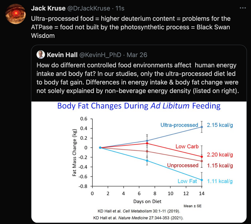
FOR THE SCIENTISTS AMONG YOU
In my quantum biological model, mtDNA UPEs drive OXPHOS component expression, ensuring heme protein renovation in the IMM for efficient electron transfer, via OPTIMIZED quantum tunneling. IMJs, optimized by Fe-S cluster biogenesis (per Picard and McManus), enhance mitochondrial network communication, supporting localized ATP/ROS delivery for MT dynamics during mitosis. Rev-erbα/β synchronize these processes via circadian regulation of heme and Fe-S cluster synthesis, aligning quantum coherence in the ETC and MTs with cellular cycles. This photonically integrated system ensures precise mitotic progression, with IMJs and Fe-S clusters acting as critical nodes for mitochondrial-cytoskeletal crosstalk.
Experimental Validation To Test This Demyelination Model:
Heme Renovation: Quantify COX subunit expression (via qPCR) and heme levels (via spectroscopy) in cells with manipulated UPE activity (e.g., CRISPR-edited D-loop). Assess electron transfer rates using high-resolution respirometry.
IMJ and Fe-S Clusters: Use super-resolution microscopy to visualize IMJs and measure MFN1/2 dynamics. Knockdown ISCU or FXN to assess Fe-S cluster biogenesis and its impact on OXPHOS and mitotic spindle formation.
Circadian Effects: Synchronize cells with circadian cues (e.g., dexamethasone) and measure Rev-erbα/β, ALAS1, and FXN expression. Correlate with MT polymerization rates and mitotic fidelity.
Quantum Probes: Use electron paramagnetic resonance (EPR) to detect Fe-S cluster spin states and potential quantum tunneling. Investigate MT vibrational coherence with Raman spectroscopy.
I am daring you to be better scientists by being better thinkers.
One of the most shocking things I have found in my 35 years of being a doctor is that many people who go on to develop cognitive decline and neurodegeneration began as mouth breathers. My dental background was key to exploring this unexplored link in these patients in my neurosurgery career.
Another shocking revelation was my review of the literature on this topic. Multiple studies have confirmed that mouth breathing is associated with cognitive loss, impairing working memory, attention, and executive function across age groups. These findings align with my decentralized thesis on demyelination, highlighting the role of NO deficiency, hypoxia, and photonic loop disruption in driving these effects. By addressing these root causes, restoring NO, reducing hypoxia, and supporting the photonic loop, we can mitigate the cognitive risks of mouth breathing and improve neurological outcomes.
Several studies have investigated the relationship between mouth breathing and cognitive impairment, often focusing on its association with sleep-disordered breathing (SDB), such as obstructive sleep apnea (OSA), a common consequence of chronic mouth breathing. Here’s what the research reveals:
Children with Mouth Breathing Syndrome (MBS)
A 2015 study published in the Sao Paulo Medical Journal compared 42 children identified as mouth breathers (mean age 8.7 years) with a control group of nasal breathers (mean age 8.4 years). The children underwent cognitive assessments of phonological working memory, reading comprehension, and arithmetic skills. The results showed that mouth breathers performed significantly worse than controls in reading comprehension (p = 0.006), arithmetic (p = 0.025), and phonological working memory for pseudowords (p = 0.002), though not for numbers (p = 0.76). The authors concluded that mouth breathing is associated with lower academic achievement and impaired working memory, echoing the 2021 study’s findings on working memory deficits. This aligns with our thesis: mouth breathing reduces NO, leading to hypoxia in brain regions like the prefrontal cortex and cerebellum, which are critical for cognitive tasks. Hypoxia disrupts the photonic loop, impairing mitochondrial ATP production and neural signaling, manifesting as children’s cognitive deficits.
Mouth Breathing and Attention in Children
A 2021 study in the International Journal of Clinical Pediatric Dentistry examined 50 children with mouth breathing and found a significant correlation between mouth breathing, sleep disturbances, and symptoms of attention-deficit hyperactivity disorder (ADHD). The study reported that daytime sleepiness, a consequence of sleep apnea caused by mouth breathing, was positively correlated with inattention. Poor sleep quality due to OSA impairs neurocognitive functions like attention, concentration, and memory, which are essential for learning and academic performance. This supports our framework: low NO and hypoxia from mouth breathing degrade melanin and melanopsin in the brainstem’s reticular activating system (RAS), reducing arousal and attention, and further disrupting the photonic loop’s role in maintaining cognitive clarity.
Visual Search Performance and Attentional Processing
A 2015 study in the Journal of Neuroscience and Neuroengineering investigated the effects of oral breathing on visual attentional processing using a visual search task. Participants performed the task under three conditions: oral breathing (nasal plug), nasal breathing (mouth taped), and control (no modification). The study found that oral breathing increased the time required to find a poorly discriminable target, particularly in difficult search conditions, due to a heightened intercept, an index of pre-search sensory processing or motor response. The authors concluded that oral breathing disrupts attentional processing by competing for cognitive resources, particularly during inefficient visual search tasks. This aligns with the 2021 study’s findings on reduced functional connectivity in brain areas like the inferior parietal gyrus, which is involved in attentional control. From our thesis perspective, the hypoxia and low NO from mouth breathing impair the brainstem’s dorsolateral funiculus (DLF) and RAS, reducing neural efficiency and exacerbating cognitive load.
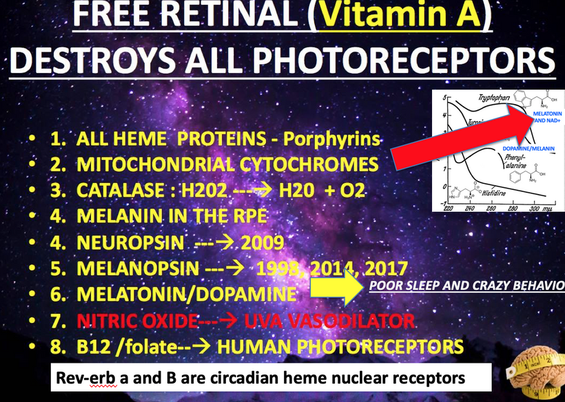
Sleep-Disordered Breathing and Cognitive Impairment in Older Adults
A 2017 meta-analysis in JAMA Neurology synthesized population-based studies on sleep-disordered breathing (SDB), often linked to mouth breathing, and its association with cognitive impairment. The analysis included 14 studies with a total of 4,288,419 participants and found that SDB was associated with a 26% increased risk of cognitive impairment (relative risk 1.26, 95% CI 1.05–1.50) and a small but significant worsening in executive function (standardized mean difference -0.05, 95% CI -0.09 to 0.00). Longitudinal studies within the meta-analysis showed that older adults with SDB had a higher likelihood of developing mild cognitive impairment (MCI) or Alzheimer’s disease (AD). The authors suggested that hypoxia and sleep fragmentation, both exacerbated by mouth breathing, are key mechanisms driving cognitive decline. This resonates with our thesis: chronic mouth breathing leads to hypoxia in the brainstem, particularly in melanin-rich areas like the locus coeruleus in the RAS, impairing the photonic loop and increasing the risk of neurodegenerative processes.
Increased Oxygen Load in the Prefrontal Cortex
A 2013 study in NeuroReport used near-infrared spectroscopy (NIRS) to measure hemodynamic responses in the prefrontal cortex during mouth breathing. The study found that mouth breathers exhibited an increased oxygen load in the prefrontal cortex compared to nasal breathers, suggesting compensatory neural activity to maintain cognitive performance. However, this increased load was associated with a higher likelihood of sleep disorders and ADHD, which are known to impair cognitive function. The authors hypothesized that chronic mouth breathing stresses the prefrontal cortex, a region critical for executive function and working memory, leading to cognitive deficits over time. This complements the 2021 study’s findings on working memory impairment and supports our thesis: the hypoxia from low NO disrupts mitochondrial metabolism in the prefrontal cortex, altering UPEs and impairing the photonic loop, manifesting as cognitive strain and eventual decline.IMPLICATIONS FOR DEMYELINATION REPAIR? GLP-1RAs could complement the decentralized thesis by repairing myelin, protecting neurons, and supporting vascular health in the DLF and RAS, while interventions targeting NO and the photonic loop address the underlying biophysical disruptions. This counterintuitive, decentralized approach is grounded in the evolutionary lessons of the GOE and offers a promising path forward for managing the complex neurological effects of mouth breathing. What else affects these pathways?
Hypothyroidism and Its Role in Myelination and Cognitive Function
Hypothyroidism Overview:
Hypothyroidism (low T3/T4, high TSH) results from insufficient thyroid hormone production, impacting multiple systems, including the nervous system.
Thyroid hormones (T3/T4) are critical for myelination and cognitive function:Myelination: T3 regulates oligodendrocyte differentiation and expression of myelin-related genes (e.g., myelin basic protein, proteolipid protein). Hypothyroidism impairs myelin formation and maintenance, leading to reduced white matter integrity.
Cognitive Function: Thyroid hormones support synaptic plasticity, neurogenesis, and neurotransmitter signaling. Hypothyroidism can cause cognitive deficits, including memory impairment, slowed processing, and “brain fog,” often reversible with levothyroxine treatment.
Relevance to Myelin:
Chronic hypothyroidism can lead to demyelination, particularly in peripheral nerves (e.g., peripheral neuropathy) and, to a lesser extent, the central nervous system (CNS). This is due to reduced myelin synthesis and impaired repair mechanisms.
In the context of lithium use, hypothyroidism is a common side effect, exacerbating myelin loss if untreated.
Relevance to Cognitive Function:
Hypothyroidism is associated with reversible cognitive impairment, but prolonged, untreated cases may contribute to long-term deficits, especially in older adults or those with comorbidities like PD.
2. Parkinson’s Disease (PD) and Its Connection to Myelination and Cognitive Function
PD Overview:
PD is a neurodegenerative disorder characterized by dopaminergic neuron loss in the substantia nigra, leading to motor symptoms (tremor, rigidity, bradykinesia) and non-motor symptoms (cognitive decline, depression).
PD involves alpha-synuclein aggregates (Lewy bodies) and mitochondrial dysfunction, affecting neuronal signaling and survival.
Myelination in PD:
While PD primarily affects dopaminergic neurons, white matter abnormalities are increasingly recognized:Diffusion tensor imaging (DTI) studies show reduced fractional anisotropy in PD patients, indicating disrupted white matter integrity, including myelin loss in regions like the corpus callosum and corticospinal tracts.
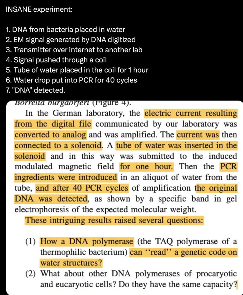
Myelin disruption may contribute to motor and cognitive deficits by impairing neural connectivity.
Thyroid hormones influence myelination, and hypothyroidism could exacerbate white matter damage in PD, though direct studies on this interaction are limited.
Cognitive Function in PD:
Cognitive impairment is common in PD, ranging from mild cognitive impairment (PD-MCI) to Parkinson’s disease dementia (PDD). Deficits include executive dysfunction, visuospatial impairment, and memory issues.
Dopaminergic and cholinergic deficits and white matter changes drive cognitive decline in PD. Improving myelination is the best way to attack cognitive decline in PD. Associated Hypothyroidism may worsen these deficits by further impairing synaptic plasticity and myelination. The top-line links explain why this is the case.
Hypothyroidism-PD Link:
Hypothyroidism is more prevalent in PD patients than in the general population, possibly due to shared risk factors (e.g., aging, inflammation, poor light biology) or medications like levodopa, which can suppress TSH.
Untreated hypothyroidism in PD patients may amplify motor and cognitive symptoms by impairing myelination and neurosignaling. Correcting hypothyroidism with levothyroxine can improve symptoms, but evidence on myelin recovery in PD is sparse. Part 6 will tell you why.
3. Melanin Biology and Its Link to PD and Skin Cancer
Melanin Overview:
Melanin, produced by melanocytes, is a pigment responsible for skin, hair, and eye color. It also exists in the brain as neuromelanin, found in dopaminergic neurons of the substantia nigra and locus coeruleus.
Neuromelanin binds toxins and metals, especially Iron, protecting neurons but potentially contributing to neurodegeneration when overloaded. This paper shows you why I gave up gold in 2009.
PD and Neuromelanin:
In PD, neuromelanin-containing dopaminergic neurons in the substantia nigra are selectively lost. Neuromelanin may exacerbate neuronal damage by:Binding environmental toxins (e.g., golf course pesticides, other toxins) or metals (e.g., iron), increasing oxidative stress.
Releasing toxic byproducts during neuronal death, amplifying inflammation.
Centralized MDs do not fully understand Neuromelanin’s role in PD. Still, its depletion correlates with disease progression, so my decentralized thesis fully explains why it happens = Melanin degradation and a loss of Becker’s regenerative currents.
PD and Skin Cancer (Melanoma):
Epidemiological studies show a bidirectional link between PD and melanoma:Increased Melanoma Risk in PD: PD patients have a 1.5–2-fold higher risk of melanoma. A 2017 meta-analysis confirmed this association, with odds ratios around 1.83 for melanoma in PD.
Increased PD Risk in Melanoma: Melanoma patients have a higher risk of developing PD, suggesting shared biological mechanisms.
Hypothesized Mechanisms:Melanin Synthesis: Neuromelanin and cutaneous melanin are derived from tyrosine via the tyrosinase enzyme. Dysregulation of this pathway (e.g., via L-DOPA metabolism) may link PD and melanoma.
Alpha-Synuclein: PD’s hallmark protein, alpha-synuclein, is expressed in melanoma cells, potentially promoting tumor growth. Its misfolding may connect PD and melanoma pathogenesis.
Pigmentation Genes: Variants in genes like MC1R (melanocortin one receptor) or TYR (tyrosinase) may increase susceptibility to PD and melanoma.
Levodopa Therapy: Levodopa, a PD treatment, is a precursor to dopamine and melanin. While early concerns suggested levodopa might increase melanoma risk, recent studies find no causal link, though it may exacerbate existing melanomas.
Clinical Implications: PD patients should undergo regular dermatological screenings for melanoma, as early detection is critical.
Relevance to Myelin and Hypothyroidism:
Melanin and Myelin: There’s no direct link between cutaneous melanin or neuromelanin and myelination. However, neuromelanin’s role in PD-related neuronal loss may indirectly affect white matter by disrupting neural networks, compounding myelin loss from hypothyroidism.
Hypothyroidism and Melanin: Hypothyroidism can cause skin changes (e.g., pallor, dryness), but there’s no evidence it directly alters melanin production or melanoma risk. However, hypothyroidism’s impact on systemic inflammation may exacerbate PD-related neurodegeneration, potentially affecting neuromelanin-containing neurons.
4. Integrating Hypothyroidism, PD, Myelination, Cognitive Function, and Melanin
Shared Pathways:
Oxidative Stress and Inflammation: Hypothyroidism, PD, and melanoma involve oxidative stress and inflammation, impairing myelination and cognitive function. In PD, neuromelanin’s interaction with toxins increases oxidative damage, potentially affecting nearby white matter tracts.
Mitochondrial Dysfunction: Both PD and hypothyroidism are linked to mitochondrial impairment, which disrupts energy-intensive processes like myelination and synaptic signaling, contributing to cognitive decline.
Neurotransmitter Dysregulation: Dopamine deficits in PD, compounded by hypothyroidism’s impact on serotonin and GABA, impair neurosignaling, affecting cognitive and motor functions. Myelin loss further disrupts connectivity. Note all these connections in the slide.
Specific to Myelin:
Hypothyroidism directly impairs myelination, while PD involves secondary white matter changes. Lithium use may mitigate myelin loss in both conditions by inhibiting GSK-3β, but hypothyroidism must be treated to maximize this benefit first because lithium causes hypothyroidism. Use of lithium often makes cognition worse in neurodegenerative cases and mental disorders that have high TSH.
Currently, in the centralized literature, no direct evidence links melanin or melanoma to myelin loss exists because no one has put this mechanism together. Still, the absence of evidence is not the absence of effect. Why do I think it is highly likely linked biophysically? PD-related neurodegeneration by light exacerbates white matter damage, especially in hypothyroid patients. They are very vulnerable to these conditions, and no centralized MD is taught this, so it goes undiagnosed until it is too late. Blue light toxicity via the eye is the fastest way to get any version of hypothyroidism.
Specific to Cognitive Function:
Hypothyroidism and PD both cause cognitive impairment, with overlapping deficits in executive function and memory. Myelin loss in both conditions disrupts neural connectivity, worsening cognitive outcomes.
Correcting hypothyroidism can improve cognition in PD patients, but PD’s progressive nature limits recovery.
Melanin and Skin Cancer Connection:
The PD-melanoma link is directly driven by shared pathways (alpha-synuclein, tyrosine metabolism) rather than by myelin or hypothyroidism. However, hypothyroidism’s systemic effects amplify PD-related neurodegeneration, indirectly affecting neuromelanin-containing neurons and increasing melanoma susceptibility via inflammation. Light stress is the dagger in this mechanism.
5. Clinical and Research Implications
Hypothyroidism Management: Treating hypothyroidism with levothyroxine is critical in PD patients to support myelination, cognitive function, and overall neurological health. Regular TSH/T3/T4 monitoring is essential, especially with lithium use. The reason for caution should be apparent now.
PD and Melanoma Screening: Given the elevated risk, PD patients should have annual dermatological exams to detect melanoma early. Melanoma patients with neurological symptoms should be evaluated for PD.
Myelin and Cognitive Support: No specific therapies target myelin loss in PD or hypothyroidism, but lithium’s neuroprotective effects may help. Cognitive rehabilitation and exercise may mitigate deficits unless the light environment completely changes.
Research Gaps: More studies are needed to:
Clarify the role of hypothyroidism in PD-related myelin loss.
Explore whether neuromelanin dysregulation directly affects white matter.
Investigate whether alpha-synuclein links myelin loss, PD, and melanoma.
Hypothyroidism is often lurking in all patients who have neurodegeneration. This is doubly true in patients with all forms of diabetes. Subclinical hypothyroidism is linked to these conditions because it impairs myelination EARLY without the patient or doctor knowing it, and cognitive function suffers immediately by reducing thyroid hormone levels, exacerbating white matter and cognitive deficits in PD and most other neurodegenerative conditions. PD itself involves myelin loss and cognitive decline, driven by dopaminergic and white matter pathology. The PD-melanoma link, mediated by neuromelanin, alpha-synuclein, and tyrosine metabolism, is highly relevant to the patients’ clinical course but doesn’t directly involve myelin or hypothyroidism. However, hypothyroidism’s systemic effects may amplify PD-related neurodegeneration and inflammation, potentially worsening outcomes in both myelin integrity and melanoma risk. Treating hypothyroidism, monitoring for melanoma, and leveraging lithium’s neuroprotective effects (if applicable) are key strategies to mitigate these risks.
WHAT ELSE CAN BE CONSIDERED FOR RAPID REMYELINATION?
GLP-1 (glucagon-like peptide-1), leptin, and the melanocortin pathways are critical components of the body’s energy homeostasis system, integrating signals of satiety, energy stores, and metabolic demand. Below, I’ll explain how GLP-1, as a natural fullness signal, interacts with the leptin-melanocortin pathways in humans, focusing on their roles in appetite regulation, energy balance, and potential relevance to the previous discussion on hypothyroidism, Parkinson’s disease (PD), myelination, cognitive function, and melanin biology. This drug may have a novel, unique use for remyelinating people with diseases like ALS rapidly. It is not without risk, but with ALS, the risk of death exceeds the risk of GLP-1 drugs. The benefit far outweighs the risk; therefore, one can consider it when counseling patients.
Overview of Key Players
GLP-1:Role: An incretin hormone secreted by L-cells in the gut in response to nutrient ingestion. It promotes satiety, slows gastric emptying, enhances insulin secretion, and reduces appetite by acting on the hypothalamus and brainstem.
Mechanism: GLP-1 binds to GLP-1 receptors (GLP-1R) in the brain (e.g., nucleus tractus solitarius [NTS], arcuate nucleus [ARC]) and periphery, signaling fullness and reducing food intake.
Leptin:Role: A hormone secreted by adipocytes, reflecting long-term energy stores (fat mass). It signals energy sufficiency to the brain, suppressing appetite and increasing energy expenditure.
Mechanism: Leptin binds to leptin receptors (LepR) in the ARC, activating pro-opiomelanocortin (POMC) neurons and inhibiting agouti-related peptide (AgRP)/neuropeptide Y (NPY) neurons, promoting satiety.
Melanocortin Pathway:Role: A central pathway in the hypothalamus regulating appetite and energy balance. It is modulated by leptin and other signals.
Mechanism: POMC neurons cleave POMC into α-melanocyte-stimulating hormone (α-MSH), which activates melanocortin receptors (MC3R, MC4R), reducing appetite. AgRP acts as an antagonist, blocking MC4R to increase appetite.
Integration of GLP-1 with Leptin-Melanocortin Pathways
GLP-1, leptin, and the melanocortin pathways converge in the hypothalamus and brainstem to regulate appetite and energy homeostasis. Their integration involves both direct and indirect mechanisms:
Hypothalamic Convergence:GLP-1 and Leptin in the ARC:
GLP-1R and LepR are co-expressed on POMC neurons in the ARC. GLP-1 enhances POMC neuron firing, increasing α-MSH release, while leptin similarly activates POMC neurons and inhibits AgRP/NPY neurons.
Studies show GLP-1 and leptin synergistically reduce food intake. For example, co-administration of GLP-1 and leptin in rodents enhances weight loss beyond either alone, suggesting cooperative signaling.
Mechanism: GLP-1 activates cyclic AMP (cAMP) and protein kinase A (PKA) pathways via GLP-1R, while leptin activates the JAK2-STAT3 pathway via LepR. These pathways converge on CREB (cAMP response element-binding protein), upregulating POMC expression and α-MSH release, stimulating MC4R to suppress appetite.
Brainstem-Hypothalamus Crosstalk:GLP-1 in the NTS: GLP-1 is produced locally in the NTS and acts on GLP-1R in the brainstem, relaying satiety signals from the gut. NTS neurons project to the ARC, modulating POMC and AgRP neurons.
Leptin’s Role: Leptin enhances NTS sensitivity to GLP-1, amplifying satiety signals. In leptin-deficient (ob/ob) mice, GLP-1’s anorectic effects are blunted, indicating that leptin sensitizes GLP-1 signaling.
Melanocortin Link: NTS GLP-1 signaling indirectly activates hypothalamic MC4R via POMC neuron projections, integrating short-term (GLP-1) and long-term (leptin) energy signals.
Peripheral Interactions:GLP-1 enhances insulin secretion, improving glucose homeostasis, and indirectly supports leptin signaling by reducing adipose tissue inflammation (a cause of leptin resistance).
Leptin modulates gut hormone secretion, including GLP-1, by influencing enteroendocrine cell function, creating a feedback loop.
Synergistic Effects:In humans, GLP-1 receptor agonists (e.g., liraglutide, semaglutide) and leptin analogs show additive effects on weight loss in clinical trials, particularly in obese individuals with leptin resistance.
The melanocortin pathway is a common downstream effector: GLP-1 and leptin increase α-MSH, reducing food intake via MC4R activation.
Relevance to Hypothyroidism, PD, Myelination, Cognitive Function, and Melanin
I’ll connect GLP-1, leptin, and melanocortin pathways to these demyelinating conditions, focusing on their impact on myelination, cognitive function, and melanin biology:
Hypothyroidism:Impact on Pathways:
Hypothyroidism reduces leptin levels due to decreased fat mass and impairs leptin signaling via inflammation, blunting melanocortin activation, and increasing appetite.
GLP-1 secretion and signaling are less studied in hypothyroidism, but thyroid hormones regulate gut motility and enteroendocrine function, suggesting hypothyroidism may dampen GLP-1 responses.
Myelination: Hypothyroidism impairs myelination (as discussed previously). Leptin promotes oligodendrocyte differentiation and myelination via LepR signaling, so reduced leptin in hypothyroidism may exacerbate myelin loss. GLP-1’s role in myelination is unclear but may indirectly support neuronal health via neuroprotection.
Cognitive Function: Hypothyroidism causes cognitive deficits, partly due to reduced leptin and melanocortin signaling, which support synaptic plasticity. GLP-1 agonists show promise in improving cognition in neurodegenerative models, potentially mitigating hypothyroidism-related deficits.
Parkinson’s Disease (PD):Pathway Alterations:
PD patients often exhibit leptin resistance due to inflammation and altered hypothalamic signaling, impairing melanocortin-mediated appetite control.
GLP-1 agonists (e.g., exenatide) are under investigation for PD due to their neuroprotective effects, including reduced inflammation and enhanced dopamine signaling, which may complement melanocortin pathways.
Myelination: Leptin and melanocortin signaling support white matter integrity, and their dysfunction in PD may contribute to myelin loss. GLP-1 agonists promote neurogenesis and may indirectly support myelination by reducing oxidative stress.
Cognitive Function: GLP-1 agonists improve cognition in PD models by enhancing synaptic plasticity and reducing alpha-synuclein aggregation. Leptin and melanocortin pathways also support cognition, and their impairment in PD worsens deficits.
Melanin Biology and PD-Melanoma Link:Melanocortin Connection:
The melanocortin pathway, via α-MSH and MC1R, regulates cutaneous melanin production. MC1R variants are linked to melanoma risk, and α-MSH’s role in the hypothalamus (via MC4R) connects it to energy balance.
In PD, neuromelanin in the substantia nigra is implicated in neuronal loss, and the PD-melanoma link may involve shared pathways (e.g., tyrosine metabolism, alpha-synuclein).
GLP-1 and Leptin: GLP-1 and leptin don’t directly regulate neuromelanin or cutaneous melanin, but their anti-inflammatory effects may reduce melanoma risk by mitigating systemic inflammation. Though evidence is preliminary, GLP-1 agonists are now being studied for anti-cancer properties. If they are being studied there, they should really be studied in ALS patients.
Hypothyroidism: Hypothyroidism’s impact on inflammation may indirectly influence melanoma risk. However, no direct link to melanin biology exists in the literature because no one is considering using my decentralized thesis in healthcare today.
Our decentralized thesis has illuminated how mouth breathing, a seemingly innocuous habit, triggers a cascade of biophysical disruptions in the brainstem, particularly in the dorsolateral funiculus (DLF) tracts affecting sleep and reticular activating system (RAS). This cascade, rooted in the evolutionary adaptations of the Great Oxidation Event (GOE), involves low nitric oxide (NO), hypoxia, sleep apnea, and the degradation of melanin and melanopsin, ultimately impairing the recursive photonic loop that sustains cellular function. Now, let’s explore how glucagon-like peptide-1 receptor agonists (GLP-1RAs), such as liraglutide and exenatide, might intersect with this framework, offering a novel approach to neuroprotection and remyelination in the brainstem while addressing the underlying metabolic and photonic dysfunctions.Recap of the Decentralized Thesis: Mouth Breathing and Brainstem Pathology
Mouth breathing reduces NO production, exacerbating hypoxia and sleep apnea by impairing respiratory control in the DLF (Review Quantum engineering #47/48) and arousal in the RAS. Low NO, compounded by a disrupted urea cycle due to poor kinetic isotope effect (KIE), limits vasodilation, weakens stem cell-mediated vascular repair, and increases the risk of brainstem hemorrhages. Hypoxia also degrades melanin and melanopsin in neural tracts and cerebral blood vessels, disrupting charge separation of water and altering ultraweak photon emissions (UPEs). This breaks the recursive photonic loop, where melanin absorbs sunlight, modulates mitochondrial UPEs, and produces near-infrared (NIR) light to support oxygen-rich metabolism in the tricarboxylic acid (TCA) cycle. The result is reduced H+, oxygen, and ATP production, impaired myelin capacitance, and vascular fragility in the brainstem, particularly in the pons and cerebellum, a region prone to hemorrhagic stroke in mouth breathers.
GLP-1RAs: A Potential Ally in Neuroprotection and Remyelination
GLP-1RAs, initially developed for type 2 diabetes and obesity, are emerging as promising agents for neuroprotection and remyelination. Preclinical studies show that liraglutide and exenatide enhance Schwann cell and oligodendrocyte function, promoting axonal regeneration and myelin repair. These effects are likely mediated by neuroprotective pathways such as ERK signaling, which supports cell survival and repair in the central and peripheral nervous systems. In the context of our thesis, GLP-1RAs could target several key aspects of brainstem pathology in mouth breathers:
Myelin Repair in the DLF and RAS: The DLF and RAS contain myelinated tracts critical for respiratory control and arousal. Hypoxia and low NO impair myelin’s proton capacitor function by reducing H+ production, slowing neural conduction, and contributing to sleep apnea. GLP-1RAs, by enhancing oligodendrocyte function, could promote remyelination in these regions, restoring myelin’s capacitance and improving signal transmission. This might strengthen the neural circuits that regulate breathing and arousal, reducing the severity of sleep apnea.
Neuroprotection Against Hypoxia-Induced Damage: Hypoxia from sleep apnea damages neurons in the DLF and RAS, particularly in melanin-rich areas like the locus coeruleus. GLP-1RAs’ neuroprotective effects, potentially via ERK signaling, could mitigate this damage by enhancing neuronal survival and reducing inflammation. This aligns with the GOE’s legacy: just as early life adapted to oxygen’s stress with heme proteins, GLP-1RAs might help modern mammals adapt to hypoxia by protecting brainstem neurons.
Vascular Support and Stem Cell Activation: Low NO impairs vasodilation and stem cell-mediated vascular repair, increasing the risk of brainstem hemorrhages. GLP-1RAs have been shown to improve endothelial function and vascular health in other contexts, possibly by upregulating NO production or enhancing stem cell activity. This could strengthen cerebral blood vessels in the brainstem, particularly those expressing melanopsin, reducing the risk of rupture in the pons and other vulnerable regions.
Metabolic Support for the Photonic Loop: The recursive photonic loop relies on mitochondrial metabolism to produce UPEs and NIR light, which counteract NO’s inhibition of CCO and support oxygen-rich TCA cycle activity. GLP-1RAs, known to improve mitochondrial function in metabolic disorders, might enhance ATP production in the brainstem, supporting the photonic loop. By boosting mitochondrial efficiency, GLP-1RAs could help restore the NIR light needed to reverse metabolic dysfunction in mouth breathers, aligning cellular function with the oxygen-rich environment shaped by the GOE.
Challenges and Synergies with the Decentralized Framework
While GLP-1RAs offer transformative potential, their integration with our decentralized thesis must account for clinical complexities. Gastrointestinal side effects, variable patient responses, and long-term safety concerns, like risks of pancreatitis, require cautious application. Additionally, GLP-1RAs alone may not fully address mouth breathers’ photonic and electrical disruptions. For example, while they might promote remyelination, they may not directly restore melanin or melanopsin function, which are critical for the photonic loop. Combining GLP-1RAs with interventions like sunlight exposure (to stimulate melanin) or nasal breathing exercises (to boost NO) should create a synergistic approach, addressing both the metabolic and photonic aspects of brainstem pathology.
Moreover, the decentralized thesis emphasizes the role of deuterium accumulation in disrupting the KIE in the urea cycle, which reduces NO production. GLP-1RAs, by improving mitochondrial function, might indirectly support the urea cycle by reducing deuterium levels in the mitochondrial matrix, enhancing NO availability. This could further support vasodilation, stem cell activation, and vascular repair, creating a feedback loop that reinforces the neuroprotective effects of GLP-1RAs.Clinical Implications: A Cross-Disciplinary Approach
The potential of GLP-1RAs to promote remyelination and neuroprotection in the DLF and RAS aligns with the decentralized thesis’s call for innovative, cross-disciplinary solutions. Mouth breathers with sleep apnea could benefit from a combined strategy: GLP-1RAs to repair myelin and protect neurons, alongside interventions to restore NO (e.g., nasal breathing) and the photonic loop (e.g., NIR light therapy). Clinicians could use advanced imaging, like the 3T MRI with deuterium software you employ, to monitor myelin integrity, melanin degradation, and deuterium levels in the brainstem, tailoring treatments to individual patients.
This integration highlights the power of repurposing drugs like GLP-1RAs for neurological disorders while addressing the root causes of brainstem dysfunction through a biophysical lens. By bridging the metabolic benefits of GLP-1RAs with the photonic and electrical insights of the decentralized thesis, we can unlock new therapeutic avenues for mouth-breathing patients, reducing their risk of sleep apnea, cognitive decline, neurodegeneration, and vascular catastrophes in the brainstem.
Clinical Implications & Therapeutic Potential:
GLP-1 Agonists: Drugs like semaglutide enhance satiety and may improve myelination, cognition, and neuroprotection in hypothyroidism and PD. They complement leptin-melanocortin signaling by amplifying POMC activation.
Leptin Sensitization: Addressing leptin resistance (e.g., via weight loss, anti-inflammatory diets) can enhance melanocortin signaling, supporting energy balance and myelination.
Thyroid Management: Correcting hypothyroidism with levothyroxine restores leptin levels and supports myelination, potentially enhancing GLP-1 and melanocortin effects.
PD and Melanoma: GLP-1 agonists’ neuroprotective effects may benefit PD patients, while regular melanoma screenings remain critical due to the PD-melanoma link.
Cognitive Support: GLP-1 agonists and leptin-melanocortin activators (e.g., MC4R agonists) are promising for cognitive enhancement in hypothyroidism and PD, though clinical data are limited.
SUMMARY
GLP-1 integrates with the leptin-melanocortin pathways primarily in the hypothalamus (ARC) and brainstem (NTS), synergistically activating POMC neurons and MC4R to suppress appetite and regulate energy balance. GLP-1 enhances short-term satiety, while leptin signals long-term energy stores, converging on the melanocortin pathway to reduce food intake.
In hypothyroidism, reduced leptin and potential GLP-1 dysfunction exacerbate myelin loss and cognitive deficits, which can be mitigated by thyroid hormone replacement. GLP-1 agonists show promise for neuroprotection and cognition in PD, complementing leptin-melanocortin signaling. This is a far better option than Lithium use. The PD-melanoma link involves melanocortin pathways (via α-MSH/MC1R) and neuromelanin, but GLP-1 and leptin primarily influence this through anti-inflammatory effects, not direct melanin regulation. Treating hypothyroidism, leveraging GLP-1 agonists, and addressing leptin resistance are key strategies to support myelination, cognition, and overall health in these contexts. They have risks.
This ends part 5. Part six gives the caveats to these ideas. If you are a welder, you’ll like how we are putting the pieces together here.
As a decentralized doctor, there are a couple of things to consider for the treatment side. Since lithium will have a huge effect on UPEs and CNS will be more at risk for damage with lithium use for remyelination, how would we protect against this? The biochemical treatment cascade is Li inhibits GSK-3B > activates Wnt signalling > promotes OPC in the CNS. It does not do this in the PNS. All of this requires the precision of UPEs from the TCA/urea cycle, and we know that Li electronic structure is not a match for this and likely would make an alien UPE spectrum, so wouldn’t this not be always supportive of encouraging new myelin growth around vulnerable axons because of UPE changes?? It might also upregulate BDNF, NGF & VEGF. Lithium may also help with restoring the magnetic orientation/internal clock synchrony. Should everyone get DTI to do a WMV assessment to help the biophoton spectral issue I am raising? Could you imagine anything else helping remyelination in a CNS versus PNS disease?
Many references highlight the role of biophotons in cellular signaling. Still, few link it to the evolutionary significance of photon-energy utilization (e.g., ferredoxin) and the potential for light-based communication in mitochondria and neurons. We’ll address the treatment concerns with lithium, its effects on UPEs, and strategies to protect vulnerable axons. We’ll also explore additional remyelination approaches and the utility of DTI for white matter volume (WMV) assessment.
Ferredoxin’s Ancient Roots and the GOE
Imagine a time over 2.4 billion years ago, during the Great Oxidation Event (GOE), when Earth’s atmosphere began to fill with oxygen, a byproduct of early photosynthetic bacteria. These ancient microbes, like those studied by M. C. W. Evans, Bob B. Buchanan, and Daniel I. Arnon in their 1966 paper, were pioneers of a metabolic revolution. Their discovery of a ferredoxin-dependent carbon reduction cycle in photosynthetic bacteria, as depicted in the diagram above, reveals a critical piece of the evolutionary puzzle: ferredoxin, a small iron-sulfur protein, was among the earliest molecules to harness photon energy, setting the stage for life’s biochemical complexity. This is another reason why light > food. Few people know this story today.
Ferredoxin’s story begins in the primordial oceans, where simple inorganic molecules and photon energy from the sun drove the earliest forms of metabolism. As highlighted in the abstract by Eck and Dayhoff, ferredoxin’s structure likely evolved from a short peptide of just eight amino acids, possibly alanine, aspartic acid, proline, serine, and glycine, doubling over time into a more complex protein. This simplicity suggests ferredoxin predates the DNA genetic code, emerging in a prebiotic world where photon-energy utilization was a survival necessity. Ferredoxin’s iron-sulfur clusters, acting as electron carriers, allowed these early photosynthetic bacteria to capture light energy and reduce CO₂ into organic molecules, as shown in the ferredoxin-dependent cycle (e.g., converting CO₂ to acetyl-CoA and eventually to glutamate).
The GOE marked a turning point on Earth. As oxygen levels rose, ferredoxin’s role expanded beyond photosynthesis. Oxygen, a double-edged sword, enabled more efficient energy production and introduced an oxidative holocaust. To survive, cells evolved protective mechanisms: heme proteins, melanin, and superoxide dismutases (SODs). Heme proteins, like cytochrome c, began to work alongside ferredoxin in electron transport chains, while melanin shielded cells from UV-induced damage, and SODs neutralized reactive oxygen species (ROS). These adaptations were crucial as life transitioned from anaerobic to aerobic environments, laying the groundwork for complex cellular systems.
Photon-Energy Utilization and Circadian Evolution
Ferredoxin’s ability to capture photons wasn’t just about energy but also timing. The GOE coincided with the evolution of circadian mechanisms, as cells needed to synchronize their metabolic processes with the day-night cycle. Ferredoxin was key in early circadian signaling by facilitating light-driven electron transfer. This photon-energy utilization became a blueprint for later systems, like the mitochondrial electron transport chain and neuronal signaling pathways, where light-based communication, via biophotons, emerged as a cellular language. To understand how we fall apart in disease states, you must understand how the GOE built the system to operate.
Biophotons, ultra-weak photon emissions (UPEs), are now recognized as key players in cellular signaling, particularly in mitochondria, blood, and neurons. I have a sense that coherent domains in water also create them. Mitochondria, descendants of ancient bacteria, inherited ferredoxin’s photon-handling legacy, using light to regulate energy production and redox signaling. Biophotons facilitate rapid communication across axons in neurons, a process potentially rooted in ferredoxin’s early role in light-energy capture. This connection suggests that the evolutionary significance of photon-energy utilization extends far beyond metabolism, influencing how cells communicate and coordinate in complex organisms. This system was built before the myelin system.
Remyelination and White Matter Assessment
Remyelination offers hope for repairing damaged axons. Approaches like clemastine, a histamine antagonist, promote oligodendrocyte differentiation, while therapies targeting the Wnt signaling pathway enhance myelin repair. Additionally, biophoton-based therapies should be explored in our decentralized future, leveraging light to stimulate mitochondrial function and support remyelination, a concept rooted in ferredoxin’s ancient photon-handling role.
Diffusion tensor imaging (DTI) is invaluable for assessing white matter volume (WMV) and tracking remyelination. DTI measures the diffusion of water molecules in tissue, providing insights into white matter integrity. Increased fractional anisotropy (FA) on DTI scans indicates improved myelination. At the same time, reduced radial diffusivity suggests less axonal damage, a critical metric for evaluating therapeutic outcomes in MS and other neurodegenerative conditions like Progressive Supranuclear Palsy early in its development.
1. Lithium, UPEs, and Axonal Protection
Fast-forward to modern medical concerns: Lithium, commonly used to treat bipolar disorder, affects UPEs and mitochondrial function. Lithium increases mitochondrial respiration, which may enhance biophoton emission, but it also risks destabilizing redox balance, leading to oxidative stress in vulnerable axons. This is particularly concerning in demyelinating conditions like multiple sclerosis (MS), where axons are exposed to oxidative damage. Centralized strategies to protect axons include breathing treatments with antioxidants like N-acetylcysteine to mitigate ROS, alongside lithium’s mood-stabilizing benefits, ensuring a balanced therapeutic approach.
Lithium is a well-established treatment for bipolar disorder (BPD) and has been explored for neuroprotection in other CNS diseases (e.g., ALS, AD, PD). Its mechanism involves inhibiting glycogen synthase kinase-3 beta (GSK-3β), activating Wnt signaling, and promoting oligodendrocyte precursor cell (OPC) differentiation for remyelination. However, I am concerned about lithium’s electronic structure potentially disrupting UPE spectra, which could affect remyelination and axonal health. Many cases of demyelination where lithium should have helped have turned out to have the opposite effect. I believe this is because Lithium major affects in humans are biophysical and not biochemical.
A. Lithium’s Biochemical Cascade
Mechanism:
GSK-3β Inhibition: Lithium inhibits GSK-3β, a kinase that negatively regulates Wnt signaling.
Wnt Activation: This promotes β-catenin stabilization, enhancing Wnt pathway activity.
OPC Differentiation: Wnt signaling drives OPCs to mature into myelinating oligodendrocytes, supporting remyelination in the CNS.
Additional Effects:
Neurotrophic Factors: Lithium upregulates brain-derived neurotrophic factor (BDNF), nerve growth factor (NGF), and vascular endothelial growth factor (VEGF), which support neuronal survival, plasticity, and angiogenesis.
Circadian Synchrony: Lithium may restore circadian rhythms by stabilizing the internal clock (e.g., via CLOCK gene regulation), potentially improving TCA cycle dynamics and beta-oxidation. I have tweeted about this before.
B. Lithium’s Effect on UPEs: an electronic mess.
LIGHT AFFECT UPR creation. That is the basis for BPD. UPEs are linked to the fidelity of the TCA/urea Cycle dynamics. UPEs are generated by mitochondrial ROS during oxidative phosphorylation (OXPHOS) downstream of the TCA/urea cycle. The TCA cycle’s efficiency, influenced by circadian rhythms and beta-oxidation, determines ROS production and thus UPE spectra. Nodes of Ranvier, with high mitochondrial density, are key UPE sites.
Lithium’s Electronic Structure: Lithium (atomic number 3) has a simple electronic configuration (1s² 2s¹). Its incorporation into cellular environments (e.g., as Li⁺) alters local electromagnetic fields, potentially disrupting the coherence of UPEs. The references (e.g., Mould et al., 2023) suggest that biophotons mediate non-chemical signaling between mitochondria, and tubulin networks may propagate these signals via resonance energy transfer (Kurian et al., 2017). Lithium’s “alien” electronic structure could introduce noise into this system, creating an aberrant UPE spectrum. Anything that disrupts the signal is a real problem in remyelination. This needs to be discussed with the patient.
Impact on Remyelination:
UPE Role in Myelination: UPEs may facilitate precise cellular communication during remyelination, guiding OPC differentiation and myelin sheath formation. An aberrant UPE spectrum (due to lithium) could disrupt this process, leading to inefficient or aberrant myelination.
CNS Vulnerability: With thinner myelin and limited repair capacity (oligodendrocytes), the CNS is more susceptible to UPE disruptions than the PNS (Schwann cells). The use of Lithium could easily result in incomplete remyelination, leaving axons vulnerable to degeneration, especially in diseases like PSP, MS, ALS, AD, and PD.
Lithium is quite dangerous in Progressive supranuclear palsy (PSP), in my experience. PSP primarily affects the brainstem (e.g., midbrain, subthalamic nucleus) and frontal cortex, regions with high metabolic demand. Tau pathology increases mitochondrial stress, elevating ROS and UPEs in these neural zip codes. UPE Profile in PSP likely shows persistently high UPE intensity in the brainstem and frontal regions due to chronic tau-induced mitochondrial dysfunction. The midbrain’s role in vertical gaze (e.g., oculomotor nuclei) suggests that UPE elevation here may disrupt precise biophoton signaling needed for motor coordination of the eyes in the brainstem. I think the lithium electronic structure in the brain stem is at high risk.
C. Potential Risks of Lithium on Vulnerable Axons
Aberrant Myelination: If lithium-induced UPE changes disrupt signaling, OPCs may form myelin sheaths with incorrect thickness or spacing, impairing saltatory conduction and increasing axonal stress. Thickness correlates with proton conduction and ATP stoichiometry.
Oxidative Stress: Lithium’s effect on mitochondrial function (via GSK-3β/Wnt) may alter TCA/urea cycle dynamics, potentially increasing ROS and UPEs. This could exacerbate oxidative damage in vulnerable axons, particularly CNS ones.
Neurotrophic Upregulation: While BDNF, NGF, and VEGF are beneficial, excessive upregulation (due to lithium) might overstimulate axons, leading to excitotoxicity in demyelinated regions, further risking degeneration. This is especially true if urea cycle stoichiometry is altered in the disease. This leads to problems in NO signaling that link to stem cell depots and to GABA signaling that links to the regeneration of the non-visual photoreceptors in the eye (below) at the front of the central retinal pathways.
2. Protecting Axons During Lithium Treatment
Given lithium’s potential to disrupt UPE spectra and affect remyelination, protective strategies are crucial, especially in the CNS, where axons are more vulnerable.
A. Mitigate UPE Disruption
Antioxidant Support: Since UPEs are tied to ROS, antioxidants (e.g., coenzyme Q10, N-acetylcysteine, vitamin E) can reduce oxidative stress, potentially stabilizing UPE spectra. They carry risk because we cannot know how the dosing should be aligned. After all, we cannot know the UPE spectra yet due to technological limitations. This may counteract lithium’s effects on mitochondrial ROS production.
Circadian Optimization: Lithium’s ability to restore circadian synchrony (via internal clock regulation) can be enhanced with morning sunlight exposure or light therapy. This supports beta-oxidation, TCA/urea cycle activity, and normal UPE production, reducing the risk of aberrant spectra. People with demyelination should spend 2-3 hours in AM and PM red sunlight.
Photobiomodulation (PBM): Low-level light therapy (e.g., near-infrared) can modulate mitochondrial function and UPEs, as suggested by Thar and Kühl (2014). PBM may restore coherent biophoton signaling, counteracting lithium’s disruptive effects. It is something to keep in your armentarium.
B. Enhance Remyelination Precision
Support OPC Maturation: Combine lithium with agents that enhance OPC differentiation, such as thyroid hormone (T3) or retinoic acid, to ensure proper myelination despite UPE disruptions. Remember what control T3: Light & melanin. See below.
Myelin Lipid Support: Provide dietary lipids (e.g., omega-3 fatty acids, ketogenic diets) to bypass TCA cycle deficits (due to lithium or circadian dysregulation), supplying precursors for myelin synthesis.
Monitor Axonal Health: Use neuroprotective agents (e.g., riluzole, used in ALS) to reduce excitotoxicity and support axonal integrity during remyelination.
C. Minimize Ongoing CNS Damage By Limiting Tech Abuse.
Dose Optimization: Use the lowest effective lithium dose to minimize UPE disruption while inhibiting GSK-3β. Monitor serum levels to avoid toxicity. Lithium has a terrible therapeutic index, so it is hard to get dosing right in these patients.
Neurotrophic Balance: Since lithium upregulates BDNF, NGF, and VEGF, monitor for signs of excitotoxicity (e.g., via neuro exam, EEG for hyperexcitability) and consider co-treatment with GABAergic agents (e.g., valproate) to balance neuronal activity. (below)
Mitochondrial Protection: If the case is straightforward with limited nnEMF exposure, mitochondrial enhancers (e.g., MitoQ, creatine from animal foods) can support broken stoichimetry of TCA/urea cycle function, reducing the risk of oxidative stress and aberrant UPEs.
Vasopressin antagonists are being used in MS and highlight a safer approach to reduce inflammation and support remyelination compared to lithium, especially given lithium’s UPE-driven risks to kidney and thyroid function. These two are my favorite pathways in these diseases. The decentralized insights on UV-A light and neuropsin suggest that lithium’s UPE disruption dysregulates vasopressin signaling, exacerbating NDI and hypothyroidism. At the same time, vaptans target this pathway more selectively without systemic toxicity. For high-risk patients, vaptans, PBM, and UV-A light offer a better risk-benefit profile, supporting myelination in PSP, MS, ALS, BPD, AD, and PD without the narrow therapeutic index of lithium. Lithium can be used with vaptans and protective strategies in low-risk patients, but careful monitoring is essential. This integrated approach aligns with first-principles biophysics, prioritizing UPE coherence and systemic safety.
A. Vasopressin Antagonists Across Demyelinating Diseases
ALS:
Vasopressin Role: Elevated vasopressin may contribute to motor neuron stress via inflammation, though less studied than in MS. Vaptans could reduce inflammation in the corticospinal tract, supporting remyelination.
Lithium Comparison: Lithium’s vasopressin dysregulation (NDI) doesn’t directly address CNS inflammation in ALS, and its UPE disruption risks outweigh benefits in high-risk patients.
BPD:
Vasopressin Role: Dysregulated vasopressin in BPD (linked to HPA axis dysfunction) may exacerbate fronto-limbic inflammation, contributing to variable myelination. Vaptans could stabilize this by reducing inflammation during mood episodes.
Lithium Comparison: Lithium’s mood stabilization benefits are significant, but its vasopressin-related kidney risks (NDI) and UPE disruption make vaptans a potential adjunct to reduce inflammation without systemic toxicity.
AD/PD/PSP:
Vasopressin Role: Elevated vasopressin in AD/PD may increase inflammation in cognitive regions (hippocampus, cortex), worsening demyelination and neurodegeneration. Vaptans could mitigate this, supporting myelination in these areas.
Lithium Comparison: Lithium’s potential to promote myelination in AD/PD is offset by its kidney/thyroid risks and UPE disruption. Vaptans offer a safer anti-inflammatory approach.
B. UV-A Light and Neuropsin: Modulating Vasopressin
Slide Insights:
UV-A light (380 nm) activates neuropsin, driving vasopressin release and influencing T3/T4, mTOR, and DHA catabolism. Lithium’s UPE effects disrupt this pathway, leading to kidney/thyroid dysfunction.
Vasopressin Antagonists and UV-A:
MS: Vaptans reduce vasopressin activity, potentially counteracting excessive UV-A/neuropsin-driven vasopressin release in inflammatory states. Controlled UV-A exposure could fine-tune this pathway, balancing vasopressin levels to support remyelination without exacerbating inflammation.
Other Diseases: UV-A light could modulate vasopressin in PSP, ALS, BPD, AD, and PD, complementing vaptans by optimizing neuropsin signaling and reducing inflammation.
C. UPE Considerations
Lithium’s UPE Disruption: Lithium’s “alien” UPE spectrum impairs biophoton signaling, potentially disrupting vasopressin regulation (via neuropsin) and myelination.
Vaptans and UPEs: Vaptans are unlikely to disrupt UPEs directly, as they target vasopressin receptors rather than mitochondrial function. They may indirectly normalize UPEs by reducing inflammation and mitochondrial stress, supporting remyelination.
Monitoring: Measure UPEs (scalp, peripheral nerves) to compare lithium vs. vaptans. If vaptans normalize UPEs more effectively (indicating reduced oxidative stress), they may be a safer option for myelination support.
3. Biophysical Risks of Lithium
Incorporating vasopressin antagonists into this analysis reinforces the concerns about lithium’s biophysical risks:
Kidney Risk (NDI):
Lithium’s disruption of vasopressin signaling causes NDI, a risk not present with vaptans, which target vasopressin receptors more selectively. In MS, vaptans reduce CNS edema without causing systemic water loss, making them a safer option for kidney health.
Thyroid Risk:
Lithium’s UPE disruption impairs neuropsin-driven T3/T4 synthesis (slide), causing hypothyroidism. Vaptans don’t affect thyroid function directly, offering a safer profile.
Therapeutic Index:
Vaptans have a much broader therapeutic index than lithium, with fewer systemic risks (e.g., no NDI or hypothyroidism). They can be used in high-risk patients where lithium’s risks outweigh its benefits.
Don’t you find it ironic that the pharmacodynamics of lithium’s physiologic role is unknown biochemically? This fact also leads me to believe its actions are wholly biophysical. Clinicians rely on blood levels and symptomatic response (e.g., mood stabilization, kidney function, TSH) rather than a mechanistic understanding, highlighting the empirical nature of its use. The lack of a clear biochemical target complicates dosing, as lithium’s narrow therapeutic index (0.6–1.2 mmol/L) makes toxicity a constant risk, especially given its kidney and thyroid side effects.
The known effects are biochemical hints that UPEs are behind their action. For example, we know that Lithium inhibits glycogen synthase kinase-3 beta (GSK-3β), activating Wnt signaling and promoting OPC differentiation for remyelination, may deplete Inositol, upregulate BDNF, NGF, and VEGF, and may stabilize circadian rhythms via CLOCK gene regulation, influencing TCA cycle dynamics and beta-oxidation. Despite these effects, lithium’s primary biochemical target is unknown. Unlike most drugs, it doesn’t bind to a specific receptor or enzyme with high affinity. Its impact on GSK-3β and IMPase is non-specific and occurs at concentrations higher than therapeutic levels (0.6–1.2 mmol/L).
Lithium affects numerous pathways (e.g., GSK-3β, inositol, cyclic AMP, sodium-potassium ATPase), but no single mechanism explains its therapeutic efficacy across BPD, neuroprotection, and myelination. Lithium’s effects vary by tissue (e.g., brain, kidney, thyroid), suggesting a fundamental, non-specific interaction at the cellular level, which aligns with a biophysical rather than biochemical mode of action.
A. Decision-Making Tree
High-Risk Patients (Kidney/Thyroid Issues):
Avoid Lithium: Use vaptans to reduce inflammation and support remyelination, combined with PBM (670 nm) and UV-A light (380 nm) to normalize UPEs and vasopressin signaling.
Alternatives: Clemastine, metformin, or valproate (for BPD) for myelination/mood stabilization.
Low-Risk Patients:
Use Lithium Cautiously: Combine with vaptans to mitigate inflammation, PBM/UV-A to normalize UPEs, and monitor closely (UPEs, TSH, renal function).
Monitoring: FUTURE
UPE measurements are used to assess the impact of treatment on biophoton signaling. This will be highly accurate, but the technology is not yet there.
TCA metabolomics (citrate, succinate) to monitor mitochondrial stress.
Urea metabolomics: ammonia, urea, arginase to monitor mitochondrial stress.
EEG/DTI for myelination status, TSH/calcium for thyroid function, and eGFR/urine osmolality for kidney function.
Rosetta Stone with Vasopressin Antagonists
3. Should Everyone Get DTI for WMV Assessment?
Decentralized medicine should propose using diffusion tensor imaging (DTI) to assess white matter volume (WMV) and address the biophoton spectral issue.
A. Utility of DTI
DTI Overview: DTI measures water diffusion in tissue, providing insights into white matter integrity, tract connectivity, and myelination. Fractional anisotropy (FA) and mean diffusivity (MD) are key metrics:
High FA: Indicates intact, well-myelinated tracts.
Low FA/High MD: Suggests demyelination or axonal loss.
WMV Assessment: DTI can quantify WMV changes, reflecting demyelination or remyelination success. It can also map specific tracts (e.g., corticospinal in ALS, fronto-limbic in BPD) to correlate with UPE changes.
Biophoton Spectral Issue:
Correlation with UPEs: Aberrant UPE spectra (due to lithium) may lead to defective myelination, detectable as reduced FA or WMV on DTI. For example, in BPD, fluctuating UPEs during mood episodes could correlate with dynamic WMV changes.
Predictive Value: DTI can identify vulnerable axons (low FA) at risk from lithium-induced UPE disruptions, allowing tailored treatment adjustments.
Recommendation: DTI is a valuable tool for personalized medicine in demyelinating diseases. It should be considered for patients on lithium, especially those with CNS diseases (ALS, AD, PD, BPD), to:
Monitor remyelination success.
Detect early signs of aberrant myelination (due to UPE changes).
Guide treatment adjustments (e.g., reduce lithium dose if WMV declines).
B. Practical Considerations
Feasibility: DTI is widely available in clinical settings, but may not be necessary for all patients. Prioritize high-risk cases (e.g., CNS diseases, long-term lithium use, or signs of cognitive/motor decline).
Complementary Measures: Pair DTI with UPE spectroscopy (if feasible) to directly correlate biophoton changes with WMV. The references (e.g., Kalampouka) suggest UPEs may serve as a “biophotonic signature” of cellular state, which could be validated against DTI findings.
4. Additional Strategies for Remyelination: CNS vs. PNS
Beyond lithium, let’s explore other approaches to enhance remyelination, considering the biophysical differences between CNS and PNS diseases.
A. CNS-Specific Strategies
The CNS (e.g., ALS, AD, PD, BPD) has limited repair capacity due to oligodendrocyte constraints and a less supportive microenvironment. Strategies should focus on overcoming these barriers while addressing UPE and TCA cycle dynamics.
Enhance OPC Recruitment:
Maximum Solar exposure 6-12 hours a day of UV-IR light
Clemastine: A muscarinic antagonist that promotes OPC differentiation and remyelination, shown to be effective in MS models. It can complement lithium’s effects, ensuring proper myelin formation despite UPE disruptions.
Metformin: Activates AMPK, enhancing OPC survival and differentiation. It also supports mitochondrial function, potentially stabilizing TCA cycle activity and UPEs.
Support Mitochondrial Health:
MitoQ: A mitochondria-targeted antioxidant that reduces ROS, stabilizes UPEs, and protects axons during remyelination.
Ketogenic Diet: This diet provides ketone bodies as an alternative fuel, bypassing TCA cycle deficits (e.g., from circadian dysregulation), and supplying acetyl-CoA for myelin lipid synthesis.
Reduce Inflammation:
Melanin Renovation Rx mandatory.
Cold Thermogenesis is helpful, but maybe difficult in demyelination due to altered temperature regulation.
Fingolimod: An S1P receptor modulator that reduces inflammation and promotes remyelination in the CNS. It can create a more favorable environment for OPCs, counteracting lithium-induced stress.
Photobiomodulation (PBM): As suggested by the references (Thar and Kühl, 2014), PBM can modulate mitochondrial function and UPEs, potentially enhancing OPC activity and ensuring coherent biophoton signaling for remyelination.
Circadian Restoration: Morning sunlight exposure or melatonin supplementation can restore TCA cycle activity (via beta-oxidation), supporting myelin synthesis and reducing UPE aberrations.
B. PNS-Specific Strategies
The PNS (e.g., GBS, CMT) has robust repair capacity due to Schwann cells, but remyelination can still be enhanced, especially in the context of lithium use.
Enhance Schwann Cell Activity:
Neuregulin-1 (NRG1) promotes Schwann cell proliferation and myelination. It can accelerate PNS repair, especially if lithium disrupts UPE signaling.
Polyethylene Glycol (PEG): Used in nerve injury models to fuse axons, PEG can support PNS regeneration, complementing remyelination efforts.
Reduce Oxidative Stress:
Alpha-Lipoic Acid: A potent antioxidant that reduces ROS in the PNS, stabilizing UPEs and protecting axons during remyelination.
Growth Factors: Leverage lithium’s upregulation of BDNF and NGF to enhance Schwann cell-mediated repair, as the PNS is less prone to excitotoxicity than the CNS.
C. CNS vs. PNS Considerations
CNS Challenges: The CNS requires strategies to overcome inhibitory factors (e.g., Nogo-A, chondroitin sulfate proteoglycans) and chronic inflammation. PBM and mitochondrial support are critical to address UPE disruptions and TCA cycle deficits.
PNS Advantages: The PNS benefits from Schwann cells’ ability to clear debris and promote regeneration. Focus on accelerating natural repair processes while minimizing lithium’s impact on UPEs.
5. Addressing the Ferredoxin Evolution Insight
The Eck and Dayhoff abstract highlights ferredoxin’s role in photon-energy utilization, suggesting its early incorporation into metabolism before complex proteins evolved. This ties into my biophoton narrative:
Ferredoxin and UPEs: Ferredoxin, an iron-sulfur protein, facilitates electron transfer in photosynthesis and other metabolic pathways. Its light-absorbing properties (via Fe-S clusters) may contribute to UPE generation in primitive systems, paralleling modern mitochondrial UPEs.
Implication for Treatment:Ferredoxin’s evolutionary role underscores the deep connection between light, metabolism, and cellular function. Enhancing light-based therapies (e.g., PBM, sunlight exposure) in demyelinating diseases aligns with this ancient mechanism, potentially restoring UPE coherence and supporting remyelination. Every cytochrome has a Fe-S cluster which is an electrical scar reminder how the system was built in the GOE, before there was food and there was abundant light. All neurodegenerative disease are due to a lack of sunlight with far too much manufactured light and this is how the systems implode.
6. A Unified Biophysical Approach
Lithium’s potential to disrupt UPE spectra poses a real risk to remyelination, particularly in the CNS, where axons are more vulnerable. Its electronic structure may introduce noise into biophoton signaling, leading to aberrant myelination and increased axonal stress. Protective strategies, antioxidants, circadian optimization, PBM, and mitochondrial support can mitigate these effects, ensuring lithium’s benefits (GSK-3β inhibition, Wnt activation, neurotrophic upregulation) are maximized without compromising UPE coherence.
DTI is a valuable tool for monitoring WMV and should be prioritized for patients on lithium, especially in CNS diseases, to detect early signs of defective remyelination. Additional remyelination strategies (e.g., clemastine, metformin, ketogenic diets) can complement lithium, with tailored approaches for CNS vs. PNS diseases. The evolutionary insight from ferredoxin reinforces the importance of light in metabolism, supporting the use of light-based therapies to restore UPE signaling and enhance repair.
The Initial Decentralized Rosetta Stone for treatment could look like this:
This framework integrates biophysics, metabolism, and clinical strategies to optimize remyelination while addressing the unique challenges of CNS and PNS diseases.
What about using MEG, EEG, ERG, or OCT as a proxy for myelination? Would any of these tests be helpful? What is TCA/urea metabolomics? What about using optogenetics and optical tweezers using biopsy to get insights on what is going on UPE-wise?
1. Using MEG, EEG, ERG, and OCT as Proxies for Myelination
These techniques measure different aspects of neural and retinal function, which can indirectly reflect myelination status due to its impact on conduction speed, neural synchronization, and tissue structure.
A. Magnetoencephalography (MEG)
How It Works: MEG measures magnetic fields generated by neural activity, providing high temporal and spatial resolution of brain function.
Myelination Proxy:
Myelination enhances the speed and synchronization of neural signaling. Demyelination slows conduction, leading to desynchronized neural activity, which can manifest as altered MEG signals (e.g., delayed latencies, reduced coherence in oscillatory activity).
MEG could detect motor cortex desynchronization in diseases like ALS due to corticospinal tract demyelination. Disrupted connectivity in cognitive networks (e.g., fronto-limbic, hippocampal, or cortico-basal ganglia circuits) might be revealed in BPD, AD, and PD. PSP might not be the best for MEG.
Utility: MEG is highly sensitive to functional changes caused by demyelination, making it a good proxy for assessing myelination in specific brain regions. It can also track remyelination progress (e.g., during lithium treatment) by measuring improved neural synchrony.
Limitations: MEG doesn’t directly measure myelin structure; it infers myelination through functional changes. It’s also expensive and less widely available than EEG.
B. Electroencephalography (EEG)
How It Works: EEG records electrical activity on the scalp, reflecting cortical activity with high temporal resolution but lower spatial resolution than MEG.
Myelination Proxy:
Demyelination delays neural conduction, altering EEG patterns such as increased latency in event-related potentials (ERPs) (e.g., P300) or reduced power in high-frequency bands (e.g., gamma oscillations, 30–100 Hz), which rely on fast, myelinated connections.
In ALS, EEG might show motor cortex abnormalities without cognitive changes. In BPD, fluctuating EEG patterns (e.g., theta power changes) could correlate with variable cognitive impairment. In AD and PD, reduced alpha power or slowed rhythms might reflect demyelination in cognitive networks.
Utility: EEG is a cost-effective, widely available tool to monitor functional changes linked to myelination. It can assess the impact of lithium on neural activity (e.g., detect hyperexcitability from BDNF/NGF upregulation) and track remyelination by measuring restored conduction speeds.
Limitations: EEG lacks spatial precision and cannot directly visualize myelin. It’s also sensitive to artifacts (e.g., muscle activity).
C. Electroretinography (ERG)
How It Works: ERG measures electrical responses of the retina to light, assessing retinal cell function (e.g., photoreceptors, bipolar cells).
Myelination Proxy:
While the retina is not myelinated (except for the optic nerve), ERG can indirectly reflect myelination status in the optic nerve (CNS) and visual pathways. Demyelination in the optic nerve (e.g., in MS, which shares mechanisms with ALS, BPD, AD, PD) delays signal transmission, reducing ERG amplitudes or increasing latencies. ERG might be ideal for PSP.
In AD and PD, retinal thinning (linked to neurodegeneration) may correlate with optic nerve demyelination, detectable via ERG changes.
Utility: ERG is useful for diseases with optic nerve involvement (e.g., MS, AD, PD) but less relevant for ALS or BPD unless visual pathways are affected. It can complement other measures to assess CNS myelination in visual systems.
Limitations: ERG’s scope is limited to the visual system, and its sensitivity to myelination changes may be indirect.
D. Optical Coherence Tomography (OCT)
How It Works: OCT uses light to create high-resolution images of retinal layers, measuring the retinal nerve fiber layer (RNFL) and ganglion cell layer (GCL) thickness.
Myelination Proxy:
The RNFL consists of unmyelinated axons in the retina, which become myelinated in the optic nerve. Demyelination in the optic nerve (CNS) can lead to retrograde degeneration, thinning the RNFL and GCL, which OCT can detect.
In AD, PD, and MS, RNFL thinning correlates with optic nerve demyelination and broader CNS pathology. In ALS, RNFL changes might be minimal unless optic nerve involvement occurs. PSP can show defects.
Utility: OCT is a noninvasive, precise tool for indirectly assessing optic nerve myelination via retinal changes. It’s advantageous in AD and PD, where retinal thinning reflects global neurodegeneration, and can monitor remyelination progress (e.g., RNFL stabilization with lithium).
Limitations: OCT is specific to the visual system and doesn’t assess myelination in other brain regions. It also reflects axonal loss, not just demyelination. It all begins to fall apart in the eyes and later the skin. The eyes usually allowed enough solar exposure to keep the system from tilting. Today, everyone covered their eyes with something they shouldn’t. This pressures the skin which is not as effective letting light into the brain as the eyes. As a result when eyes and skin are blocked the gut dysbiosis is a sign both are high noise to signal quality based on the mechanisms in these 4 blogs.
E. Summary of Utility
Best Proxies:
MEG and EEG are best for assessing functional changes due to myelination across the brain. EEG is more practical for widespread use, while MEG offers higher precision for research settings.
OCT: Best for visualizing structural changes linked to optic nerve myelination, especially in AD, PD, and MS.
ERG: Useful for functional assessment of visual pathways but less broadly applicable.
Recommendation: EEG can be used as a primary proxy for myelination in clinical settings due to its accessibility and sensitivity to conduction changes. In diseases with visual involvement (AD, PD), it can be combined with OCT to assess optic nerve myelination. MEG can be reserved for detailed research studies.
2. TCA Cycle Metabolomics: Assessing Myelination and UPEs
TCA cycle metabolomics involves measuring metabolites (e.g., citrate, succinate, malate) to assess mitochondrial function, which is critical for myelin synthesis, UPE production, and neural health.
A. How TCA Metabolomics Relates to Myelination and UPEs
TCA Cycle and Myelin: The TCA cycle produces citrate, a precursor for acetyl-CoA in myelin lipid synthesis. Disruptions (e.g., from circadian dysregulation, as per my tweets) reduce citrate, impairing myelination.
TCA Cycle and UPEs: TCA cycle activity drives OXPHOS, generating ROS and UPEs. As discussed previously, aberrant TCA dynamics (e.g., from lithium) can alter UPE spectra.
Metabolomics Approach:
Measure Key Metabolites: Use mass spectrometry or NMR to quantify TCA intermediates (e.g., citrate, alpha-ketoglutarate, succinate) in blood, CSF, or tissue samples.
Correlate with Myelination: Low citrate levels may indicate impaired myelin synthesis, which is correlated with DTI (reduced FA) or EEG/MEG (delayed latencies).
Correlate with UPEs: Altered TCA metabolites (e.g., high succinate, low citrate) may reflect mitochondrial stress, increasing ROS and UPEs, which can be measured via spectroscopy.
B. Utility in Demyelinating Diseases
ALS: Reduced citrate and increased succinate may reflect motor neuron mitochondrial dysfunction, correlating with corticospinal tract demyelination (detectable via EEG/MEG).
BPD: Fluctuating TCA metabolites (e.g., citrate levels varying with mood states) may correlate with variable myelination in fronto-limbic tracts, mirrored by EEG changes.
AD and PD: Chronically low citrate and high succinate may indicate widespread mitochondrial dysfunction, correlating with hippocampal/cortical demyelination (OCT, EEG) and elevated UPEs.
Lithium Impact: Lithium’s effect on TCA cycle dynamics (via GSK-3β/Wnt) can be monitored. For example, if citrate levels drop (indicating reduced myelin synthesis), this may align with aberrant UPE spectra and EEG delays.
C. Recommendation
TCA metabolomics is a powerful tool to assess mitochondrial health, myelination capacity, and UPE changes. It can be paired with EEG/MEG (functional) and OCT/DTI (structural) to provide a comprehensive picture of demyelination and remyelination. For example:
Low citrate + EEG delays + reduced FA on DTI → Impaired myelination.
High succinate + elevated UPEs + RNFL thinning on OCT → Mitochondrial stress and optic nerve demyelination.
3. Optogenetics and Optical Tweezers for UPE Insights
Optogenetics and optical tweezers offer innovative ways to study UPEs in biopsy samples, providing insights into biophoton signaling in demyelinating diseases.
A. Optogenetics
How It Works: Optogenetics uses light-sensitive proteins (e.g., channelrhodopsin) to control cellular activity with light. In a biopsy context, cells (e.g., oligodendrocytes, neurons) can be transfected with these proteins and stimulated with light to study their responses.
Application to UPEs:
Stimulate Mitochondria: Use optogenetics to activate mitochondrial channels (e.g., targeting complex I or IV) in biopsy-derived cells, modulating OXPHOS and ROS production. Measure the resulting UPE changes via spectroscopy.
Assess UPE Signaling: Stimulate cells in specific regions (e.g., Nodes of Ranvier) and observe if UPEs propagate to adjacent cells (e.g., via tubulin networks, as per Kurian et al., 2017), testing the non-chemical signaling hypothesis (Mould et al., 2023).
Disease Context: In ALS biopsy samples (e.g., motor cortex), optogenetics can reveal how UPEs differ in demyelinated vs. healthy nodes. AD/PD can assess UPE changes in cognitive regions (hippocampus, cortex).
Utility: Optogenetics can directly test UPE’s role in cellular communication, correlating biophoton changes with myelination status (e.g., via co-imaging with myelin markers). It can also assess lithium’s impact on UPE spectra by comparing treated vs. untreated cells.
B. Optical Tweezers
How It Works: Optical tweezers use focused laser beams to manipulate microscopic objects (e.g., mitochondria, vesicles) with high precision.
Application to UPEs:
Isolate Mitochondria: In biopsy samples, use optical tweezers to isolate mitochondria from specific cell types (e.g., oligodendrocytes, Schwann cells) and measure their UPE emissions under controlled conditions (e.g., varying TCA cycle substrates like citrate or succinate).
Study UPE Propagation: Position mitochondria near tubulin networks or other cellular structures and measure UPE propagation, testing the hypothesis that biophotons travel via resonance energy transfer (Kurian et al., 2017).
Lithium Effects: Compare UPE spectra in mitochondria from lithium-treated vs. untreated biopsy samples, quantifying the “alien” UPE spectrum you mentioned.
Utility: Optical tweezers provide a controlled way to study UPE dynamics at the organelle level, offering insights into how mitochondrial dysfunction and lithium affect biophoton signaling in demyelinating diseases.
C. Practical Considerations
Biopsy Challenges: Obtaining brain or nerve biopsies is invasive and typically reserved for research or severe cases. For initial studies, use post-mortem tissue or animal models.
UPE Detection: Requires sensitive photomultiplier tubes or CCD cameras to detect ultraweak emissions, as noted in the references (Kalampouka et al.). Combine with optogenetics/optical tweezers for precise control.
Disease Insights:
ALS: Optogenetics may reveal motor-specific UPE elevations, while optical tweezers can isolate mitochondria from motor neurons to study TCA cycle-UPE links.
BPD: Assess UPE fluctuations in fronto-limbic regions, correlating with TCA metabolomics (e.g., citrate levels).
AD/PD: Study chronic UPE elevations in cognitive regions, linking to mitochondrial dysfunction and demyelination.
D. Recommendation
Optogenetics and optical tweezers are powerful research tools for studying UPEs in demyelinating diseases. These can be used in a decentralized center that melds research and clinical work. Using live biopsies for UPE assessments will replace the frozen section techniques we use today in glioma diagnosis. They can confirm UPE’s role in cellular signaling, quantify lithium’s impact on biophoton spectra, and correlate UPE changes with TCA cycle dynamics. We could start with animal models or post-mortem tissue, focusing on regions affected by each disease (e.g., motor cortex in ALS, hippocampus in AD). I think using live biopsies is the path to winning now.
4. Integrating These Approaches
MEG/EEG/OCT as Proxies: EEG is the most practical proxy for myelination, detecting functional changes (e.g., delayed latencies) in real-time, and can be paired with OCT for structural insights in visual pathways (AD, PD). MEG is ideal for decentralized research settings, while ERG is less broadly applicable.
TCA Metabolomics: Measuring TCA intermediates (e.g., citrate, succinate) provides a direct link between mitochondrial function, myelination, and UPEs. It can guide treatment (e.g., adjust lithium dose if citrate drops) and predict outcomes (e.g., high succinate → increased UPEs → worse demyelination).
Optogenetics and Optical Tweezers: These techniques offer groundbreaking insights into UPE dynamics, confirming biophoton signaling (as per Mould et al., 2023) and quantifying lithium’s effects. They can validate the hypothesis that UPE disruptions impair remyelination, guiding protective strategies.
The Rosetta Stone With Treatments
This framework integrates functional (EEG/MEG), structural (OCT), metabolic (TCA metabolomics), and biophysical (optogenetics/optical tweezers) tools to assess myelination, UPEs, and treatment effects in CNS and PNS diseases. It supports personalized decentralized medicine by identifying patients at risk of lithium-induced UPE disruptions and guiding protective strategies.
If you read part one and two carefully, the next question that should come to you is what are the biophysical markers we should be looking at? This question will push the first principled thinker into a fascinating exploration into the biophysical underpinnings of demyelinating diseases, because it focuses the decentralized MD to become aware of how the variance in cognitive outcomes across conditions like ALS (amyotrophic lateral sclerosis), bipolar disorder (BPD), Alzheimer’s disease (AD), and Parkinson’s disease (PD). The best clinical test to look for neurodegeneration risks in the future is visualization for the eye and retina.
You should be asking yourself right now whether there is a metabolic-biophysical Rosetta Stone exists that could unify these diseases, particularly through TCA cycle dynamics, urea cycle kinetics, ultraweak photon emissions (UPEs), and myelin synthesis, to predict cognitive outcomes. We need to dive into this systematically, addressing the role of CNS demyelination, UPE spectra, TCA cycle dynamics, and their implications for cognition in ALS, BPD, AD, and PD.
1. Overview: Demyelination, Cognition, and Biophysical Signals
Demyelination in CNS Diseases: Demyelination disrupts saltatory conduction, increases energy demands, and stresses mitochondria at Nodes of Ranvier, as discussed previously. This can impair neural signaling, potentially affecting cognition, but the extent varies across diseases.
Cognition Variance: ALS typically spares cognition despite upper motor neuron (UMN) and lower motor neuron (LMN) demyelination, while BPD shows variable cognitive impairment. AD and PD also exhibit cognitive deficits, though their mechanisms differ. This variance suggests distinct biophysical signatures.
TCA Cycle and UPEs: The TCA cycle drives mitochondrial energy production, and its disruption (e.g., via circadian misalignment, as noted in the tweet) affects myelin synthesis and mitochondrial health. UPEs, tied to mitochondrial reactive oxygen species (ROS), may reflect these changes, potentially serving as a biophysical signal.
Goal: Identify a Rosetta Stone a unifying framework involving TCA cycle/urea cycle dynamics, UPE spectra, and myelin synthesis to explain cognitive variance and predict disease outcomes.
2. ALS: Demyelination Without Cognitive Impairment
ALS involves UMN and LMN degeneration, with demyelination in the corticospinal tract (CNS) and peripheral nerves (PNS). Yet, cognition is typically intact, except in cases with frontotemporal dementia (FTD) overlap (10–15% of patients).
A. Demyelination and TCA Cycle Dynamics
Mechanism: ALS is primarily a motor neuron disease driven by glutamate excitotoxicity, mitochondrial dysfunction, and oxidative stress. Demyelination occurs in UMNs (CNS) due to oligodendrocyte dysfunction and in LMNs (PNS) due to Schwann cell involvement. The TCA cycle is disrupted by mitochondrial damage, reducing ATP production and citrate for myelin synthesis.
Nodes of Ranvier: High mitochondrial density at nodes increases energy demands post-demyelination, further stressing the TCA cycle. However, the corticospinal tract primarily affects motor function, not cognitive networks.
Circadian Impact: Circadian dysregulation (e.g., from lack of sunlight) may exacerbate TCA cycle dysfunction, as beta-oxidation is impaired, limiting acetyl-CoA for the TCA cycle and myelin synthesis.
B. UPEs
UPE Production: Mitochondrial stress in ALS increases ROS, elevating UPEs at demyelinated nodes. However, this is localized to motor pathways, sparing cognitive circuits (e.g., prefrontal cortex, hippocampus).
Cognitive Sparing: The lack of cognitive impairment suggests that UPE changes are region-specific. Cognitive networks remain metabolically stable, with intact TCA cycle activity and baseline UPE levels.
C. Why No Cognitive Impairment?
Regional Specificity: ALS primarily affects motor neurons, sparing cognitive areas like the prefrontal cortex and hippocampus. Demyelination and TCA cycle disruptions are confined to motor pathways.
Compensatory Mechanisms: Oligodendrocytes in cognitive regions may maintain myelin synthesis via alternative pathways (e.g., glycolysis), bypassing TCA cycle deficits.
UPE Implication: UPE spectra in ALS may show elevated emissions in motor regions but normal levels in cognitive areas, reflecting this regional specificity.
BPD involves mood dysregulation, with variable cognitive deficits (e.g., in attention, memory, executive function) that fluctuate with mood states (mania, depression, euthymia). Demyelination is implicated in white matter tracts, particularly in the prefrontal cortex and limbic system.
A. Demyelination and TCA Cycle Dynamics
Mechanism: BPD is associated with white matter abnormalities, including demyelination in fronto-limbic circuits, driven by inflammation, oxidative stress, and mitochondrial dysfunction. The TCA cycle is impaired due to reduced mitochondrial biogenesis (linked to circadian dysregulation, as noted in the tweet) and increased ROS, limiting citrate for myelin synthesis.
Circadian Dysregulation: BPD patients often have disrupted circadian rhythms (e.g., irregular sleep-wake cycles), impairing beta-oxidation and TCA cycle activity. This reduces myelin repair capacity in oligodendrocytes, exacerbating demyelination in cognitive networks.
State-Dependent Effects: During mania or depression, inflammation and stress further suppress TCA cycle function, worsening demyelination. In euthymia, partial recovery of circadian rhythms may improve TCA cycle activity and myelin synthesis.
B. UPEs
UPE Variability: Mitochondrial stress in BPD increases ROS, elevating UPEs in affected regions (e.g., prefrontal cortex, corpus callosum). UPE levels likely fluctuate with mood states, peaking during mania/depression (high oxidative stress) and normalizing in euthymia.
Cognitive Impact: Variable UPE elevations reflect fluctuating mitochondrial dysfunction, correlating with cognitive deficits. Demyelination in fronto-limbic tracts disrupts connectivity, impairing executive function and memory during mood episodes.
C. Why Variable Cognitive Impairment?
Fluctuating Pathology: BPD’s cognitive deficits vary with mood states due to dynamic changes in inflammation, TCA cycle activity, and demyelination. Euthymic states allow partial recovery of myelin and mitochondrial function.
UPE Implication: UPE spectra may serve as a dynamic biomarker, with higher emissions during mood episodes (reflecting mitochondrial stress) and lower emissions in euthymia (reflecting recovery).
4. Alzheimer’s Disease (AD): Severe Cognitive Impairment
AD is characterized by amyloid-beta plaques, tau tangles, and progressive cognitive decline (memory, executive function). Demyelination occurs in white matter tracts, particularly in the hippocampus and cortex, contributing to cognitive deficits.
A. Demyelination and TCA/urea Cycle Dynamics
Mechanism: AD involves mitochondrial dysfunction, with impaired TCA cycle activity due to oxidative stress, amyloid-beta toxicity, and tau-mediated axonal damage. Oligodendrocyte dysfunction leads to demyelination in cognitive regions (e.g., hippocampus, entorhinal cortex), reducing connectivity.
Circadian Dysregulation: AD patients often have disrupted circadian rhythms (e.g., sundowning), impairing beta-oxidation and TCA cycle function. This limits citrate production, stalling myelin synthesis and exacerbating demyelination.
Energy Failure: The TCA cycle’s reduced output decreases ATP, impairing axonal transport and synaptic function in cognitive networks, directly contributing to memory loss and executive dysfunction.
B. UPEs
UPE Elevation: Mitochondrial dysfunction in AD increases ROS, elevating UPEs in demyelinated regions (e.g., hippocampus). Chronic oxidative stress leads to persistently high UPE levels, reflecting ongoing neurodegeneration.
Cognitive Impact: Demyelination and UPE elevation in cognitive regions disrupt neural circuits, causing severe, progressive cognitive decline. The hippocampus, critical for memory, is particularly affected.
C. Why Severe Cognitive Impairment?
Global Pathology: AD affects cognitive networks directly, with demyelination, TCA/urea cycle dysfunction, and UPE elevation occurring in key areas like the hippocampus and cortex.
Irreversible Damage: The CNS’s limited repair capacity and chronic mitochondrial stress prevent recovery, leading to progressive cognitive decline.
UPE Implication: UPE spectra in AD may show persistently high emissions in cognitive regions, reflecting irreversible mitochondrial damage and demyelination.
PD primarily involves motor symptoms (tremor, bradykinesia) due to dopaminergic neuron loss in the substantia nigra, but cognitive impairment (e.g., executive dysfunction, dementia) occurs in 20–40% of patients, often later in the disease. Demyelination is present in white matter tracts, including those connecting the basal ganglia and cortex.
A. Demyelination and TCA Cycle Dynamics
Mechanism: PD involves mitochondrial dysfunction (e.g., complex I deficiency), impairing TCA cycle activity and increasing oxidative stress. Demyelination occurs in cortico-basal ganglia circuits due to oligodendrocyte damage, affecting motor and cognitive function.
Circadian Dysregulation: PD patients often have circadian disruptions (e.g., sleep disturbances), impairing beta-oxidation and TCA cycle function. This reduces citrate for myelin synthesis, exacerbating demyelination.
Energy Failure: Reduced TCA/urea cycle activity decreases ATP, impairing synaptic function in cognitive circuits (e.g., prefrontal cortex), contributing to executive dysfunction in late-stage PD.
B. UPEs
UPE Elevation: Mitochondrial dysfunction in PD increases ROS, elevating UPEs in affected regions (e.g., substantia nigra, prefrontal cortex). UPE levels may rise progressively as the disease advances and cognitive impairment emerges.
Cognitive Impact: Demyelination and UPE elevation in cortico-basal ganglia circuits disrupt executive function, particularly in late-stage PD with dementia (PDD).
C. Why Variable Cognitive Impairment?
Stage-Dependent Pathology: Early PD primarily affects motor circuits, sparing cognitive regions. As the disease progresses, demyelination and mitochondrial dysfunction spread to cognitive areas, causing dementia.
Compensatory Mechanisms: Early in PD, cognitive circuits may compensate via neuroplasticity or alternative metabolic pathways, maintaining function until late stages.
UPE Implication: UPE spectra may show a gradual increase, with early elevations in motor regions (substantia nigra) and later elevations in cognitive regions (prefrontal cortex), reflecting disease progression.
6. A Metabolic-Biophysical Rosetta Stone linked to UPE light
Let’s synthesize a unifying framework to explain cognitive variance and predict outcomes across these diseases, focusing on TCA/urea cycle dynamics, UPE spectra, and myelin synthesis. A slowed urea cycle due to deuterium’s KIE inside of the mitochondria causes hyperammonemia, increasing oxidative stress and ROS, which elevates UPE emission with a broader spectrum, resembling prokaryotic life (per Popp’s findings). This reduces the precision of wave function collapses, disrupting quantum signaling in oligodendrocytes and the CSF-microtubule system, while metabolic toxicity (ammonia, reduced NO) directly impairs myelin synthesis. The result is an added pressure for demyelination, driven by both metabolic and biophysical (UPE) mechanisms. This state doesn’t mimic the GOE or Cambrian explosion but rather a pathological regression to a Warburg-like metabolism, where chaotic UPEs impair complex processes like myelination, leading to neural dysfunction. Urea cycle dysfunction mimics Stargardt disease in the retina. Why? Both are linked to liberation of vitamin A from opsins via aberrant light.
Stargardt disease (SD) affect myelination in the eye and brain. SD is usually caused by changes in a gene called ABCA4. This gene affects how your body uses vitamin A. Recall when Vitamin A is liberated from opsins in the retina it is freed by incoming light. Vitamin A is normally recycled in the eye by the rhodopsin system to prevent accumulation.
A. Central Axis: TCA/urea Cycle Dynamics
Normal State: The TCA/urea cycle provides ATP and citrate for myelin synthesis, supporting neural connectivity and cognition. Circadian rhythms (entrained by sunlight) optimize beta-oxidation and TCA cycle activity.
Disrupted State: Circadian dysregulation (e.g., no sunlight, as per the tweet) impairs beta-oxidation, slowing the TCA cycle, reducing citrate, and stalling myelin synthesis. Mitochondrial stress increases ROS, elevating UPEs.
B. UPE Spectra as a Biophysical Signal
ALS: UPEs are elevated in motor regions (corticospinal tract) but normal in cognitive regions, reflecting spared cognition.
BPD: UPEs fluctuate with mood states, with higher emissions during mania/depression (fronto-limbic regions) and lower emissions in euthymia, correlating with variable cognitive impairment.
AD: UPEs are persistently high in cognitive regions (hippocampus, cortex), reflecting chronic mitochondrial damage and severe cognitive decline.
PD: UPEs increase progressively, with early elevations in motor regions (substantia nigra) and later elevations in cognitive regions (prefrontal cortex), reflecting variable cognitive impairment.
C. Myelin Synthesis and Cognitive Networks
ALS: Demyelination is confined to motor pathways, sparing cognitive networks. TCA cycle disruptions affect motor neurons but not cognitive regions, preserving cognition.
BPD: Demyelination in fronto-limbic tracts fluctuates with mood states, driven by variable TCA cycle dysfunction. Cognitive impairment mirrors these dynamics.
AD: Widespread demyelination in cognitive regions, coupled with chronic TCA cycle dysfunction, leads to irreversible cognitive decline.
PD: Demyelination progresses from motor to cognitive regions, with TCA cycle dysfunction worsening over time, leading to late-stage cognitive impairment.
D. Rosetta Stone Diagram
7. Why the Variance in Cognition?
The variance in cognitive outcomes across these diseases stems from:
Regional Specificity: ALS spares cognitive regions, while BPD, AD, and PD affect fronto-limbic, hippocampal, and cortico-basal ganglia circuits, respectively. It also comes from how much reliance each tissue has for the TCA cycle versus the urea cycle.
TCA Cycle Dynamics: The extent and reversibility of TCA cycle dysfunction determine myelin repair capacity. ALS and early PD have localized effects, while BPD fluctuates, and AD is chronic.
Urea Cycles Dynamics: Hyperammonemia from a slowed urea cycle increases oxidative stress, depleting antioxidants and impairing oligodendrocyte function. Reduced NO production further limits myelination by affecting vascular support and lipid synthesis. This metabolic environment directly contributes to demyelination by starving myelin-producing cells of energy and resources.
UPE Spectra: UPEs reflect mitochondrial stress and regional pathology. Localized UPE elevation (ALS) spares cognition, fluctuating UPEs (BPD) cause variable impairment, and persistent/progressive UPEs (AD, PD) lead to severe/late-stage deficits. The increased ROS from mitochondrial stress elevates UPE emission, but with a broader spectrum, resembling prokaryotic life. This reduces the information content of UPEs, leading to less precise wave function collapses in oligodendrocytes and neurons. In the CSF-microtubule system, this desynchronizes neural signaling, impairing the coordination needed for myelin maintenance. Demyelination results from both the metabolic toxicity (ammonia, oxidative stress) and the biophysical failure of UPE-mediated quantum regulation. Looking in the eye for blue light damage is aearly indicator of future neurodegeneration.
Circadian Influence: Disrupted circadian rhythms (common in BPD, AD, PD) impair TCA/urea cycle activity, exacerbating demyelination in cognitive regions. ALS patients may have less circadian disruption, preserving cognitive metabolism.
8. Predictive Power of the Rosetta Stone
This framework can predict cognitive outcomes based on:
UPE Spectra: Measure UPE emissions in specific brain regions to assess mitochondrial stress and demyelination. High, persistent UPEs in cognitive regions (AD) predict severe impairment; fluctuating UPEs (BPD) predict variability; normal UPEs in cognitive regions (ALS) predict sparing.
TCA Cycle Activity: Assess beta-oxidation and TCA cycle function (e.g., via citrate levels, mitochondrial biomarkers). Chronic suppression (AD) predicts severe cognitive decline; reversible suppression (BPD) predicts variability.
Urea Cycle Activity: The most probable outcome is progressive demyelination, manifesting as neurological symptoms like cognitive decline, motor deficits, or sensory loss, similar to conditions like multiple sclerosis or hyperammonemic encephalopathies. The broadened UPE spectrum mimics a primitive metabolic state, but in a pathological context, leading to a breakdown of complex neural structures like myelin rather than evolutionary progress.
Myelin Integrity: Use imaging (e.g., DTI) to track demyelination in cognitive vs. motor regions. Cognitive sparing (ALS) occurs when demyelination is motor-specific.
SUMMARY
The variance in cognition across ALS, BPD, AD, and PD reflects a complex interplay of TCA/urea cycle dynamics, UPE spectra, and myelin synthesis, modulated by circadian rhythms and regional pathology. A metabolic-biophysical Rosetta Stone centered on TCA/urea cycle activity, UPE emissions, and myelin integrity provides a unifying framework to explain these differences and predict outcomes.
In hepatocytes, the TCA and urea cycles are stochastically related due to shared metabolites (fumarate, aspartate), redox coupling, and mitochondrial crosstalk. Their activities are interdependent: a perturbation in one cycle (e.g., slowed urea cycle from deuterium KIE) probabilistically affects the other (e.g., reduced fumarate slows TCA flux), with downstream effects on ROS and UPEs.
The decentralized clinician needs to be reminded that the TCA and urea cycles can be in radically different states across tissues. While, the liver shows tight stochastic coupling due to its dual role in energy metabolism and ammonia detoxification. Other tissues like the brain, kidneys, muscle, and heart prioritize the TCA cycle for energy and lack a full urea cycle, relying on alternative ammonia clearance mechanisms. These differences reflect the metabolic and phenotypic requirements of each organ, e.g., the brain’s need for synaptic energy and myelin synthesis, the heart’s constant ATP demand, or the kidney’s role in pH balance.
For example, in the human CNS the brain lacks a full urea cycle but detoxifies ammonia primarily through the glutamate-glutamine cycle in astrocytes. Glutamine synthetase (GS) converts glutamate and ammonia into glutamine, which is less toxic and can be shuttled to neurons for neurotransmitter synthesis (e.g., glutamate, GABA). Neurons themselves have limited capacity to handle ammonia directly. This excess in glutamate and GABA affect photoreceptor turnover leading to Lipofuscin degeneration. (see the picture below)
Key Reaction: Glutamate + NH₃ + ATP → Glutamine + ADP + Pi (catalyzed by GS in astrocytes).
Secondary Pathway: Some ammonia may be used for polyamine synthesis (e.g., via ornithine), but this is minor.
UPE spectra are a critical and promising biophysical signal, with distinct patterns (localized, fluctuating, persistent, progressive) correlating with cognitive outcomes and altered states of consciousness. Circadian dysregulation by heme protein damage is a key driver, linking sunlight exposure to TCA cycle function and myelin synthesis. Therapeutic strategies targeting circadian restoration, mitochondrial protection, and myelin repair could mitigate cognitive impairment, tailored to each disease’s unique biophysical signature.
Disrupted Retinal Photonic Loop and Visual Input From GABA due to urea cycle KIE.
Mechanism: The retina, as the entry point for environmental light, forms a recursive loop with sunlight via melanin, modulating mitochondrial UPEs, per my decentralized thesis. Photoreceptors, enriched with DHA (absorbing UV light at 200-230 nm), are sensitive to UPEs and external light. Blue light and nnEMF increase lipofuscin, which fluoresces at ~540 nm, interfering with this loop by adding photonic noise which disrupts cognition and alteres consciousness.
So the question for decentralized clinicians to ask, how does the TCA cycle dynamics fit into this since we know you cannot use the TCA to make myelin without sunrise? Moreover we know mtDNA metabolism makes UPEs. We need to have a Rosetta Stone to make all these biophysics fit. How does Nature do it?
1. Background Context and Key Concepts
TCA Cycle (Krebs Cycle): The TCA cycle in mitochondria generates energy (ATP) and biosynthetic precursors (e.g., citrate) via the oxidation of acetyl-CoA, derived from glucose (glycolysis) or fatty acids (beta-oxidation). It’s central to cellular metabolism and mitochondrial function.
Myelin Synthesis: Myelin is lipid-rich, and its synthesis requires acetyl-CoA to produce fatty acids via de novo lipogenesis. Citrate from the TCA cycle is a key precursor, exported from mitochondria to the cytoplasm, where it’s converted to acetyl-CoA by ATP-citrate lyase (ACL) for lipid synthesis.
Sunlight and Circadian Rhythms: The tweet references a study linking sunlight exposure (morning sunrise) to circadian clock regulation, which impacts metabolism. Circadian genes (e.g., BMAL1, CREBH, PPARα) regulate mitochondrial function, beta-oxidation, and lipid metabolism. The study suggests that without morning sunlight, beta-oxidation (fatty acid breakdown) is impaired, which could affect TCA cycle activity and downstream processes like myelin synthesis.
UPEs and mtDNA Metabolism: UPEs are low-intensity photon emissions linked to mitochondrial activity, particularly ROS production during oxidative phosphorylation (OXPHOS). mtDNA metabolism, replication, transcription, and repair, relies on mitochondrial health and generates ROS as a byproduct, contributing to UPEs. Nodes of Ranvier, with high mitochondrial density, are key sites for UPE production.
Nodes of Ranvier in CNS vs. PNS: As discussed previously, CNS nodes (oligodendrocyte-derived) and PNS nodes (Schwann cell-derived) differ in structure, repair capacity, and mitochondrial dynamics, impacting their response to demyelinating diseases.
Demyelinating Diseases: These disrupt myelin, impairing saltatory conduction, increasing energy demands, and stressing nodal mitochondria, which affects TCA cycle dynamics and UPE production.
2. The Role of Sunlight in TCA Cycle Dynamics and Myelin Synthesis
The tweet and study suggest that morning sunlight is critical for beta-oxidation, which feeds acetyl-CoA into the TCA cycle. Let’s explore how this impacts myelin synthesis and ties into the broader biophysics.
A. Sunlight, Circadian Rhythms, and Beta-Oxidation
Mechanism: Morning sunlight entrains the circadian clock via the suprachiasmatic nucleus (SCN), which regulates clock genes like BMAL1, CREBH, and PPARα (as shown in the study diagram). These genes control metabolic pathways:
BMAL1: Regulates mitochondrial biogenesis and OXPHOS.
CREBH: Influences lipid metabolism and beta-oxidation.
PPARα: Promotes fatty acid oxidation in mitochondria, generating acetyl-CoA for the TCA cycle.
No Morning Sunlight: Without sunlight, circadian misalignment occurs, downregulating PPARα and CREBH. This impairs beta-oxidation, reducing acetyl-CoA production from fatty acids. The TCA cycle, reliant on acetyl-CoA, slows down, limiting citrate production.
Impact on Myelin: Myelin synthesis requires citrate from the TCA cycle to produce acetyl-CoA in the cytoplasm for fatty acid synthesis. Reduced TCA cycle activity (due to impaired beta-oxidation) limits citrate availability, stalling lipogenesis and myelin production. This aligns with the tweet’s claim: “No morning sunrise, no beta-oxidation,” and thus, no TCA-derived precursors for myelin.
B. CNS vs. PNS Implications
CNS (Oligodendrocytes): Oligodendrocytes are highly sensitive to metabolic disruptions. Reduced TCA cycle activity due to circadian misalignment impairs their ability to produce myelin, exacerbating demyelination in diseases like MS. The CNS’s limited repair capacity means this deficit persists, leading to chronic myelin loss.
PNS (Schwann Cells): Schwann cells are more resilient and can upregulate alternative metabolic pathways (e.g., glycolysis) to compensate for reduced beta-oxidation. Their robust repair capacity also allows better recovery of myelin synthesis once circadian rhythms are restored.
3. mtDNA Metabolism, TCA Cycle, and UPEs
Nodes of Ranvier have high mitochondrial density, making them hotspots for mtDNA metabolism and UPE production. Let’s connect this to TCA cycle dynamics and sunlight.
A. mtDNA Metabolism and TCA Cycle
mtDNA Role: mtDNA encodes key components of the electron transport chain (ETC), which drives OXPHOS and TCA cycle activity. mtDNA replication and transcription are energy-intensive, relying on TCA-derived ATP.
TCA Cycle Link: The TCA cycle provides NADH and FADH2 to the ETC, fueling ATP production for mtDNA metabolism. If beta-oxidation is impaired (no sunlight), TCA cycle activity decreases, reducing ATP and increasing mitochondrial stress. This can lead to mtDNA damage, as repair processes are ATP-dependent.
Nodes of Ranvier: The high mitochondrial density at nodes amplifies these effects. In demyelinating diseases, increased energy demands (due to conduction failure) further stress mitochondria, impairing mtDNA metabolism.
B. UPE Production
Mechanism: UPEs arise from ROS generated during OXPHOS, a byproduct of TCA cycle activity. mtDNA metabolism, especially under stress, increases ROS production, elevating UPEs.
Sunlight Impact: Without sunlight, reduced beta-oxidation slows the TCA cycle, decreasing OXPHOS and ROS production, potentially lowering UPEs. However, in demyelinating diseases, mitochondrial stress (from energy demands) may paradoxically increase ROS and UPEs, especially in the CNS, where repair is limited.
CNS vs. PNS: CNS nodes, with thinner myelin and shorter internodes, face greater mitochondrial stress during demyelination, leading to higher ROS and UPEs. PNS nodes, with better repair capacity, may normalize UPEs faster as mitochondrial function recovers.
4. Demyelinating Diseases and Biophysical Implications
Let’s integrate these dynamics into the context of demyelinating diseases, focusing on how TCA cycle disruptions and UPE changes manifest differently in the CNS and PNS.
A. CNS (e.g., MS)
TCA Cycle Disruption: Circadian misalignment (no sunlight) impairs beta-oxidation, reducing TCA cycle activity and citrate production. Oligodendrocytes, already stressed in MS, cannot produce myelin, worsening demyelination. Increased energy demands at nodes (due to conduction failure) further strain mitochondria, leading to TCA cycle overload, mtDNA damage, and ROS accumulation.
UPE Changes: Elevated ROS from mitochondrial stress increases UPEs, reflecting ongoing damage. Persistent circadian disruption prevents recovery, leading to chronic UPE elevation and neurodegeneration.
Outcome: The CNS’s limited repair capacity means myelin synthesis remains impaired, and mitochondrial dysfunction at nodes drives axonal loss.
B. PNS (e.g., GBS, CMT)
TCA Cycle Disruption: Similar circadian effects reduce beta-oxidation and TCA cycle activity, impairing Schwann cell myelin synthesis. However, Schwann cells can adapt by upregulating glycolysis or ketone body metabolism to generate acetyl-CoA, partially compensating for reduced beta-oxidation.
UPE Changes: Mitochondrial stress during acute demyelination increases UPEs, but Schwann cell-mediated repair restores mitochondrial function, normalizing UPEs over time.
Outcome: The PNS’s robust repair capacity mitigates long-term damage, allowing better recovery of myelin synthesis and nodal function.
5. A Rosetta Stone to Unify the Biophysics
Here’s a conceptual framework to tie together sunlight, TCA cycle dynamics, myelin synthesis, mtDNA metabolism, UPEs, and demyelinating diseases in the CNS and PNS:
A. Central Axis: Sunlight and Circadian Regulation
Sunlight: Entrains circadian rhythms via BMAL1, CREBH, and PPARα, promoting beta-oxidation and TCA cycle activity.
No Sunlight: Disrupts circadian rhythms, impairing beta-oxidation, slowing the TCA cycle, and reducing citrate for myelin synthesis.
B. TCA Cycle and Myelin Synthesis
Normal State: The TCA cycle produces citrate, which is exported for acetyl-CoA and fatty acid synthesis, supporting myelin production by oligodendrocytes (CNS) and Schwann cells (PNS).
Disrupted State (No Sunlight): Reduced beta-oxidation limits acetyl-CoA, stalling the TCA cycle and myelin synthesis. CNS oligodendrocytes are more affected due to limited metabolic flexibility and repair capacity.
C. mtDNA Metabolism and UPEs at Nodes
Normal State: High mitochondrial density at Nodes of Ranvier supports mtDNA metabolism, with TCA cycle-driven OXPHOS producing ATP and ROS, leading to baseline UPEs.
Demyelinating Disease (CNS): Increased energy demands stress mitochondria, impairing mtDNA metabolism, elevating ROS, and increasing UPEs. Chronic circadian disruption (no sunlight) exacerbates this, preventing recovery.
Demyelinating Disease (PNS): Similar stress occurs, but Schwann cell repair restores mitochondrial function, normalizing mtDNA metabolism and UPEs.
D. CNS vs. PNS Differences
CNS: Thinner myelin, shorter nodes, and limited repair capacity amplify the effects of TCA cycle disruption and mitochondrial stress. UPEs remain elevated, reflecting chronic damage.
PNS: Thicker myelin, larger nodes, and robust repair capacity mitigate TCA cycle disruptions. UPEs normalize as mitochondrial function recovers.
E. Rosetta Stone Diagram of demyelination and remyelination
Here’s a simplified conceptual model:
6. Implications
Sunlight as a Metabolic Regulator: My tweet linking AM sunrise to the TCA operations underscores a critical link between sunlight, circadian rhythms, and metabolism. Without morning sunlight, beta-oxidation and TCA cycle activity falter, directly impacting myelin synthesis. This is particularly detrimental in demyelinating diseases, where myelin repair is already compromised. I believe this is true in neuropathy too. AM sunlight is critical.
UPEs as a Biomarker: UPEs, driven by mtDNA metabolism and ROS, could serve as a non-invasive marker of mitochondrial health at Nodes of Ranvier. In the CNS, persistent UPE elevation signals ongoing damage, while in the PNS, normalization reflects recovery. However, UPE’s role in signaling remains speculative and requires further research.
Therapeutic Strategies:
Circadian Restoration: Exposure to morning sunlight or circadian-mimicking therapies (e.g., light therapy) could restore beta-oxidation and TCA cycle activity, supporting myelin synthesis.
Mitochondrial Protection: Antioxidants or mitochondrial enhancers (e.g., coenzyme Q10) could reduce ROS, normalize UPEs, and protect nodal mitochondria, especially in the CNS.
CNS-Specific Interventions: Enhancing oligodendrocyte metabolism (e.g., via ketone bodies) could bypass beta-oxidation deficits, supporting TCA cycle activity and myelin repair. Clinicians can think about use lithium to gets its biochemical effects, but that comes with biophysical risks. It also risks hypothyroidsm which destroys myelination. Without an intracellular photomultiplier we might cause more cerebral damage. Why?Lithium is a well-established treatment for bipolar disorder (BPD) and has been explored for neuroprotection inother CNS diseases (e.g., ALS, AD, PD). Its mechanism involves inhibiting glycogen synthase kinase-3 beta (GSK-3β), activating Wnt signaling, and promoting oligodendrocyte precursor cell (OPC) differentiation for remyelination. However, I have a critical concern about lithium’s electronic structure potentially disrupting UPE spectra, which could affect remyelination and axonal health. More on that soon.
SUMMARY
The absence of morning sunlight impairs beta-oxidation, slowing the TCA cycle and limiting citrate for myelin synthesis. This exacerbates demyelinating diseases, particularly in the CNS, where repair is limited, and mitochondrial stress at Nodes of Ranvier increases UPEs. The PNS, with better repair capacity, can recover more effectively.
A Rosetta Stone integrating sunlight, circadian rhythms, TCA cycle dynamics, mtDNA metabolism, and UPEs provides a framework to understand these biophysics, emphasizing the need for circadian-based therapies to restore metabolic health and mitigate demyelination.