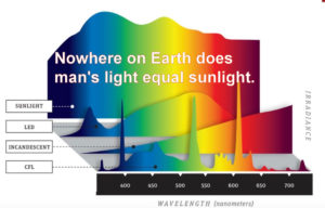
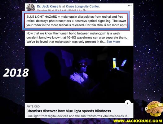
Several of my members went to a bio-hacking conference in California sponsored by Dave Apsrey and were told blue light exposure during the day was NOT harmful at all in 2016, and quite helpful for the human eye. In fact, blue light had benefits of alerting people and improving cognition. When they returned and told me about the event, I responded that this advice was probably the most damaging advice ever given at any event I’ve reviewed.
Then two years later, in 2018 the study from the University of Toledo proved my response to Asprey’s claims were fraudulent. See the we have many PEER reviewed article pointing out just how bad man-made blue light from screens are for the human retina.

Blue light is the number one health issue facing all modern humans even in the age when 5G is here because of what blue light does the retina. It causes irreversible photoreceptor damage permanently.
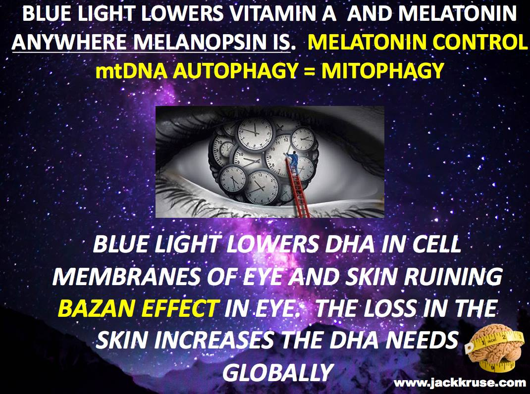
Chronic man-made blue light cause hypoxia to develop and lowers NAD+, while also dehydrates cells because of damage to cytochrome C oxidase. Liberated retinol from melanopsin is how the damage begins and progresses to cause disease. This occurs because artificial blue light decreases cause hypoxia and lowers the amount of water and CO2 a mitochondrion makes in the retina and central retinal pathways. Blue light is a form of nnEMF and all nnEMF cause elevation of blood glucose, pseudohypoxia, and dehydration of our retinal cells just as if we placed a piece of steak being heated up in a microwave oven does.

MOST DO NOT BELIEVE IT. MOST THINK IT IS HYPERBOLE.
Listen to the video before going forward in this blog.
Without retinal pigmented epithelial cells (RPE), our vision would not be sustainable. Blue light destroys the melanin (tyrosine) in the RPE to cause blindness. The RPE is a monolayer of highly polarized, quasi-hexagonal, epithelial cells. The apical membrane of RPE cells lies adjacent to the photoreceptor OS, whereas the RPE cell basal surface faces Bruch’s membrane which in some species changes shape and elongates when the eye is exposed to light. About 20–40 photoreceptor cells project toward a single RPE cell. In humans, this ratio depends on the location of these cells in the eye because the gradient of rod/cone cells and also slight differences in the dimension of photoreceptors dictates their packing density. In the periphery, the rod to RPE cell ratio is 29 whereas, in the fovea, the cones to RPE cells ratio is 22. Functions of the RPE cell layer are diverse, but from the perspective, the two roles are the most critical.
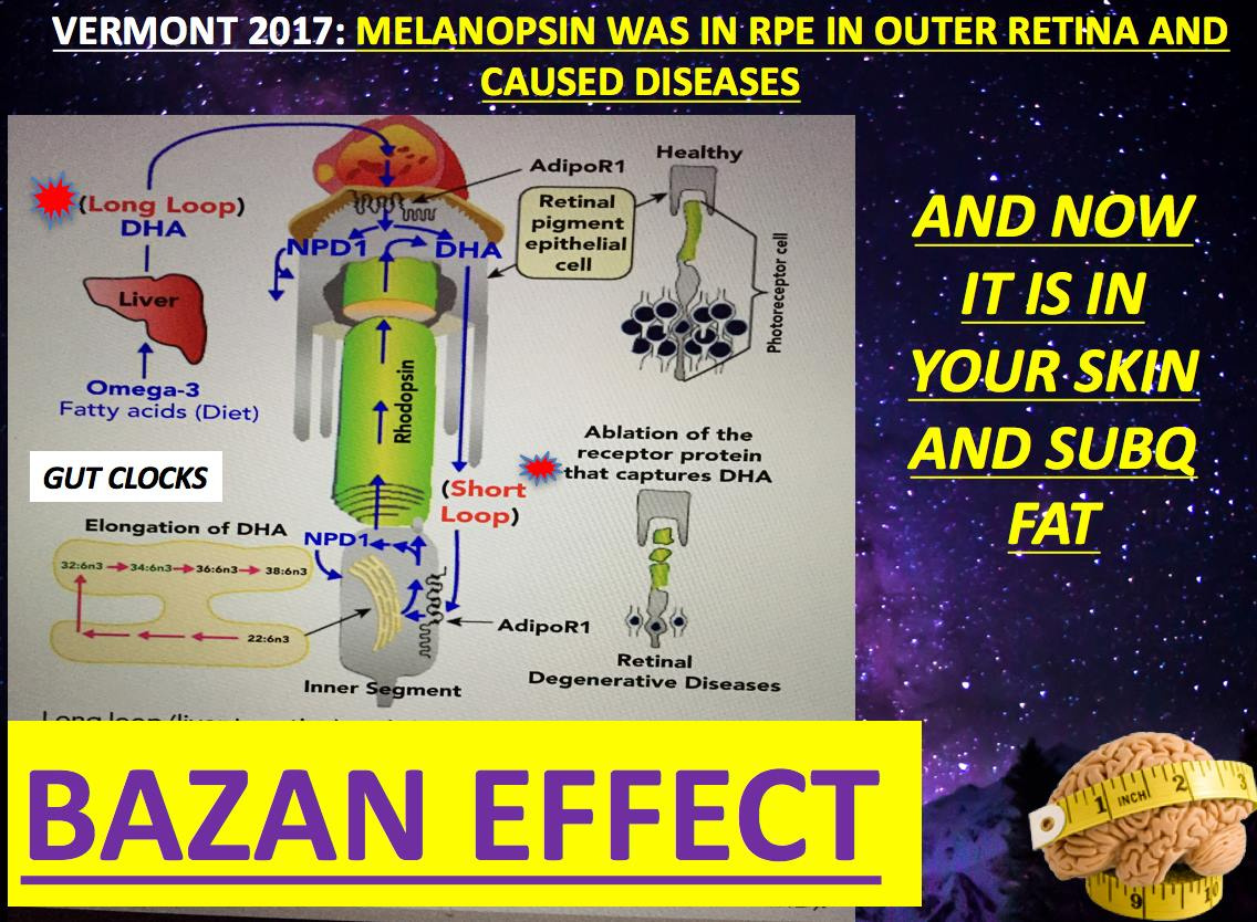
The first function of the RPE is to facilitate photoreceptor cell renewal. To provide optimal signal amplification, membranes of ROS and COS (rod/cone outer segments (ROS/COS) contain high levels of unsaturated lipids (DHA). These lipids are prone to oxidation in the presence of light, (photo)reactive retinal, and high oxygen tension, which are physiological conditions encountered in the retina. Photochemical reactions induced by light in transparent retinal tissue require a delicate balance between protein, lipid, and metabolite renewal and damaged component disposal, which when disturbed can lead to rapid and massive retinal degeneration. Impressively, postmitotic rod and cone photoreceptor cells undergo a daily regeneration process wherein ∼10% of their OS volume is shed, subsequently phagocytosed by adjacent RPE cells and replaced with newly formed outer segments. Thus, RPE cells dispose of but also accumulate an immense amount of oxidized cellular debris. Indeed it was estimated that each RPE cell phagocytoses hundreds of thousands of OS disks over a human lifetime. Several potentially toxic byproducts are condensation compounds derived from all-trans-retinal. Dysfunction of such processes as phagocytosis, lysosomal degradation, and removal of waste products by the RPE can lead to severe retinopathies, including age-related macular degeneration (AMD). This causes blindness mentioned in the video above.

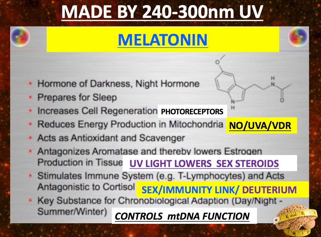
The photosensitive active retinoid, 11-cis-retinal, is produced in the RPE and delivered to the photoreceptors. The RPE is loaded with melanin. Tyrosine is the aromatic amino acid that makes melanin in the RPE. Melanin production is linked to UV light exposure of the retina and is codified by the nitric oxide levels in the eye. When anything blocks UV light exposure in the eye NO levels are altered and things made by tyrosine in the eye diminish. This lowered melanin, dopamine, melatonin, NE, and thyroid hormones in the granules of the central retinal pathways that innervate the pituitary system and the hypothalamus.

In 2018 I wrote this on my forum: ” UV radiation promotes melanin synthesis in epidermal melanocytes. UVA causes ROS/RNS generation and leads to immediate pigment darkening (IPD) within minutes, via an unknown mechanism. I believe this mechanism is tied to the blue light UVA/UVB transition and not UVA light at all because of all the things I mentioned in the When Sept ends blog/webinar on ocular melanoma. My most likely target is NO release by the RPE from full spectrum UV light.” Today in 2020 I have proof that my hypothesis was correct.
I went on to write in 2018: “Exposure of primary human epidermal melanocytes (HEMs) to high powered blue and UVA causes calcium mobilization and early melanin synthesis in human cells.
The visual photopigment rhodopsin is expressed in HEMs and contributes to UVR phototransduction. I believe melanopsin is as well with blue light. Upon UVR exposure, significant melanin production was measured within one hour; cellular melanin continued to increase in a retinal- and calcium-dependent manner up to five-fold after 24 hours.
This is why I think AMD is caused by a lack of sunlight and too much screen blue light. The reasoning is simple: Melanin synthesis via the UVB/DNA damage pathway occurs >12 h after exposure and requires de novo generation of tyrosinase, the key enzyme required for synthesis. In contrast, the mechanism underlying the UVA- and retinal-dependent melanogenesis reported causes synthesis within 1 – 4 h of exposure. This relatively rapid time course suggests that early synthesis occurs through a novel mechanism, which may use existing tyrosinase that becomes enzymatically active downstream of receptor activation.” This novel mechanism is nitric oxide production from UVB radiation inside the retina.


Sunglasses block UV light from entering the pineal gland through the optic nerves in the eyes via the central retinal pathway I spoke about in my Vermont 2017 talk on YouTube.

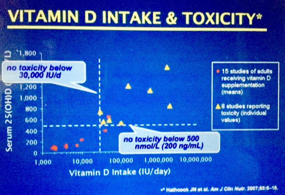
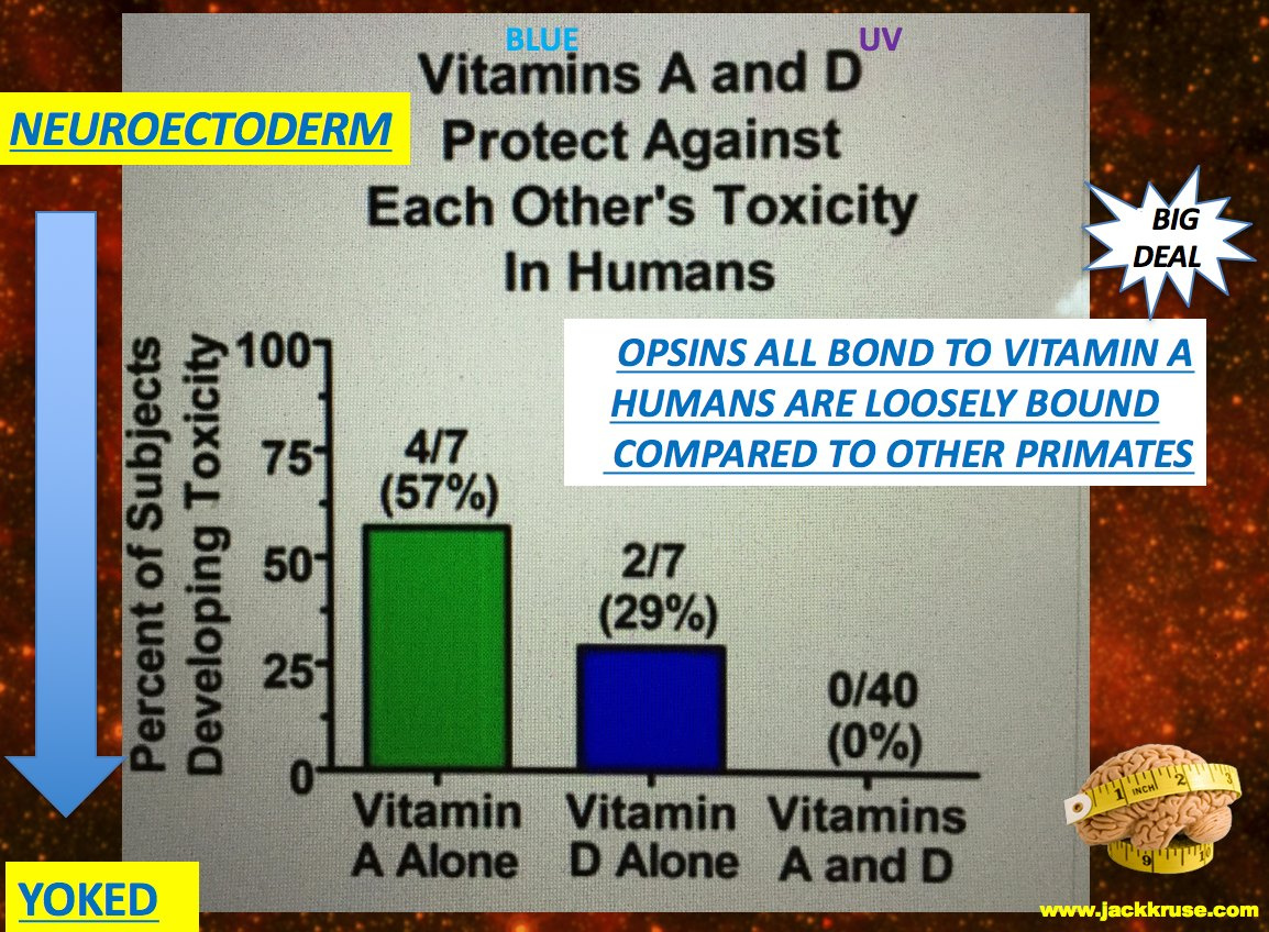
This prevents the brain from sending the signal to the pituitary gland to produce melanin, the pigment that tans the skin and protects the skin from UV radiation. Excess Vitamin A in the blood also causes a reduction of melanin and lowers its photochemical abilities as the picture above shows. This implies blocking solar frequencies from the eyes lowers melanin in the skin and the RPE. Both become more susceptible to solar damage. This is an inducible event because of wearing glasses and/or use of sun creams.

BLUE LIGHT AND KIDS: WHERE DID THE BELIEF THAT UV LIGHT IS DANGEROUS COME FROM?
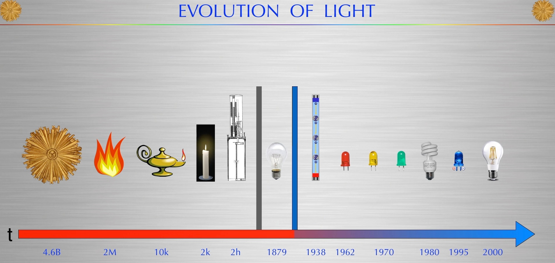
People have no idea that blue light can affect their baby born with jaundice in the hospital. Did you know today’s light specifically use blue light to get rid of jaundice? The reason is simple……yellow is the color of jaundice from the breakdown of hemoglobin and blue is yellow’s complimentary color; therefore phototherapy can be used breaking yellow pigments down using specific wavelengths of light. What is not well known is that blue light in children stimulates melanogenesis (melanocytes) and hyperpigmentation and that lowers the ability to handle UV light. This happens because cell become hypoxic and because it dehydrates (low NAD+) their cells because it raises their heteroplasmy rate. It is even associated with more nevi and more melanoma longer term in kids placed under blue light for jaundice. This is new data we have gotten over the last 30 years.
Phototherapy induces isomerization of bilirubin rendering it extractable because it becomes water-soluble by altering the charge of the yellow pigment in the kidney’s basement membrane allowing its easy clearance via the urine and hence it is used as a routine treatment of neonatal jaundice. What most people do not tell you is that pre-1950’s full spectrum lights were used for jaundice and it was a more effective choice than today’s blue lights. Why did we change from using sunlight to blue light to treat babies with jaundice in the 1950s?
In 1959 a paper on retrolental hyperplasia of the eye in jaundiced children showed up in the literature and caused all pediatricians to begin avoiding UV light in jaundice because of this one flawed paper. They linked UV light used in bulbs to the retrolental hyperplasia incorrectly. They believed the UV light in the bulbs were equivalent to the UV light in sunlight and they are not. This one paper is why today the trend in modern medicine believes/thinks UV light is always toxic. This includes the UV light that is present in the sun. None of them seem to realize that UV light has the protection of red light in sunlight at all times. No where is UV light ever present without red light in sunlight.

John Ott talked about this paper in his book “Heath and Light”. Fake artificial UV light used in those kids is not equivalent to full spectrum bulbs used prior to 1960 and it is also not equivalent to sunlight. You might want to read the paper and his book to see those details for yourself. Today’s literature now links uveal melanoma to early blue light exposure in jaundice cases. Kids with jaundice are almost always born to mothers with high heteroplasmy rates in the germ line from nnEMF exposure pre-pregnancy. These kids are all baked under blue light in NICU’s now.
Unlike cutaneous melanoma, however, ultraviolet radiation does not figure prominently among the risk factors for ocular melanoma. The reason should be obvious if you read the top half of this blog correctly and understand the implications. Moms who wear eye covering should expect jaundiced babies. You might be shocked to find out that man-made blue light exposure does seem to be linked to this new fast-growing cancer. Eye melanoma is the fastest growing cancer of the eye today. Guess why? We now abuse blue light via tech screens.
SUMMARY
Blue light use in technology/TV/mobiles is behind it. Its use is now ubiquitous globally. The effect is linked to places where humans do not get enough sunlight exposure. This is why ladies in the UK have a high incidence or melasma, melanogenesis, and jaundiced births. Studies have now described the development of an ocular tumor in animals following blue light exposure (434–475 nm).
This is the range of the melanopsin receptor in the human eye known to control melatonin production in the eye and DHA turnover in cell membranes to control the entire central retinal pathways to the SCN. When melanopsin is damage freed retinol is liberated and it permanently destroys all photoreceptors. Melatonin and COX are two such photoreceptors in the eye. In December 2017 we found out melanopsin is now in the skin and subcutaneous fat……..SO IS THIS LIKELY WHY MELANOMA AND SKIN CANCER is exploding in places like OZ. When you block melanin ability to work you sensitize the skin and eyes to damage. The sun is not the problem. How we cover our eyes and the light we now abuse is the issue that cause photoreceptor hypoxia and permanent death to cells. Liberated Vitamin A from melanopsin is the pathway to cell death via hypoxia.
Retinoid Toxicity Associated with the Evolution of Vitamin A Functions
All that glitters is not gold with Vitamin A. Many might think that Blue light hazard can be easily offset by talking exogenous Vitamin A to counteract the falling Vitamin A in the plasma. You’d
be wise to avoid people who try this or recommend it.
Virtually all vitamin A in vertebrate blood is bound to retinol bonding protein (RBP).
A broadened biological activity of vitamin A is a double-edged sword that also leads to broader toxicity caused by excessive vitamin A or its derivatives. Retinoid toxicity can be caused by physical properties of retinoid (e.g., acting like a detergent at sufficient concentrations), chemical reactivity of retinoid (e.g., modification of random proteins by free retinal), or inappropriate biological
activities (e.g., retinoic acid activating or suppressing gene expression at the wrong cell type or at the wrong time). These ideas show that proper timing is the critical aspect of Vitamin A physiology to get right. Taking exogenous Vitamin A is dangerous. Vitamin A levels in the eye, skin, brain, and blood must be tightly coupled to the circadian mechanisms to operate properly.

MELANOPSIN COUPLES TO VITAMIN A in the retina, so does this imply that all CIRCADIAN DISEASES CAN BE TRACKED BY ALTERATIONS IN THE state of electrons and protons in the RETINA FOR MOST DISEASES IN BIOLOGY????
Does it mean it might be true for the skin too?
I am going to tell you the answer now appears to be, YES.

CITES:
https://www.sciencedirect.com/science/article/abs/pii/S0306987716304169
https://www.sciencedirect.com/science/article/pii/S0022202X15301160


