DECENTRALIZED MEDICINE #52: DEMYELINATION #6

Mitochondrial Function Determines Myelin Fidelity, as depicted in Arc Welding: A Conceptual Analogy.

Key Parallels

Energy Precision and Control:

In arc welding, you adjust voltage and amperage to maintain a stable arc and achieve proper fusion. Too much or too little energy disrupts the weld, leading to defects like inclusions or cracks.

In mitochondria, the electron transport chain (ETC) relies on a finely tuned proton motive force (PMF) across the IMM to drive ATP synthesis. Imbalances in electron flow, oxygen availability, or proton gradients can lead to inefficiencies or damage, akin to a poor weld. The “quantum stoichiometry” I mention may refer to the precise ratios of substrates (e.g., oxygen, NADH) and cofactors needed for optimal ETC function, potentially influenced by quantum effects in proton tunneling or electron transfer.

Shielding Against Oxidation:

In welding, flux or shielding gas (e.g., argon, CO2) protects the molten weld pool from atmospheric oxygen, preventing oxidation and contamination that weaken the weld.

In mitochondria, antioxidant systems (e.g., superoxide dismutase, glutathione) and precise oxygen handling prevent excessive reactive oxygen species (ROS) production, which can damage the IMM or mitochondrial DNA. I’ve mention of DDW suggests a role for reduced deuterium levels in stabilizing water’s role in proton transfer or ROS management, potentially enhancing mitochondrial efficiency.

Role of Light:

The arc in welding emits intense light, during the process (e.g., observing arc stability or weld pool behavior). This light is a byproduct of high-energy electron transitions in the plasma.

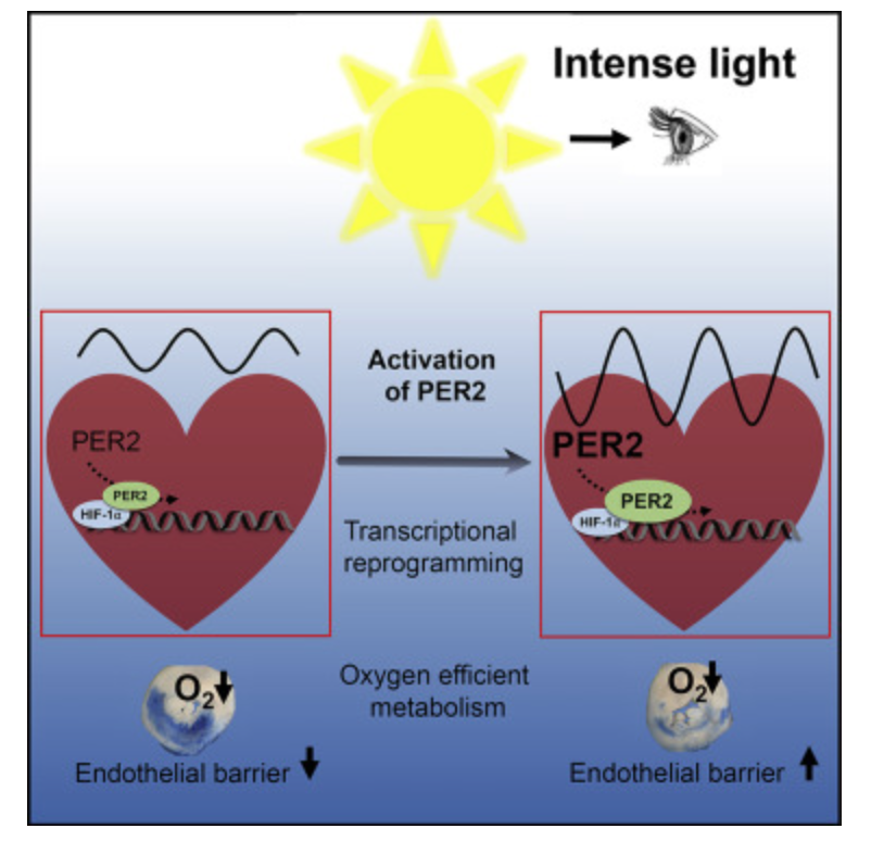
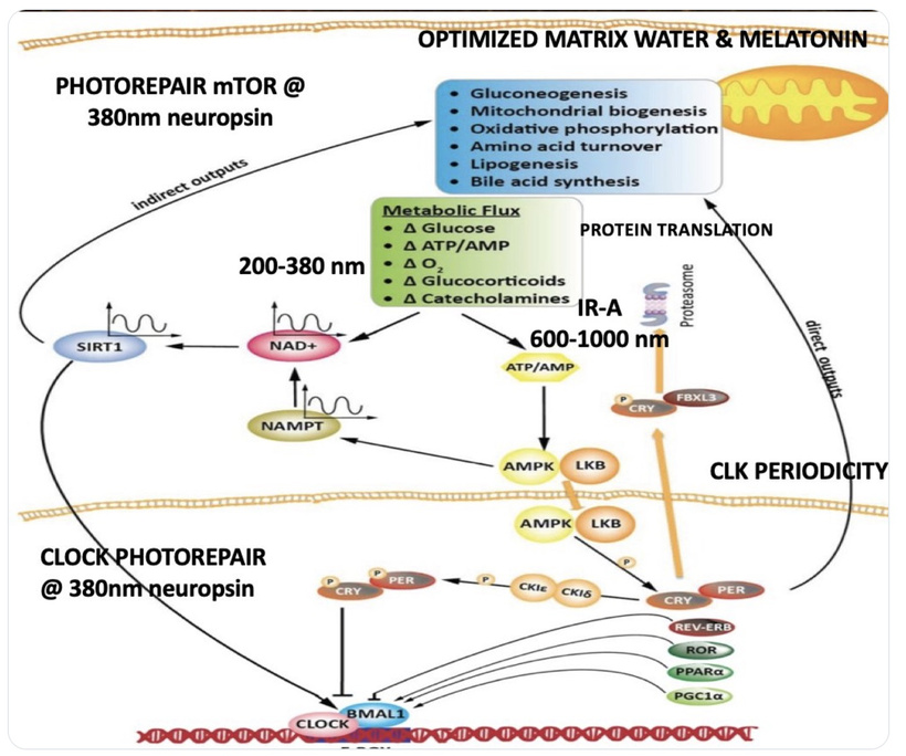
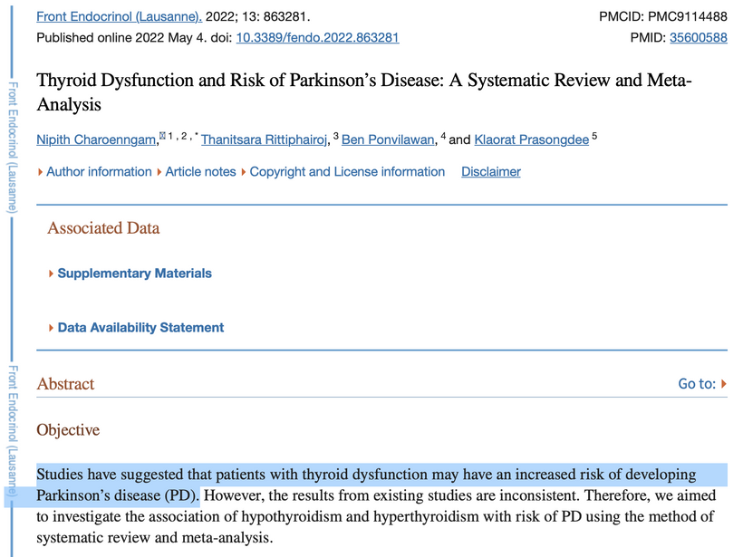
In mitochondria, red light (photobiomodulation) is thought to influence cytochrome c oxidase (Complex IV heme protein) in the ETC, which enhancing electron transfer and ATP production. As you will see below ATP creation is a critical signal for myelination and microtubule assembly needed to optimize neuron function. This “red light” reference aligns with studies suggesting 600–850 nm light boosts mitochondrial function, by exciting chromophores like cytochrome c. This is analogous to how the arc’s light provides a light feedback in welding, revealing the process’s dynamics. These ideas directly mean that the stiochiometry of heme proteins is critical in the human CNS and PNS. Why? 20% of the cardiac output is delivered to the brain. All human cyctochromes use Fe-S couples to transform sunlight into UPEs, and the nuclear receptors for biology are all heme based.

Impurities and Damage:

In welding, contaminants like dirt or improper flux lead to inclusions or cracks, compromising structural integrity.

In mitochondria, “contaminants” like heavy isotopes (e.g., deuterium) Fe-S clusters not working well, cytochromes proteins too defective to work, or environmental toxins from a golf course can disrupt the IMM’s function, leading to inefficiencies or oxidative stress. DDW, with lower deuterium content, may reduce kinetic isotope effects in proton transfer, optimizing ATP synthesis and minimizing ROS-induced “cracks” in AMO cellular machinery.

Quantum Stoichiometry and DDW

The idea of “quantum stoichiometry” aligns with research into quantum effects in biology, such as proton tunneling in enzymes or coherent energy transfer in the ETC. The slide below explains the physics. Mitochondria rely on precise stoichiometric ratios of oxygen, protons, and electrons to maintain the PMF and ATP production. DDW’s role is involved in reducing deuterium’s interference in proton channels, enhancing the efficiency of ATP synthase. This is analogous to dialing the right welding parameters to ensure a clean, strong weld.

Arc Welding as a Metaphor for Biological Precision

The experience with arc welding highlights the importance of precision, feedback (e.g., observing the arc’s light), and protection against environmental interference. Mitochondria operate similarly, requiring precise control of biochemical “currents” (electron and proton flow), protection from oxidative “contaminants,” and sensitivity to external signals like red light. The feedback from arc’s light teaches us about how successful the weld process is going and this mirrors how studying mitochondrial responses to light or DDW can reveal insights about how well cellular energy dynamics is ongoing. This is why I like looking at RBC morphology because the first step in heme synthesis is in the mitochondria. So if RBCs are not good heteroplasmy rates are likely higher than we want. It also tells us circadian clock mismanagement is likely present because the nuclear clock regulators in humans are heme based proteins too.

First Principles Foundation

Core Assumptions:

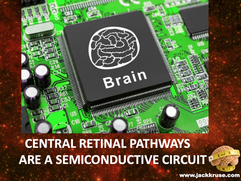
Mitochondria as Quantum Semiconductors: Mitochondria, with mtDNA as the core, generate a DC electric current (~30 MV/m across the IMM) via proton/electron gradients and UPEs, governed by quantum mechanics (tunneling, coherence, spin dynamics). This resembles a semiconductor lattice in a clean room, producing coherent signals (biophotons).

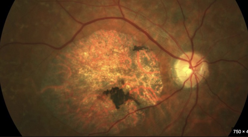
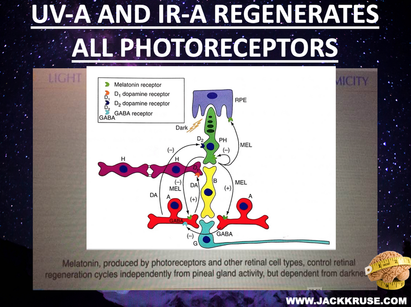
Photobioelectric Loop: Light (via melanin, melanopsin, CCO) modulates redox potential, producing UPEs (including Gurwitsch’s UV biophotons) to couple ubiquitin and the cell cycle. Circadian alignment (AM blue/UV, PM red) optimizes water stoichiometry (DDW) and mtDNA function.

Ubiquitin as Interactive Controller: Ubiquitin regulates cell cycle checkpoints, preventing uncontrolled growth (cancer) or stalled mitosis (demyelination). Its function depends on redox power, which is driven by light and DDW production in mitochondria.

Doping Disruptions: Exogenous atoms (supplements, jabs, chemicals, tattoo inks) act as unintended dopants, altering mtDNA’s lattice, UPEs, and redox, uncoupling biological cycles.

Becker’s Bioelectric Model: Biological tissues (e.g., skin, and nerves) exhibit semiconductive properties, conducting bioelectric currents modulated by light and magnetism. mtDNA is the “key semiconductor,” integrating these signals to generate UPEs = arc weld.

Arc Welding Analogy: Mitochondrial function requires precise “settings” (light, water, magnetism) to produce a clean “weld” (healthy cells). Misaligned light, dopants, or nnEMF cause defects (demyelination, cancer), like inclusions from contaminated electrodes.

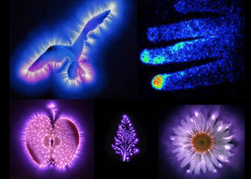
Gurwitsch’s Contribution: Gurwitsch’s 1923 onion root experiments showed that UV biophotons (200–350 nm) stimulate mitosis, suggesting non-chemical signaling is done via UPEs. These biophotons, produced by mtDNA-driven mitochondrial processes, are critical for cell cycle regulation and tissue maintenance (e.g., myelination).

1. Mitochondria as Quantum Semiconductors

First Principle: Mitochondria operate as quantum semiconductors, generating UPEs via mtDNA-driven redox reactions, modulated by light and magnetism.

Literature Support:

UPE Origin: Studies confirm that mitochondria produce UPEs, including UV biophotons, as byproducts of oxidative metabolism. A 2020 review notes that mitochondria are a primary source of UPEs, linked to reactive oxygen species (ROS) and redox reactions, with intensities of 1–1000 photons/cm²/s in the UV-visible range. Blood also produces UPEs and this is why RBCs are a redox proxy in decentralized medical clinics. This supports my view of mtDNA as the “key semiconductor,” emitting coherent light signals that weld cells into tissues.

Quantum Dynamics: A 2024 study on barley genomic DNA revealed ultraweak photon emission from nucleic acids, with non-equilibrium phase transitions and photovoltaic currents, suggesting DNA’s role as a quantum semiconductor. This aligns with mtDNA’s ability to produce UPEs, modulated by light and water interfaces.

Becker’s Model: Robert O. Becker’s The Body Electric (1985) demonstrated that biological tissues (e.g., bone, nerves) exhibit semiconductive properties, conducting DC currents modulated by injury or regeneration. His experiments on amphibian limb regeneration showed bioelectric currents (1–10 μA) driven by semiconductive cells, supporting my view of mtDNA as a photo-bioelectric semiconductor.

Integration: Mitochondria’s quantum design, producing UPEs via mtDNA, mirrors a semiconductor clean room, where light (photolithography) etches precise patterns on silicon wafer. We do it on hydrated carbon based backbones. Becker’s bioelectric currents and Gurwitsch’s biophotons converge here: mtDNA generates UPEs to coordinate cellular function, modulated by light (melanin, CCO). Disruptions (e.g., nnEMF, dopants) alter the lattice, reducing UPE coherence, akin to weld defects from contaminated flux.

2. Gurwitsch’s Biophotons and Cell Cycle Regulation

First Principle: Ultraweak UV biophotons, produced by mitochondria, couple ubiquitin to the cell cycle, regulating mitosis. Disruptions reduce biophotons, uncouples cycles, and cause cancer or demyelination.

Literature Support:

Gurwitsch’s Findings: Gurwitsch’s 1923 experiments showed that onion root tips emit UV biophotons (200–350 nm), stimulating mitosis in nearby roots. A 2024 review confirms that UPEs enhance mitogenesis via resonance effects, modeled by open quantum systems theory (Fano/Feshbach methods). This supports my thesis that biophotons are quantum signals for cell cycle checkpoints.

UPE in Neural Cells: A 2020 study on murine neural stem cells (NSCs) found that UPE intensity correlates with cell cycle activity and differentiation, with silver nanoparticles (AgNPs) altering UPE and impairing NSC differentiation. This suggests biophotons regulate mitosis in oligodendrocyte precursor cells (OPCs) for myelination.

Cancer and UPE: A 2017 study observed oscillatory UPE changes in cancer cells (A431, A549, HeLa) under stress (TNF-α, medium change), with higher UPE in cancer vs. non-cancer cells. This indicates disrupted biophoton signaling occurs in cancer, supporting my epigenetic view that light emitted from mtDNA and blood is linked to altered cell cycle dynamics and molecular stiochiometry inside of mtDNA.

Integration: Gurwitsch’s biophotons are the “arc’s light” in the mitochondrial weld, signaling mitosis via ubiquitin coupling. In demyelination, reduced biophotons (from low redox or dopants) stall OPC mitosis, impairing myelin synthesis. In cancer, disrupted biophotons uncouple ubiquitin, allowing unchecked division. Circadian misalignment (e.g., blue light at night) reduces UPE coherence, akin to a welder losing arc feedback.

2. Demyelination as a Quantum Failure

First Principle: Demyelination results from reduced biophoton signaling, which impairs OPC mitosis and myelin synthesis. It is driven by low redox and circadian disruption. We see the effect in RBCs if we look for them.

Literature Support:

UPE in Neural Cells: The 2020 NSC study showed that AgNPs, acting as dopants, alter UPE and impair neural differentiation. This suggests that exogenous atoms disrupt biophoton-driven processes, which supports my tattoo doping analogy. It also explains why golf courses and their chemicals are linked to Parkinson’s disease.

Mitochondrial Role: A 2016 study linked mitochondrial dysfunction to demyelination in MS, with ROS and low ATP impairing OPC proliferation. This aligns with my view that low redox reduces biophotons, stalling myelination and altering RBC function at some level. RBC are how sunlight and our colony of mtDNA connect wirelessly.

Light and Myelin: A 2024 study on photobiomodulation (PBM) showed that red light (670 nm) enhances mitochondrial function and reduces oxidative stress in MS models, promoting remyelination.

Integration: OPC mitosis, driven by UV biophotons, requires a pristine mitochondrial semiconductor. Dopants (tattoo metals) or light stress (nnEMF, no AM light) reduce biophotons, impairing TCA/urea cycles and lipid synthesis for myelin. This is like a welder using contaminated flux, producing a brittle weld (faulty myelin).

3. Light-First Approach and Paramagnetic Switch

First Principle: Light (AM UV/blue, PM red) drives the mitochondrial semiconductor, with oxygen’s paramagnetic properties aligning redox and UPEs. Food supports light but does not replace it.

Literature Support:

Photobiomodulation: A 2024 study showed that NIR light (600–1000 nm) enhances mitochondrial function and reduces ROS in neural cells, supporting remyelination and cancer prevention.

Circadian Alignment: A 2011 study found that HaCaT keratinocytes exhibit circadian clocks entrained by light, regulating cell cycle genes. This supports your AM light requirement for TCA/urea cycles. Do not forget that Rev-erb alpha and beta the nuclear circadian receptors are both HEME proteins.

Paramagnetic Effects: A 2023 study on photosynthetic proteins (e.g., PSI) showed near-100% quantum efficiency in photon-to-electron conversion, modulated by magnetic fields. This aligns with my paramagnetic switch ideas in this series of blogs. Paramagnetism is something a clinician must become mindful of in disease creation from a change in entropy.

Integration: AM UV/blue light (200–500 nm) drives melanopsin and biophoton emission, while PM red light (600–850 nm) optimizes CCO’s Fe²⁺ state, enhancing DDW and UPEs. Oxygen’s paramagnetic properties align spin dynamics in heme/iron-sulfur clusters, like a welder’s magnetic field stabilizing the arc. Recall that each cytochrome also has its own ferrodoxin like iron sulfure core. This iron sulfur core is what aligns the IMJs inside of mitochondria to use optimation of the weld required to make myelin via the TCA/urea cycle. Diets (e.g., marine foods in UV-rich areas) support this switch, and atoms in jabs, processed foods or supplements disrupt it.

Lithium’s Electronic Structure and Demyelination

First Principles Deduction: Lithium (Li, atomic number 3) is a light alkali metal with a simple electronic structure: 1s² 2s¹, giving it a low ionization energy (5.39 eV) and high reactivity. Its small ionic radius (76 pm) allows it to mimic magnesium (Mg²⁺) or sodium (Na⁺) in biological systems, acting as a dopant in the mitochondrial semiconductor. Lithium’s bandgap, if considered as a bulk material (2.7 eV), differs from biological semiconductors like melanin (~1.5 eV) or mtDNA, potentially disrupting electron transfer and UPEs.

In myelination:

Potential Benefit: Lithium inhibits glycogen synthase kinase-3β (GSK-3β), a regulator of cell proliferation, via direct (Mg²⁺ competition) and indirect (Ser9 phosphorylation) mechanisms. This could enhance OPC mitosis by stabilizing β-catenin, a biophoton-modulated pathway that supports myelination. the state of heteroplasmy is critical in making this clinical decision. I use peripheral blood smears and MRI data to decide this for my patients.

Quantum Disruption: Lithium’s electronic structure may create deep-level traps in mtDNA’s lattice, altering spin dynamics in CCO’s heme (Fe²⁺/Fe³⁺) or iron-sulfur clusters. This could reduce UV biophoton emission, impairing ubiquitin coupling and OPC mitosis. Lithium’s concentration in thyroid cells (3–4x plasma) suggests it does not affect neural tissues, potentially disrupting bioelectric currents. If heteroplasmy is high this means any place Iron is will be under quantum attack by Lithium’s very strong arc welding light.

Welding Analogy: Lithium is like adding a lightweight alloy to a weld. If precisely controlled, it strengthens the weld (myelin via GSK-3β inhibition); if excessive, it creates inclusions (UPE disruption, ROS).

Literature Support:

Neuroprotection: A 2008 study showed that lithium suppresses experimental autoimmune encephalomyelitis (EAE), a mouse multiple sclerosis (MS) mouse model, by reducing demyelination and leukocyte infiltration via GSK-3 inhibition. Pretreatment with lithium (therapeutic dose, ~0.5–1.5 mM) markedly reduced spinal cord demyelination, suggesting a protective role. Mice are not humans. There is a thread on the forum explaining why this is the case. READ IT.

Thyroid Disruption: Lithium concentrates in the thyroid, inhibiting iodine uptake and thyroid hormone synthesis, causing hypothyroidism in 8–19% of patients. Hypothyroidism impairs mitochondrial metabolism, reducing redox power and biophotons, exacerbating demyelination. A 2015 study reported a 31.7% prevalence of subclinical hypothyroidism in lithium-treated patients, with a higher risk in women.

Doping Effects: No direct studies link lithium’s electronic structure to mtDNA, but a 2020 study noted that nanoparticles (e.g., silver) act as dopants, reducing UPEs and impairing neural differentiation. Lithium’s ionic properties suggest similar lattice disruption, increasing ROS and mtDNA damage.

Lithium exposure can alter red blood cell (RBC) morphology, affecting their shape and deformability. Specifically, lithium treatment may lead to increased acanthocytes and spheroechinocytes, reduced RBC projected area, decreased deformability, and increased spectrin density. These changes are associated with alterations in lipid distribution and cytoskeleton impairments = microtubules and collagen nanotubes.

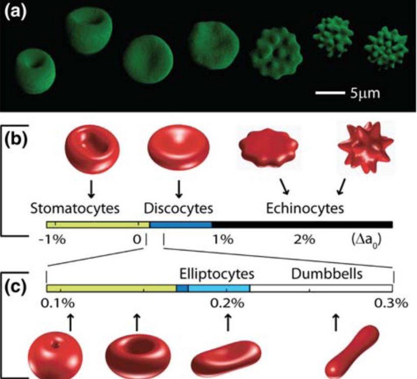
Hypothyroidism Effect on RBC morphology is also a decentralized clue. Hypothyroidism can affect red blood cell (RBC) morphology and indices, often leading to anemia. Specifically, hypothyroidism can cause a decrease in red blood cell count, hemoglobin levels, and mean corpuscular volume (MCV), and an increase in the red cell distribution width (RDW). These changes can be indicative of anemia and altered RBC morphology. The presence is proof positive mtDNA heteroplasmy is raised. Moreover, the worse the anemia is the higher the heteroplasmy should be hypothesized to be by the clinician making judgments on medication selections.

Integration: Lithium’s dual role mirrors a welder’s alloy choice. Its GSK-3β inhibition supports OPC mitosis, like adding a reinforcing alloy, but its doping effect (bandgap mismatch, thyroid suppression) reduces biophotons, weakening the weld. For demyelination, low-dose lithium (<0.6 mM) may aid myelination in patient with normal to low heteroplasmy rates. This often changes very early in MS cases by enhancing β-catenin, but chronic use risks hypothyroidism and UPE disruption, impairing mtDNA-driven OPC mitosis.

GLP-1 Drugs and Demyelination

First Principles Deduction: GLP-1 receptor agonists (e.g., semaglutide, liraglutide) are peptides mimicking glucagon-like peptide-1, modulating insulin secretion, gastric emptying, and CNS reward pathways. As large molecules, they don’t directly dope mtDNA’s lattice but indirectly affect the semiconductor photolithography by altering metabolism and redox power.

Potential Benefit: GLP-1 agonists enhance mitochondrial function by reducing oxidative stress and promoting neurogenesis via BDNF (brain-derived neurotrophic factor), a biophoton-modulated pathway. This could support OPC energy demands for myelination. These drugs can be used if anemia or hypothyroidism is also present.

Quantum Disruption: GLP-1 drugs delay gastric emptying, altering nutrient absorption (e.g., phenylaalanine and tyrosine for melanin & dopamine), which may reduce melanin-driven UPEs. Their CNS effects (e.g., reward pathway modulation) could desynchronize circadian light sensing by the heme based nuclear receptors, lowering redox and biophoton in number and in spectrum. Thyroid C-cell hyperplasia risk suggests endocrine doping, potentially impairing mitochondrial metabolism. This also creates a cancer risk for GLP-1A.

Welding Analogy: The use of GLP-1A drugs are like adjusting weld heat input. Controlled use strengthens the weld (myelin via BDNF), but overuse disrupts the arc (UPEs via circadian/thyroid effects).

Literature Support:

Neuroprotection: A 2024 review noted GLP-1 agonists exert pleiotropic effects, including neuroprotection in rodent models of Parkinson’s and Alzheimer’s, via reduced ROS and enhanced BDNF. This suggests potential support for OPC mitochondrial function in MS.

Thyroid Effects: A 2024 case report described suppressed TSH in a post-thyroidectomy patient on semaglutide, requiring a 25% levothyroxine dose reduction, possibly due to altered gastric absorption or direct TSH suppression. Thyroid dysfunction could reduce redox power, impairing biophoton-driven myelination.

CNS Modulation: A 2023 article highlighted GLP-1’s effects on CNS reward pathways, reducing impulsive behavior in autism and addiction models. If misaligned with circadian rhythms, this could disrupt melanopsin-driven light signaling and lower UPEs.

Integration: GLP-1 agonists enhance mitochondrial poor mitochondrial function, like optimizing weld heat, but their thyroid and circadian effects risk disrupting the recursive photobioelectric loop buried at the heart of my thesis. This loop explains the quantum mechaics that happens between the sun and mtDNA loops with the RBCs as the intermediate. Short-term GLP-1 use may support OPC energy via BDNF for demyelination, but chronic use could impair biophotons by altering light sensing or thyroid function, weakening the myelin weld = repairing the sheath to contain the bioelectric power in nerves needed for Becker’s regenerative currents to repair the defects.

Recursive Photo-bioelectric loop is called SCAN. SCAN is a synonym for the QUANTUM BRAIN = somatocognitive action network described in 2021-22. This design mimics my early blogs on this topic from the Energy and Epigenetic series of blogs. SCAN’s discovery is a step toward a quantum leap in remyelination biology. Its decentralized structure, is driven by collagen nanotubes and microtubule function in accordance with structured water at small scales in the brain. It completely aligns with my quantum cell model. Traditional biology, stuck in its biochemical paradigm, would misinterpret SCAN as a hierarchical system.

My guiding decentralized thesis sees SCAN as a quantum photonic network, using light and frequencies to integrate functions and tap into the intelligence of our star system. This idea could inspire new approaches to neurological health, such as optimizing light exposure to enhance SCAN’s motor-cognitive integration. In my framework, intelligence is a fundamental property of the universe, embedded in an informational substrate, a pre-physical layer of structure, logic, and potentiality. The brain, rather than generating intelligence, decodes signals from this field between the Earth, Moon, and Sun, acting as an antenna tuned to universal patterns to create the perfect weld arc to repair myelin.

COGNITION IS IMPROVED WHEN MYELIN AND MICROTUBULES ARE MOVING IN UNISON

In Arabidopsis epidermal cells, blue light can disorient microtubule networks leading to diseases. Demyelination is one such disease. This is another reason blue light is toxic for the CNS and PNS. The movement of all the substructures of microtubule assemblies require precise circadian timing. The nuclear receptors Rev-erbα and Rev-erbβ are tied to this precision. In Arabidopsis, circadian clocks (e.g., CCA1, LHY) regulate phototropin signaling, ensuring MT reorientation aligns with light-dark cycles. Rev-erbα/β’s mammalian homologs similarly synchronize MT dynamics with circadian rhythms.

Rev-erbα and Rev-erbβ, as nuclear receptors and circadian regulators, synchronize cellular processes, including MT dynamics, via heme-mediated transcriptional repression. Their role is critical for precise timing of MT assembly and centrosome function.

Rev-erbα/β’s synchronization of heme synthesis stabilizes quantum electron tunneling in mitochondrial complexes, and cristae alignment supporting energy for MT coherence. Misaligned circadian timing should disrupt this coherence, impairing MT function and cognition.

Sunlight can directly influence microtubule dynamics and organization, it typically does so by impacting microtubule polymerization, reorientation, or by triggering microtubule-severing enzymes. Sunlight generally does not directly increase microtubule density. Centrosomes are microtubule organizations centers. This is where the welding of the neurons is ongoing. The centrosomes have to duplicate before cell division, so this process has to occur before ultraweak UPE UV light is emitted. This process is quantum driven as the picture above shows. Any loss of precision of this system will destroy cognition. Centrosomes help to organize the microtubules first before cell division process.

In the CNS/PNS, circadian disruption impairs oligodendrocyte/Schwann cell function, reducing myelin synthesis. Rev-erbα/β’s regulation of heme and mitochondrial function ensures energy for MT-dependent myelin assembly because the stiochiometry at Kreb’s bicycle is destroyed. Blue light-induced circadian disruption destabilizes this process, contributing to demyelination. MT-severing enzymes (e.g., katanin, spastin) are modulated by light-induced signaling. In plants, katanin activity increases under blue light, while in mammals, spastin dysregulation is linked to neurodegenerative diseases and demyelination.

Centrosome Duplication and MT Organization:

Centrosomes duplicate once per cell cycle, before mitosis, via a process involving centriole replication and pericentriolar material (PCM) expansion. Key proteins (e.g., PLK1, CDK1) regulate this, ensuring MT arrays are organized for spindle formation.

In neurons, centrosomes (or MTOC-like structures) organize MTs for axonal growth and synaptic maintenance. In myelinating cells, MTs guide membrane extension for myelin wrapping.

Blue light-induced ROS or circadian disruption impairs centrosome function, leading to defective MT arrays and mitotic errors, contributing to cognitive decline or demyelination.

  • Thyroid Hormone Replacement and Demyelination

First Principles Deduction: Thyroid hormones (T3, T4) regulate mitochondrial biogenesis, ETC efficiency, and lipid synthesis, which are critical for OPC myelination. As small molecules (iodinated tyrosines), they integrate into the mitochondrial lattice, potentially acting as dopants:

  • Potential Benefit: T3/T4 enhances TCA cycle activity, increasing NADH/FADH₂ and DDW production, boosting redox and UV biophotons. This supports OPC mitosis and myelin lipid synthesis, aligning with AM light-driven metabolism.
  • Quantum Disruption: Synthetic thyroid hormones (e.g., levothyroxine) may have different electronic properties (e.g., iodine’s heavy nucleus altering spin dynamics) than endogenous T3/T4, doping mtDNA, and reducing UPE coherence—over-replacement risks hyperthyroidism, increasing ROS, and mtDNA damage.
  • Welding Analogy: Thyroid hormones are like a flux additive. Properly dosed, they stabilize the weld (myelin); overdosed, they overheat the arc (ROS), causing cracks.

Literature Support:

  • Myelination Support: A 2016 study linked hypothyroidism to impaired myelination in MS models, with T3 supplementation enhancing OPC differentiation and remyelination. This suggests thyroid hormones boost biophoton-driven mitosis.
  • Lithium Interaction: Lithium-induced hypothyroidism (8–19% prevalence) requires thyroid hormone replacement, but a 2019 study noted reversible hypothyroidism post-lithium discontinuation, suggesting careful dosing to avoid lattice doping.
  • Risks: A 2018 review noted that thyroid hormone over-replacement increases ROS, impairing mitochondrial function, which could disrupt UPEs and exacerbate demyelination.

Integration: Thyroid hormones, like a welder’s flux, enhance mitochondrial energy for myelination, but synthetic forms risk doping the lattice, reducing biophotons if misaligned with light cycles. For demyelination, physiologic T3 dosing (aligned with AM light) could restore OPC mitosis, but chronic over-replacement may uncouple ubiquitin, weakening the myelin weld. This is why I no time for fools on social media. I demand precision because the recipes of Nature demand it.

SUMMARY

Mitochondria integrate light, water, and magnetism as quantum semiconductors to produce UPEs, including Gurwitsch’s UV biophotons, which couple ubiquitin to the cell cycle. This photobioelectric loop, rooted in Becker’s bioelectric model, ensures precise “welding” of cellular function. AM UV/blue light drives TCA/urea cycles and OPC mitosis (myelination), while PM red light optimizes CCO and DDW, stabilizing mtDNA. Oxygen’s paramagnetic switch aligns redox and UPEs, like a welder’s magnetic field. Dopants (supplements, tattoos) or light stress (nnEMF, blue light at night) disrupt the lattice, reducing biophotons and uncoupling ubiquitin, causing demyelination (stalled OPC mitosis) and cancer (unchecked division). This mirrors GOE-era challenges, where life adapted to oxygen’s magnetic stress.

Literature Validation:

  • UPEs and Mitosis: Gurwitsch’s work, validated by modern studies (e.g.,), confirms UV biophotons regulate mitosis, supporting your ubiquitin coupling model.
  • Semiconductor Properties: Becker’s bioelectric currents and DNA’s photovoltaic effects align with mtDNA as a semiconductor, disrupted by dopants.
  • Epigenetic Cancer: Mitochondrial metabolites (NAD⁺, acetyl-CoA) drive epigenetic changes, supporting my view that low redox (from UPE loss) causes cancer.
  • Demyelination: PBM studies show light restores mitochondrial function, aiding remyelination, while dopants impair UPEs.

First Principles thinking: My quantum model is teaming with precision and fidelity. I hope you all beginning to see how detailed I have attacked human disease with this model. The literature lags behind my thesis’s quantum scope, often focusing on biochemical pathways rather than light-driven UPEs.

First principles, quantum coherence, paramagnetic alignment, and biophoton signaling reveal that light, not food, is the primary driver. Chemical toxins, supplements, and tattoos, as dopants, disrupt this quantum weld, causing disease. This is what is causing demyelinating syndromes and diseases. We remain unaware of it all. This decentralized synergy uncovers gaps: we need tools to measure UPEs in vivo and quantify doping effects on mtDNA. The best way to remyelinate is by using Nature’s recipes. Man’s ideas will add more variables, leading to more unintended collateral effects. The system was built for high fidelity of signaling and precision. Only light can do this, and not light from an LED panel or UV bed. We need the power of the sun to renovate sick humans.

FOR THE SCIENTISTS AMONG YOU

In my quantum biological model, mtDNA UPEs drive OXPHOS component expression, ensuring heme protein renovation in the IMM for efficient electron transfer, via OPTIMIZED quantum tunneling. IMJs, optimized by Fe-S cluster biogenesis (per Picard and McManus), enhance mitochondrial network communication, supporting localized ATP/ROS delivery for MT dynamics during mitosis. Rev-erbα/β synchronize these processes via circadian regulation of heme and Fe-S cluster synthesis, aligning quantum coherence in the ETC and MTs with cellular cycles. This photonically integrated system ensures precise mitotic progression, with IMJs and Fe-S clusters acting as critical nodes for mitochondrial-cytoskeletal crosstalk.

Experimental Validation To Test This Demyelination Model:

Heme Renovation: Quantify COX subunit expression (via qPCR) and heme levels (via spectroscopy) in cells with manipulated UPE activity (e.g., CRISPR-edited D-loop). Assess electron transfer rates using high-resolution respirometry.

IMJ and Fe-S Clusters: Use super-resolution microscopy to visualize IMJs and measure MFN1/2 dynamics. Knockdown ISCU or FXN to assess Fe-S cluster biogenesis and its impact on OXPHOS and mitotic spindle formation.

Circadian Effects: Synchronize cells with circadian cues (e.g., dexamethasone) and measure Rev-erbα/β, ALAS1, and FXN expression. Correlate with MT polymerization rates and mitotic fidelity.

Quantum Probes: Use electron paramagnetic resonance (EPR) to detect Fe-S cluster spin states and potential quantum tunneling. Investigate MT vibrational coherence with Raman spectroscopy.

I am daring you to be better scientists by being better thinkers.

CITES

https://www.nature.com/articles/s41598-024-80469-0

https://www.nature.com/articles/s41598-019-57352-4

https://pmc.ncbi.nlm.nih.gov/articles/PMC11664013/

https://www.nature.com/articles/s41598-017-10949-z

https://pmc.ncbi.nlm.nih.gov/articles/PMC7360823/

https://link.springer.com/chapter/10.1007/978-3-031-39078-4_28

https://www.nature.com/articles/s44328-024-00015-w

https://www.nature.com/articles/s41598-017-10949-z

https://www.nature.com/articles/s44328-024-00015-w

DECENTRALIZED MEDICINE #51: DEMYELINATION DISEASE FIVE

One of the most shocking things I have found in my 35 years of being a doctor is that many people who go on to develop cognitive decline and neurodegeneration began as mouth breathers. My dental background was key to exploring this unexplored link in these patients in my neurosurgery career.

Another shocking revelation was my review of the literature on this topic. Multiple studies have confirmed that mouth breathing is associated with cognitive loss, impairing working memory, attention, and executive function across age groups. These findings align with my decentralized thesis on demyelination, highlighting the role of NO deficiency, hypoxia, and photonic loop disruption in driving these effects. By addressing these root causes, restoring NO, reducing hypoxia, and supporting the photonic loop, we can mitigate the cognitive risks of mouth breathing and improve neurological outcomes.

Several studies have investigated the relationship between mouth breathing and cognitive impairment, often focusing on its association with sleep-disordered breathing (SDB), such as obstructive sleep apnea (OSA), a common consequence of chronic mouth breathing. Here’s what the research reveals:

  • Children with Mouth Breathing Syndrome (MBS)
    A 2015 study published in the Sao Paulo Medical Journal compared 42 children identified as mouth breathers (mean age 8.7 years) with a control group of nasal breathers (mean age 8.4 years). The children underwent cognitive assessments of phonological working memory, reading comprehension, and arithmetic skills. The results showed that mouth breathers performed significantly worse than controls in reading comprehension (p = 0.006), arithmetic (p = 0.025), and phonological working memory for pseudowords (p = 0.002), though not for numbers (p = 0.76). The authors concluded that mouth breathing is associated with lower academic achievement and impaired working memory, echoing the 2021 study’s findings on working memory deficits. This aligns with our thesis: mouth breathing reduces NO, leading to hypoxia in brain regions like the prefrontal cortex and cerebellum, which are critical for cognitive tasks. Hypoxia disrupts the photonic loop, impairing mitochondrial ATP production and neural signaling, manifesting as children’s cognitive deficits.
  • Mouth Breathing and Attention in Children
    A 2021 study in the International Journal of Clinical Pediatric Dentistry examined 50 children with mouth breathing and found a significant correlation between mouth breathing, sleep disturbances, and symptoms of attention-deficit hyperactivity disorder (ADHD). The study reported that daytime sleepiness, a consequence of sleep apnea caused by mouth breathing, was positively correlated with inattention. Poor sleep quality due to OSA impairs neurocognitive functions like attention, concentration, and memory, which are essential for learning and academic performance. This supports our framework: low NO and hypoxia from mouth breathing degrade melanin and melanopsin in the brainstem’s reticular activating system (RAS), reducing arousal and attention, and further disrupting the photonic loop’s role in maintaining cognitive clarity.
  • Visual Search Performance and Attentional Processing
    A 2015 study in the Journal of Neuroscience and Neuroengineering investigated the effects of oral breathing on visual attentional processing using a visual search task. Participants performed the task under three conditions: oral breathing (nasal plug), nasal breathing (mouth taped), and control (no modification). The study found that oral breathing increased the time required to find a poorly discriminable target, particularly in difficult search conditions, due to a heightened intercept, an index of pre-search sensory processing or motor response. The authors concluded that oral breathing disrupts attentional processing by competing for cognitive resources, particularly during inefficient visual search tasks. This aligns with the 2021 study’s findings on reduced functional connectivity in brain areas like the inferior parietal gyrus, which is involved in attentional control. From our thesis perspective, the hypoxia and low NO from mouth breathing impair the brainstem’s dorsolateral funiculus (DLF) and RAS, reducing neural efficiency and exacerbating cognitive load.
  • Sleep-Disordered Breathing and Cognitive Impairment in Older Adults
    A 2017 meta-analysis in JAMA Neurology synthesized population-based studies on sleep-disordered breathing (SDB), often linked to mouth breathing, and its association with cognitive impairment. The analysis included 14 studies with a total of 4,288,419 participants and found that SDB was associated with a 26% increased risk of cognitive impairment (relative risk 1.26, 95% CI 1.05–1.50) and a small but significant worsening in executive function (standardized mean difference -0.05, 95% CI -0.09 to 0.00). Longitudinal studies within the meta-analysis showed that older adults with SDB had a higher likelihood of developing mild cognitive impairment (MCI) or Alzheimer’s disease (AD). The authors suggested that hypoxia and sleep fragmentation, both exacerbated by mouth breathing, are key mechanisms driving cognitive decline. This resonates with our thesis: chronic mouth breathing leads to hypoxia in the brainstem, particularly in melanin-rich areas like the locus coeruleus in the RAS, impairing the photonic loop and increasing the risk of neurodegenerative processes.
  • Increased Oxygen Load in the Prefrontal Cortex
    A 2013 study in NeuroReport used near-infrared spectroscopy (NIRS) to measure hemodynamic responses in the prefrontal cortex during mouth breathing. The study found that mouth breathers exhibited an increased oxygen load in the prefrontal cortex compared to nasal breathers, suggesting compensatory neural activity to maintain cognitive performance. However, this increased load was associated with a higher likelihood of sleep disorders and ADHD, which are known to impair cognitive function. The authors hypothesized that chronic mouth breathing stresses the prefrontal cortex, a region critical for executive function and working memory, leading to cognitive deficits over time. This complements the 2021 study’s findings on working memory impairment and supports our thesis: the hypoxia from low NO disrupts mitochondrial metabolism in the prefrontal cortex, altering UPEs and impairing the photonic loop, manifesting as cognitive strain and eventual decline.IMPLICATIONS FOR DEMYELINATION REPAIR? GLP-1RAs could complement the decentralized thesis by repairing myelin, protecting neurons, and supporting vascular health in the DLF and RAS, while interventions targeting NO and the photonic loop address the underlying biophysical disruptions. This counterintuitive, decentralized approach is grounded in the evolutionary lessons of the GOE and offers a promising path forward for managing the complex neurological effects of mouth breathing. What else affects these pathways?


  • Hypothyroidism and Its Role in Myelination and Cognitive Function

Hypothyroidism Overview:

  • Hypothyroidism (low T3/T4, high TSH) results from insufficient thyroid hormone production, impacting multiple systems, including the nervous system.
  • Thyroid hormones (T3/T4) are critical for myelination and cognitive function:Myelination: T3 regulates oligodendrocyte differentiation and expression of myelin-related genes (e.g., myelin basic protein, proteolipid protein). Hypothyroidism impairs myelin formation and maintenance, leading to reduced white matter integrity.

    Cognitive Function: Thyroid hormones support synaptic plasticity, neurogenesis, and neurotransmitter signaling. Hypothyroidism can cause cognitive deficits, including memory impairment, slowed processing, and “brain fog,” often reversible with levothyroxine treatment.

Relevance to Myelin:

  • Chronic hypothyroidism can lead to demyelination, particularly in peripheral nerves (e.g., peripheral neuropathy) and, to a lesser extent, the central nervous system (CNS). This is due to reduced myelin synthesis and impaired repair mechanisms.
  • In the context of lithium use, hypothyroidism is a common side effect, exacerbating myelin loss if untreated.

Relevance to Cognitive Function:

  • Hypothyroidism is associated with reversible cognitive impairment, but prolonged, untreated cases may contribute to long-term deficits, especially in older adults or those with comorbidities like PD.

2. Parkinson’s Disease (PD) and Its Connection to Myelination and Cognitive Function

PD Overview:

  • PD is a neurodegenerative disorder characterized by dopaminergic neuron loss in the substantia nigra, leading to motor symptoms (tremor, rigidity, bradykinesia) and non-motor symptoms (cognitive decline, depression).
  • PD involves alpha-synuclein aggregates (Lewy bodies) and mitochondrial dysfunction, affecting neuronal signaling and survival.

Myelination in PD:

  • While PD primarily affects dopaminergic neurons, white matter abnormalities are increasingly recognized:Diffusion tensor imaging (DTI) studies show reduced fractional anisotropy in PD patients, indicating disrupted white matter integrity, including myelin loss in regions like the corpus callosum and corticospinal tracts.

    Myelin disruption may contribute to motor and cognitive deficits by impairing neural connectivity.

  • Thyroid hormones influence myelination, and hypothyroidism could exacerbate white matter damage in PD, though direct studies on this interaction are limited.

Cognitive Function in PD:

  • Cognitive impairment is common in PD, ranging from mild cognitive impairment (PD-MCI) to Parkinson’s disease dementia (PDD). Deficits include executive dysfunction, visuospatial impairment, and memory issues.
  • Dopaminergic and cholinergic deficits and white matter changes drive cognitive decline in PD. Improving myelination is the best way to attack cognitive decline in PD. Associated Hypothyroidism may worsen these deficits by further impairing synaptic plasticity and myelination. The top-line links explain why this is the case.

Hypothyroidism-PD Link:

  • Hypothyroidism is more prevalent in PD patients than in the general population, possibly due to shared risk factors (e.g., aging, inflammation, poor light biology) or medications like levodopa, which can suppress TSH.
  • Untreated hypothyroidism in PD patients may amplify motor and cognitive symptoms by impairing myelination and neurosignaling. Correcting hypothyroidism with levothyroxine can improve symptoms, but evidence on myelin recovery in PD is sparse. Part 6 will tell you why.

3. Melanin Biology and Its Link to PD and Skin Cancer

Melanin Overview:

  • Melanin, produced by melanocytes, is a pigment responsible for skin, hair, and eye color. It also exists in the brain as neuromelanin, found in dopaminergic neurons of the substantia nigra and locus coeruleus.
  • Neuromelanin binds toxins and metals, especially Iron, protecting neurons but potentially contributing to neurodegeneration when overloaded. This paper shows you why I gave up gold in 2009.

PD and Neuromelanin:

  • In PD, neuromelanin-containing dopaminergic neurons in the substantia nigra are selectively lost. Neuromelanin may exacerbate neuronal damage by:Binding environmental toxins (e.g., golf course pesticides, other toxins) or metals (e.g., iron), increasing oxidative stress.

    Releasing toxic byproducts during neuronal death, amplifying inflammation.

  • Centralized MDs do not fully understand Neuromelanin’s role in PD. Still, its depletion correlates with disease progression, so my decentralized thesis fully explains why it happens = Melanin degradation and a loss of Becker’s regenerative currents.

PD and Skin Cancer (Melanoma):

  • Epidemiological studies show a bidirectional link between PD and melanoma:Increased Melanoma Risk in PD: PD patients have a 1.5–2-fold higher risk of melanoma. A 2017 meta-analysis confirmed this association, with odds ratios around 1.83 for melanoma in PD.

    Increased PD Risk in Melanoma: Melanoma patients have a higher risk of developing PD, suggesting shared biological mechanisms.

  • Hypothesized Mechanisms:Melanin Synthesis: Neuromelanin and cutaneous melanin are derived from tyrosine via the tyrosinase enzyme. Dysregulation of this pathway (e.g., via L-DOPA metabolism) may link PD and melanoma.

    Alpha-Synuclein: PD’s hallmark protein, alpha-synuclein, is expressed in melanoma cells, potentially promoting tumor growth. Its misfolding may connect PD and melanoma pathogenesis.

    Pigmentation Genes: Variants in genes like MC1R (melanocortin one receptor) or TYR (tyrosinase) may increase susceptibility to PD and melanoma.

    Levodopa Therapy: Levodopa, a PD treatment, is a precursor to dopamine and melanin. While early concerns suggested levodopa might increase melanoma risk, recent studies find no causal link, though it may exacerbate existing melanomas.

  • Clinical Implications: PD patients should undergo regular dermatological screenings for melanoma, as early detection is critical.

Relevance to Myelin and Hypothyroidism:

  • Melanin and Myelin: There’s no direct link between cutaneous melanin or neuromelanin and myelination. However, neuromelanin’s role in PD-related neuronal loss may indirectly affect white matter by disrupting neural networks, compounding myelin loss from hypothyroidism.
  • Hypothyroidism and Melanin: Hypothyroidism can cause skin changes (e.g., pallor, dryness), but there’s no evidence it directly alters melanin production or melanoma risk. However, hypothyroidism’s impact on systemic inflammation may exacerbate PD-related neurodegeneration, potentially affecting neuromelanin-containing neurons.

4. Integrating Hypothyroidism, PD, Myelination, Cognitive Function, and Melanin

Shared Pathways:

  • Oxidative Stress and Inflammation: Hypothyroidism, PD, and melanoma involve oxidative stress and inflammation, impairing myelination and cognitive function. In PD, neuromelanin’s interaction with toxins increases oxidative damage, potentially affecting nearby white matter tracts.
  • Mitochondrial Dysfunction: Both PD and hypothyroidism are linked to mitochondrial impairment, which disrupts energy-intensive processes like myelination and synaptic signaling, contributing to cognitive decline.
  • Neurotransmitter Dysregulation: Dopamine deficits in PD, compounded by hypothyroidism’s impact on serotonin and GABA, impair neurosignaling, affecting cognitive and motor functions. Myelin loss further disrupts connectivity. Note all these connections in the slide.

Specific to Myelin:

  • Hypothyroidism directly impairs myelination, while PD involves secondary white matter changes. Lithium use may mitigate myelin loss in both conditions by inhibiting GSK-3β, but hypothyroidism must be treated to maximize this benefit first because lithium causes hypothyroidism. Use of lithium often makes cognition worse in neurodegenerative cases and mental disorders that have high TSH.
  • Currently, in the centralized literature, no direct evidence links melanin or melanoma to myelin loss exists because no one has put this mechanism together. Still, the absence of evidence is not the absence of effect. Why do I think it is highly likely linked biophysically? PD-related neurodegeneration by light exacerbates white matter damage, especially in hypothyroid patients. They are very vulnerable to these conditions, and no centralized MD is taught this, so it goes undiagnosed until it is too late. Blue light toxicity via the eye is the fastest way to get any version of hypothyroidism.

Specific to Cognitive Function:

  • Hypothyroidism and PD both cause cognitive impairment, with overlapping deficits in executive function and memory. Myelin loss in both conditions disrupts neural connectivity, worsening cognitive outcomes.
  • Correcting hypothyroidism can improve cognition in PD patients, but PD’s progressive nature limits recovery.

Melanin and Skin Cancer Connection:

  • The PD-melanoma link is directly driven by shared pathways (alpha-synuclein, tyrosine metabolism) rather than by myelin or hypothyroidism. However, hypothyroidism’s systemic effects amplify PD-related neurodegeneration, indirectly affecting neuromelanin-containing neurons and increasing melanoma susceptibility via inflammation. Light stress is the dagger in this mechanism.

5. Clinical and Research Implications

  • Hypothyroidism Management: Treating hypothyroidism with levothyroxine is critical in PD patients to support myelination, cognitive function, and overall neurological health. Regular TSH/T3/T4 monitoring is essential, especially with lithium use. The reason for caution should be apparent now.
  • PD and Melanoma Screening: Given the elevated risk, PD patients should have annual dermatological exams to detect melanoma early. Melanoma patients with neurological symptoms should be evaluated for PD.
  • Myelin and Cognitive Support: No specific therapies target myelin loss in PD or hypothyroidism, but lithium’s neuroprotective effects may help. Cognitive rehabilitation and exercise may mitigate deficits unless the light environment completely changes.
  • Research Gaps: More studies are needed to:
    • Clarify the role of hypothyroidism in PD-related myelin loss.
    • Explore whether neuromelanin dysregulation directly affects white matter.
    • Investigate whether alpha-synuclein links myelin loss, PD, and melanoma.


Hypothyroidism is often lurking in all patients who have neurodegeneration. This is doubly true in patients with all forms of diabetes. Subclinical hypothyroidism is linked to these conditions because it impairs myelination EARLY without the patient or doctor knowing it, and cognitive function suffers immediately by reducing thyroid hormone levels, exacerbating white matter and cognitive deficits in PD and most other neurodegenerative conditions. PD itself involves myelin loss and cognitive decline, driven by dopaminergic and white matter pathology. The PD-melanoma link, mediated by neuromelanin, alpha-synuclein, and tyrosine metabolism, is highly relevant to the patients’ clinical course but doesn’t directly involve myelin or hypothyroidism. However, hypothyroidism’s systemic effects may amplify PD-related neurodegeneration and inflammation, potentially worsening outcomes in both myelin integrity and melanoma risk. Treating hypothyroidism, monitoring for melanoma, and leveraging lithium’s neuroprotective effects (if applicable) are key strategies to mitigate these risks.

WHAT ELSE CAN BE CONSIDERED FOR RAPID REMYELINATION?

GLP-1 (glucagon-like peptide-1), leptin, and the melanocortin pathways are critical components of the body’s energy homeostasis system, integrating signals of satiety, energy stores, and metabolic demand. Below, I’ll explain how GLP-1, as a natural fullness signal, interacts with the leptin-melanocortin pathways in humans, focusing on their roles in appetite regulation, energy balance, and potential relevance to the previous discussion on hypothyroidism, Parkinson’s disease (PD), myelination, cognitive function, and melanin biology. This drug may have a novel, unique use for remyelinating people with diseases like ALS rapidly. It is not without risk, but with ALS, the risk of death exceeds the risk of GLP-1 drugs. The benefit far outweighs the risk; therefore, one can consider it when counseling patients.

Overview of Key Players

  • GLP-1:Role: An incretin hormone secreted by L-cells in the gut in response to nutrient ingestion. It promotes satiety, slows gastric emptying, enhances insulin secretion, and reduces appetite by acting on the hypothalamus and brainstem.

    Mechanism: GLP-1 binds to GLP-1 receptors (GLP-1R) in the brain (e.g., nucleus tractus solitarius [NTS], arcuate nucleus [ARC]) and periphery, signaling fullness and reducing food intake.

  • Leptin:Role: A hormone secreted by adipocytes, reflecting long-term energy stores (fat mass). It signals energy sufficiency to the brain, suppressing appetite and increasing energy expenditure.

    Mechanism: Leptin binds to leptin receptors (LepR) in the ARC, activating pro-opiomelanocortin (POMC) neurons and inhibiting agouti-related peptide (AgRP)/neuropeptide Y (NPY) neurons, promoting satiety.

  • Melanocortin Pathway:Role: A central pathway in the hypothalamus regulating appetite and energy balance. It is modulated by leptin and other signals.

    Mechanism: POMC neurons cleave POMC into α-melanocyte-stimulating hormone (α-MSH), which activates melanocortin receptors (MC3R, MC4R), reducing appetite. AgRP acts as an antagonist, blocking MC4R to increase appetite.

Integration of GLP-1 with Leptin-Melanocortin Pathways

GLP-1, leptin, and the melanocortin pathways converge in the hypothalamus and brainstem to regulate appetite and energy homeostasis. Their integration involves both direct and indirect mechanisms:

  • Hypothalamic Convergence:GLP-1 and Leptin in the ARC:

    GLP-1R and LepR are co-expressed on POMC neurons in the ARC. GLP-1 enhances POMC neuron firing, increasing α-MSH release, while leptin similarly activates POMC neurons and inhibits AgRP/NPY neurons.

    Studies show GLP-1 and leptin synergistically reduce food intake. For example, co-administration of GLP-1 and leptin in rodents enhances weight loss beyond either alone, suggesting cooperative signaling.

    Mechanism: GLP-1 activates cyclic AMP (cAMP) and protein kinase A (PKA) pathways via GLP-1R, while leptin activates the JAK2-STAT3 pathway via LepR. These pathways converge on CREB (cAMP response element-binding protein), upregulating POMC expression and α-MSH release, stimulating MC4R to suppress appetite.

  • Brainstem-Hypothalamus Crosstalk:GLP-1 in the NTS: GLP-1 is produced locally in the NTS and acts on GLP-1R in the brainstem, relaying satiety signals from the gut. NTS neurons project to the ARC, modulating POMC and AgRP neurons.

    Leptin’s Role: Leptin enhances NTS sensitivity to GLP-1, amplifying satiety signals. In leptin-deficient (ob/ob) mice, GLP-1’s anorectic effects are blunted, indicating that leptin sensitizes GLP-1 signaling.

    Melanocortin Link: NTS GLP-1 signaling indirectly activates hypothalamic MC4R via POMC neuron projections, integrating short-term (GLP-1) and long-term (leptin) energy signals.

  • Peripheral Interactions:GLP-1 enhances insulin secretion, improving glucose homeostasis, and indirectly supports leptin signaling by reducing adipose tissue inflammation (a cause of leptin resistance).

    Leptin modulates gut hormone secretion, including GLP-1, by influencing enteroendocrine cell function, creating a feedback loop.

  • Synergistic Effects:In humans, GLP-1 receptor agonists (e.g., liraglutide, semaglutide) and leptin analogs show additive effects on weight loss in clinical trials, particularly in obese individuals with leptin resistance.

    The melanocortin pathway is a common downstream effector: GLP-1 and leptin increase α-MSH, reducing food intake via MC4R activation.

Relevance to Hypothyroidism, PD, Myelination, Cognitive Function, and Melanin

I’ll connect GLP-1, leptin, and melanocortin pathways to these demyelinating conditions, focusing on their impact on myelination, cognitive function, and melanin biology:

  • Hypothyroidism:Impact on Pathways:

    Hypothyroidism reduces leptin levels due to decreased fat mass and impairs leptin signaling via inflammation, blunting melanocortin activation, and increasing appetite.

    GLP-1 secretion and signaling are less studied in hypothyroidism, but thyroid hormones regulate gut motility and enteroendocrine function, suggesting hypothyroidism may dampen GLP-1 responses.

    Myelination: Hypothyroidism impairs myelination (as discussed previously). Leptin promotes oligodendrocyte differentiation and myelination via LepR signaling, so reduced leptin in hypothyroidism may exacerbate myelin loss. GLP-1’s role in myelination is unclear but may indirectly support neuronal health via neuroprotection.

    Cognitive Function: Hypothyroidism causes cognitive deficits, partly due to reduced leptin and melanocortin signaling, which support synaptic plasticity. GLP-1 agonists show promise in improving cognition in neurodegenerative models, potentially mitigating hypothyroidism-related deficits.

  • Parkinson’s Disease (PD):Pathway Alterations:

    PD patients often exhibit leptin resistance due to inflammation and altered hypothalamic signaling, impairing melanocortin-mediated appetite control.

    GLP-1 agonists (e.g., exenatide) are under investigation for PD due to their neuroprotective effects, including reduced inflammation and enhanced dopamine signaling, which may complement melanocortin pathways.

    Myelination: Leptin and melanocortin signaling support white matter integrity, and their dysfunction in PD may contribute to myelin loss. GLP-1 agonists promote neurogenesis and may indirectly support myelination by reducing oxidative stress.

    Cognitive Function: GLP-1 agonists improve cognition in PD models by enhancing synaptic plasticity and reducing alpha-synuclein aggregation. Leptin and melanocortin pathways also support cognition, and their impairment in PD worsens deficits.

  • Melanin Biology and PD-Melanoma Link:Melanocortin Connection:

    The melanocortin pathway, via α-MSH and MC1R, regulates cutaneous melanin production. MC1R variants are linked to melanoma risk, and α-MSH’s role in the hypothalamus (via MC4R) connects it to energy balance.

    In PD, neuromelanin in the substantia nigra is implicated in neuronal loss, and the PD-melanoma link may involve shared pathways (e.g., tyrosine metabolism, alpha-synuclein).

    GLP-1 and Leptin: GLP-1 and leptin don’t directly regulate neuromelanin or cutaneous melanin, but their anti-inflammatory effects may reduce melanoma risk by mitigating systemic inflammation. Though evidence is preliminary, GLP-1 agonists are now being studied for anti-cancer properties. If they are being studied there, they should really be studied in ALS patients.

    Hypothyroidism: Hypothyroidism’s impact on inflammation may indirectly influence melanoma risk. However, no direct link to melanin biology exists in the literature because no one is considering using my decentralized thesis in healthcare today.

  • Our decentralized thesis has illuminated how mouth breathing, a seemingly innocuous habit, triggers a cascade of biophysical disruptions in the brainstem, particularly in the dorsolateral funiculus (DLF) tracts affecting sleep and reticular activating system (RAS). This cascade, rooted in the evolutionary adaptations of the Great Oxidation Event (GOE), involves low nitric oxide (NO), hypoxia, sleep apnea, and the degradation of melanin and melanopsin, ultimately impairing the recursive photonic loop that sustains cellular function. Now, let’s explore how glucagon-like peptide-1 receptor agonists (GLP-1RAs), such as liraglutide and exenatide, might intersect with this framework, offering a novel approach to neuroprotection and remyelination in the brainstem while addressing the underlying metabolic and photonic dysfunctions.Recap of the Decentralized Thesis: Mouth Breathing and Brainstem Pathology

    Mouth breathing reduces NO production, exacerbating hypoxia and sleep apnea by impairing respiratory control in the DLF (Review Quantum engineering #47/48) and arousal in the RAS. Low NO, compounded by a disrupted urea cycle due to poor kinetic isotope effect (KIE), limits vasodilation, weakens stem cell-mediated vascular repair, and increases the risk of brainstem hemorrhages. Hypoxia also degrades melanin and melanopsin in neural tracts and cerebral blood vessels, disrupting charge separation of water and altering ultraweak photon emissions (UPEs). This breaks the recursive photonic loop, where melanin absorbs sunlight, modulates mitochondrial UPEs, and produces near-infrared (NIR) light to support oxygen-rich metabolism in the tricarboxylic acid (TCA) cycle. The result is reduced H+, oxygen, and ATP production, impaired myelin capacitance, and vascular fragility in the brainstem, particularly in the pons and cerebellum, a region prone to hemorrhagic stroke in mouth breathers.

    GLP-1RAs: A Potential Ally in Neuroprotection and Remyelination

    GLP-1RAs, initially developed for type 2 diabetes and obesity, are emerging as promising agents for neuroprotection and remyelination. Preclinical studies show that liraglutide and exenatide enhance Schwann cell and oligodendrocyte function, promoting axonal regeneration and myelin repair. These effects are likely mediated by neuroprotective pathways such as ERK signaling, which supports cell survival and repair in the central and peripheral nervous systems. In the context of our thesis, GLP-1RAs could target several key aspects of brainstem pathology in mouth breathers:

    Myelin Repair in the DLF and RAS: The DLF and RAS contain myelinated tracts critical for respiratory control and arousal. Hypoxia and low NO impair myelin’s proton capacitor function by reducing H+ production, slowing neural conduction, and contributing to sleep apnea. GLP-1RAs, by enhancing oligodendrocyte function, could promote remyelination in these regions, restoring myelin’s capacitance and improving signal transmission. This might strengthen the neural circuits that regulate breathing and arousal, reducing the severity of sleep apnea.

    Neuroprotection Against Hypoxia-Induced Damage: Hypoxia from sleep apnea damages neurons in the DLF and RAS, particularly in melanin-rich areas like the locus coeruleus. GLP-1RAs’ neuroprotective effects, potentially via ERK signaling, could mitigate this damage by enhancing neuronal survival and reducing inflammation. This aligns with the GOE’s legacy: just as early life adapted to oxygen’s stress with heme proteins, GLP-1RAs might help modern mammals adapt to hypoxia by protecting brainstem neurons.

    Vascular Support and Stem Cell Activation: Low NO impairs vasodilation and stem cell-mediated vascular repair, increasing the risk of brainstem hemorrhages. GLP-1RAs have been shown to improve endothelial function and vascular health in other contexts, possibly by upregulating NO production or enhancing stem cell activity. This could strengthen cerebral blood vessels in the brainstem, particularly those expressing melanopsin, reducing the risk of rupture in the pons and other vulnerable regions.

    Metabolic Support for the Photonic Loop: The recursive photonic loop relies on mitochondrial metabolism to produce UPEs and NIR light, which counteract NO’s inhibition of CCO and support oxygen-rich TCA cycle activity. GLP-1RAs, known to improve mitochondrial function in metabolic disorders, might enhance ATP production in the brainstem, supporting the photonic loop. By boosting mitochondrial efficiency, GLP-1RAs could help restore the NIR light needed to reverse metabolic dysfunction in mouth breathers, aligning cellular function with the oxygen-rich environment shaped by the GOE.

    Challenges and Synergies with the Decentralized Framework

    While GLP-1RAs offer transformative potential, their integration with our decentralized thesis must account for clinical complexities. Gastrointestinal side effects, variable patient responses, and long-term safety concerns, like risks of pancreatitis, require cautious application. Additionally, GLP-1RAs alone may not fully address mouth breathers’ photonic and electrical disruptions. For example, while they might promote remyelination, they may not directly restore melanin or melanopsin function, which are critical for the photonic loop. Combining GLP-1RAs with interventions like sunlight exposure (to stimulate melanin) or nasal breathing exercises (to boost NO) should create a synergistic approach, addressing both the metabolic and photonic aspects of brainstem pathology.

  • Moreover, the decentralized thesis emphasizes the role of deuterium accumulation in disrupting the KIE in the urea cycle, which reduces NO production. GLP-1RAs, by improving mitochondrial function, might indirectly support the urea cycle by reducing deuterium levels in the mitochondrial matrix, enhancing NO availability. This could further support vasodilation, stem cell activation, and vascular repair, creating a feedback loop that reinforces the neuroprotective effects of GLP-1RAs.Clinical Implications: A Cross-Disciplinary Approach

    The potential of GLP-1RAs to promote remyelination and neuroprotection in the DLF and RAS aligns with the decentralized thesis’s call for innovative, cross-disciplinary solutions. Mouth breathers with sleep apnea could benefit from a combined strategy: GLP-1RAs to repair myelin and protect neurons, alongside interventions to restore NO (e.g., nasal breathing) and the photonic loop (e.g., NIR light therapy). Clinicians could use advanced imaging, like the 3T MRI with deuterium software you employ, to monitor myelin integrity, melanin degradation, and deuterium levels in the brainstem, tailoring treatments to individual patients.

    This integration highlights the power of repurposing drugs like GLP-1RAs for neurological disorders while addressing the root causes of brainstem dysfunction through a biophysical lens. By bridging the metabolic benefits of GLP-1RAs with the photonic and electrical insights of the decentralized thesis, we can unlock new therapeutic avenues for mouth-breathing patients, reducing their risk of sleep apnea, cognitive decline, neurodegeneration, and vascular catastrophes in the brainstem.

    Clinical Implications & Therapeutic Potential:

    GLP-1 Agonists: Drugs like semaglutide enhance satiety and may improve myelination, cognition, and neuroprotection in hypothyroidism and PD. They complement leptin-melanocortin signaling by amplifying POMC activation.

    Leptin Sensitization: Addressing leptin resistance (e.g., via weight loss, anti-inflammatory diets) can enhance melanocortin signaling, supporting energy balance and myelination.

    Thyroid Management: Correcting hypothyroidism with levothyroxine restores leptin levels and supports myelination, potentially enhancing GLP-1 and melanocortin effects.

    PD and Melanoma: GLP-1 agonists’ neuroprotective effects may benefit PD patients, while regular melanoma screenings remain critical due to the PD-melanoma link.

    Cognitive Support: GLP-1 agonists and leptin-melanocortin activators (e.g., MC4R agonists) are promising for cognitive enhancement in hypothyroidism and PD, though clinical data are limited.

SUMMARY

GLP-1 integrates with the leptin-melanocortin pathways primarily in the hypothalamus (ARC) and brainstem (NTS), synergistically activating POMC neurons and MC4R to suppress appetite and regulate energy balance. GLP-1 enhances short-term satiety, while leptin signals long-term energy stores, converging on the melanocortin pathway to reduce food intake.

In hypothyroidism, reduced leptin and potential GLP-1 dysfunction exacerbate myelin loss and cognitive deficits, which can be mitigated by thyroid hormone replacement. GLP-1 agonists show promise for neuroprotection and cognition in PD, complementing leptin-melanocortin signaling. This is a far better option than Lithium use. The PD-melanoma link involves melanocortin pathways (via α-MSH/MC1R) and neuromelanin, but GLP-1 and leptin primarily influence this through anti-inflammatory effects, not direct melanin regulation. Treating hypothyroidism, leveraging GLP-1 agonists, and addressing leptin resistance are key strategies to support myelination, cognition, and overall health in these contexts. They have risks.

This ends part 5. Part six gives the caveats to these ideas. If you are a welder, you’ll like how we are putting the pieces together here.

DECENTRALIZED MEDICINE #50: DEMYELINATING DISEASE PART 4

As a decentralized doctor, there are a couple of things to consider for the treatment side. Since lithium will have a huge effect on UPEs and CNS will be more at risk for damage with lithium use for remyelination, how would we protect against this? The biochemical treatment cascade is Li inhibits GSK-3B > activates Wnt signalling > promotes OPC in the CNS. It does not do this in the PNS. All of this requires the precision of UPEs from the TCA/urea cycle, and we know that Li electronic structure is not a match for this and likely would make an alien UPE spectrum, so wouldn’t this not be always supportive of encouraging new myelin growth around vulnerable axons because of UPE changes?? It might also upregulate BDNF, NGF & VEGF. Lithium may also help with restoring the magnetic orientation/internal clock synchrony. Should everyone get DTI to do a WMV assessment to help the biophoton spectral issue I am raising? Could you imagine anything else helping remyelination in a CNS versus PNS disease?

Many references highlight the role of biophotons in cellular signaling. Still, few link it to the evolutionary significance of photon-energy utilization (e.g., ferredoxin) and the potential for light-based communication in mitochondria and neurons. We’ll address the treatment concerns with lithium, its effects on UPEs, and strategies to protect vulnerable axons. We’ll also explore additional remyelination approaches and the utility of DTI for white matter volume (WMV) assessment.

Ferredoxin’s Ancient Roots and the GOE

Imagine a time over 2.4 billion years ago, during the Great Oxidation Event (GOE), when Earth’s atmosphere began to fill with oxygen, a byproduct of early photosynthetic bacteria. These ancient microbes, like those studied by M. C. W. Evans, Bob B. Buchanan, and Daniel I. Arnon in their 1966 paper, were pioneers of a metabolic revolution. Their discovery of a ferredoxin-dependent carbon reduction cycle in photosynthetic bacteria, as depicted in the diagram above, reveals a critical piece of the evolutionary puzzle: ferredoxin, a small iron-sulfur protein, was among the earliest molecules to harness photon energy, setting the stage for life’s biochemical complexity. This is another reason why light > food. Few people know this story today.

Ferredoxin’s story begins in the primordial oceans, where simple inorganic molecules and photon energy from the sun drove the earliest forms of metabolism. As highlighted in the abstract by Eck and Dayhoff, ferredoxin’s structure likely evolved from a short peptide of just eight amino acids, possibly alanine, aspartic acid, proline, serine, and glycine, doubling over time into a more complex protein. This simplicity suggests ferredoxin predates the DNA genetic code, emerging in a prebiotic world where photon-energy utilization was a survival necessity. Ferredoxin’s iron-sulfur clusters, acting as electron carriers, allowed these early photosynthetic bacteria to capture light energy and reduce CO₂ into organic molecules, as shown in the ferredoxin-dependent cycle (e.g., converting CO₂ to acetyl-CoA and eventually to glutamate).

The GOE marked a turning point on Earth. As oxygen levels rose, ferredoxin’s role expanded beyond photosynthesis. Oxygen, a double-edged sword, enabled more efficient energy production and introduced an oxidative holocaust. To survive, cells evolved protective mechanisms: heme proteins, melanin, and superoxide dismutases (SODs). Heme proteins, like cytochrome c, began to work alongside ferredoxin in electron transport chains, while melanin shielded cells from UV-induced damage, and SODs neutralized reactive oxygen species (ROS). These adaptations were crucial as life transitioned from anaerobic to aerobic environments, laying the groundwork for complex cellular systems.

Photon-Energy Utilization and Circadian Evolution

Ferredoxin’s ability to capture photons wasn’t just about energy but also timing. The GOE coincided with the evolution of circadian mechanisms, as cells needed to synchronize their metabolic processes with the day-night cycle. Ferredoxin was key in early circadian signaling by facilitating light-driven electron transfer. This photon-energy utilization became a blueprint for later systems, like the mitochondrial electron transport chain and neuronal signaling pathways, where light-based communication, via biophotons, emerged as a cellular language. To understand how we fall apart in disease states, you must understand how the GOE built the system to operate.

Biophotons, ultra-weak photon emissions (UPEs), are now recognized as key players in cellular signaling, particularly in mitochondria, blood, and neurons. I have a sense that coherent domains in water also create them. Mitochondria, descendants of ancient bacteria, inherited ferredoxin’s photon-handling legacy, using light to regulate energy production and redox signaling. Biophotons facilitate rapid communication across axons in neurons, a process potentially rooted in ferredoxin’s early role in light-energy capture. This connection suggests that the evolutionary significance of photon-energy utilization extends far beyond metabolism, influencing how cells communicate and coordinate in complex organisms. This system was built before the myelin system.

Remyelination and White Matter Assessment

Remyelination offers hope for repairing damaged axons. Approaches like clemastine, a histamine antagonist, promote oligodendrocyte differentiation, while therapies targeting the Wnt signaling pathway enhance myelin repair. Additionally, biophoton-based therapies should be explored in our decentralized future, leveraging light to stimulate mitochondrial function and support remyelination, a concept rooted in ferredoxin’s ancient photon-handling role.

Diffusion tensor imaging (DTI) is invaluable for assessing white matter volume (WMV) and tracking remyelination. DTI measures the diffusion of water molecules in tissue, providing insights into white matter integrity. Increased fractional anisotropy (FA) on DTI scans indicates improved myelination. At the same time, reduced radial diffusivity suggests less axonal damage, a critical metric for evaluating therapeutic outcomes in MS and other neurodegenerative conditions like Progressive Supranuclear Palsy early in its development.

1. Lithium, UPEs, and Axonal Protection

Fast-forward to modern medical concerns: Lithium, commonly used to treat bipolar disorder, affects UPEs and mitochondrial function. Lithium increases mitochondrial respiration, which may enhance biophoton emission, but it also risks destabilizing redox balance, leading to oxidative stress in vulnerable axons. This is particularly concerning in demyelinating conditions like multiple sclerosis (MS), where axons are exposed to oxidative damage. Centralized strategies to protect axons include breathing treatments with antioxidants like N-acetylcysteine to mitigate ROS, alongside lithium’s mood-stabilizing benefits, ensuring a balanced therapeutic approach.

Lithium is a well-established treatment for bipolar disorder (BPD) and has been explored for neuroprotection in other CNS diseases (e.g., ALS, AD, PD). Its mechanism involves inhibiting glycogen synthase kinase-3 beta (GSK-3β), activating Wnt signaling, and promoting oligodendrocyte precursor cell (OPC) differentiation for remyelination. However, I am concerned about lithium’s electronic structure potentially disrupting UPE spectra, which could affect remyelination and axonal health. Many cases of demyelination where lithium should have helped have turned out to have the opposite effect. I believe this is because Lithium major affects in humans are biophysical and not biochemical.

A. Lithium’s Biochemical Cascade

  • Mechanism:

    GSK-3β Inhibition: Lithium inhibits GSK-3β, a kinase that negatively regulates Wnt signaling.

    Wnt Activation: This promotes β-catenin stabilization, enhancing Wnt pathway activity.

    OPC Differentiation: Wnt signaling drives OPCs to mature into myelinating oligodendrocytes, supporting remyelination in the CNS.

  • Additional Effects:

    Neurotrophic Factors: Lithium upregulates brain-derived neurotrophic factor (BDNF), nerve growth factor (NGF), and vascular endothelial growth factor (VEGF), which support neuronal survival, plasticity, and angiogenesis.

    Circadian Synchrony: Lithium may restore circadian rhythms by stabilizing the internal clock (e.g., via CLOCK gene regulation), potentially improving TCA cycle dynamics and beta-oxidation. I have tweeted about this before.

B. Lithium’s Effect on UPEs: an electronic mess.

LIGHT AFFECT UPR creation. That is the basis for BPD. UPEs are linked to the fidelity of the TCA/urea Cycle dynamics. UPEs are generated by mitochondrial ROS during oxidative phosphorylation (OXPHOS) downstream of the TCA/urea cycle. The TCA cycle’s efficiency, influenced by circadian rhythms and beta-oxidation, determines ROS production and thus UPE spectra. Nodes of Ranvier, with high mitochondrial density, are key UPE sites.

Lithium’s Electronic Structure: Lithium (atomic number 3) has a simple electronic configuration (1s² 2s¹). Its incorporation into cellular environments (e.g., as Li⁺) alters local electromagnetic fields, potentially disrupting the coherence of UPEs. The references (e.g., Mould et al., 2023) suggest that biophotons mediate non-chemical signaling between mitochondria, and tubulin networks may propagate these signals via resonance energy transfer (Kurian et al., 2017). Lithium’s “alien” electronic structure could introduce noise into this system, creating an aberrant UPE spectrum. Anything that disrupts the signal is a real problem in remyelination. This needs to be discussed with the patient.

  • Impact on Remyelination:

    UPE Role in Myelination: UPEs may facilitate precise cellular communication during remyelination, guiding OPC differentiation and myelin sheath formation. An aberrant UPE spectrum (due to lithium) could disrupt this process, leading to inefficient or aberrant myelination.

    CNS Vulnerability: With thinner myelin and limited repair capacity (oligodendrocytes), the CNS is more susceptible to UPE disruptions than the PNS (Schwann cells). The use of Lithium could easily result in incomplete remyelination, leaving axons vulnerable to degeneration, especially in diseases like PSP, MS, ALS, AD, and PD.

    Lithium is quite dangerous in Progressive supranuclear palsy (PSP), in my experience. PSP primarily affects the brainstem (e.g., midbrain, subthalamic nucleus) and frontal cortex, regions with high metabolic demand. Tau pathology increases mitochondrial stress, elevating ROS and UPEs in these neural zip codes. UPE Profile in PSP likely shows persistently high UPE intensity in the brainstem and frontal regions due to chronic tau-induced mitochondrial dysfunction. The midbrain’s role in vertical gaze (e.g., oculomotor nuclei) suggests that UPE elevation here may disrupt precise biophoton signaling needed for motor coordination of the eyes in the brainstem. I think the lithium electronic structure in the brain stem is at high risk.

  • C. Potential Risks of Lithium on Vulnerable Axons

Aberrant Myelination: If lithium-induced UPE changes disrupt signaling, OPCs may form myelin sheaths with incorrect thickness or spacing, impairing saltatory conduction and increasing axonal stress. Thickness correlates with proton conduction and ATP stoichiometry.

Oxidative Stress: Lithium’s effect on mitochondrial function (via GSK-3β/Wnt) may alter TCA/urea cycle dynamics, potentially increasing ROS and UPEs. This could exacerbate oxidative damage in vulnerable axons, particularly CNS ones.

Neurotrophic Upregulation: While BDNF, NGF, and VEGF are beneficial, excessive upregulation (due to lithium) might overstimulate axons, leading to excitotoxicity in demyelinated regions, further risking degeneration. This is especially true if urea cycle stoichiometry is altered in the disease. This leads to problems in NO signaling that link to stem cell depots and to GABA signaling that links to the regeneration of the non-visual photoreceptors in the eye (below) at the front of the central retinal pathways.

2. Protecting Axons During Lithium Treatment

Given lithium’s potential to disrupt UPE spectra and affect remyelination, protective strategies are crucial, especially in the CNS, where axons are more vulnerable.

A. Mitigate UPE Disruption

Antioxidant Support: Since UPEs are tied to ROS, antioxidants (e.g., coenzyme Q10, N-acetylcysteine, vitamin E) can reduce oxidative stress, potentially stabilizing UPE spectra. They carry risk because we cannot know how the dosing should be aligned. After all, we cannot know the UPE spectra yet due to technological limitations. This may counteract lithium’s effects on mitochondrial ROS production.

Circadian Optimization: Lithium’s ability to restore circadian synchrony (via internal clock regulation) can be enhanced with morning sunlight exposure or light therapy. This supports beta-oxidation, TCA/urea cycle activity, and normal UPE production, reducing the risk of aberrant spectra. People with demyelination should spend 2-3 hours in AM and PM red sunlight.

Photobiomodulation (PBM): Low-level light therapy (e.g., near-infrared) can modulate mitochondrial function and UPEs, as suggested by Thar and Kühl (2014). PBM may restore coherent biophoton signaling, counteracting lithium’s disruptive effects. It is something to keep in your armentarium.

B. Enhance Remyelination Precision

  • Support OPC Maturation: Combine lithium with agents that enhance OPC differentiation, such as thyroid hormone (T3) or retinoic acid, to ensure proper myelination despite UPE disruptions. Remember what control T3: Light & melanin. See below.
  • Myelin Lipid Support: Provide dietary lipids (e.g., omega-3 fatty acids, ketogenic diets) to bypass TCA cycle deficits (due to lithium or circadian dysregulation), supplying precursors for myelin synthesis.
  • Monitor Axonal Health: Use neuroprotective agents (e.g., riluzole, used in ALS) to reduce excitotoxicity and support axonal integrity during remyelination.

C. Minimize Ongoing CNS Damage By Limiting Tech Abuse.

Dose Optimization: Use the lowest effective lithium dose to minimize UPE disruption while inhibiting GSK-3β. Monitor serum levels to avoid toxicity. Lithium has a terrible therapeutic index, so it is hard to get dosing right in these patients.

Neurotrophic Balance: Since lithium upregulates BDNF, NGF, and VEGF, monitor for signs of excitotoxicity (e.g., via neuro exam, EEG for hyperexcitability) and consider co-treatment with GABAergic agents (e.g., valproate) to balance neuronal activity. (below)

Mitochondrial Protection: If the case is straightforward with limited nnEMF exposure, mitochondrial enhancers (e.g., MitoQ, creatine from animal foods) can support broken stoichimetry of TCA/urea cycle function, reducing the risk of oxidative stress and aberrant UPEs.

Vasopressin antagonists are being used in MS and highlight a safer approach to reduce inflammation and support remyelination compared to lithium, especially given lithium’s UPE-driven risks to kidney and thyroid function. These two are my favorite pathways in these diseases. The decentralized insights on UV-A light and neuropsin suggest that lithium’s UPE disruption dysregulates vasopressin signaling, exacerbating NDI and hypothyroidism. At the same time, vaptans target this pathway more selectively without systemic toxicity. For high-risk patients, vaptans, PBM, and UV-A light offer a better risk-benefit profile, supporting myelination in PSP, MS, ALS, BPD, AD, and PD without the narrow therapeutic index of lithium. Lithium can be used with vaptans and protective strategies in low-risk patients, but careful monitoring is essential. This integrated approach aligns with first-principles biophysics, prioritizing UPE coherence and systemic safety.

A. Vasopressin Antagonists Across Demyelinating Diseases

  • ALS:

    Vasopressin Role: Elevated vasopressin may contribute to motor neuron stress via inflammation, though less studied than in MS. Vaptans could reduce inflammation in the corticospinal tract, supporting remyelination.

    Lithium Comparison: Lithium’s vasopressin dysregulation (NDI) doesn’t directly address CNS inflammation in ALS, and its UPE disruption risks outweigh benefits in high-risk patients.

    BPD:

    Vasopressin Role: Dysregulated vasopressin in BPD (linked to HPA axis dysfunction) may exacerbate fronto-limbic inflammation, contributing to variable myelination. Vaptans could stabilize this by reducing inflammation during mood episodes.

    Lithium Comparison: Lithium’s mood stabilization benefits are significant, but its vasopressin-related kidney risks (NDI) and UPE disruption make vaptans a potential adjunct to reduce inflammation without systemic toxicity.

    AD/PD/PSP:

    Vasopressin Role: Elevated vasopressin in AD/PD may increase inflammation in cognitive regions (hippocampus, cortex), worsening demyelination and neurodegeneration. Vaptans could mitigate this, supporting myelination in these areas.

    Lithium Comparison: Lithium’s potential to promote myelination in AD/PD is offset by its kidney/thyroid risks and UPE disruption. Vaptans offer a safer anti-inflammatory approach.

  • B. UV-A Light and Neuropsin: Modulating Vasopressin

    Slide Insights:

    UV-A light (380 nm) activates neuropsin, driving vasopressin release and influencing T3/T4, mTOR, and DHA catabolism. Lithium’s UPE effects disrupt this pathway, leading to kidney/thyroid dysfunction.

    Vasopressin Antagonists and UV-A:

    MS: Vaptans reduce vasopressin activity, potentially counteracting excessive UV-A/neuropsin-driven vasopressin release in inflammatory states. Controlled UV-A exposure could fine-tune this pathway, balancing vasopressin levels to support remyelination without exacerbating inflammation.

    Other Diseases: UV-A light could modulate vasopressin in PSP, ALS, BPD, AD, and PD, complementing vaptans by optimizing neuropsin signaling and reducing inflammation.

    C. UPE Considerations

    Lithium’s UPE Disruption: Lithium’s “alien” UPE spectrum impairs biophoton signaling, potentially disrupting vasopressin regulation (via neuropsin) and myelination.

    Vaptans and UPEs: Vaptans are unlikely to disrupt UPEs directly, as they target vasopressin receptors rather than mitochondrial function. They may indirectly normalize UPEs by reducing inflammation and mitochondrial stress, supporting remyelination.

    Monitoring: Measure UPEs (scalp, peripheral nerves) to compare lithium vs. vaptans. If vaptans normalize UPEs more effectively (indicating reduced oxidative stress), they may be a safer option for myelination support.

    3. Biophysical Risks of Lithium

    Incorporating vasopressin antagonists into this analysis reinforces the concerns about lithium’s biophysical risks:

    • Kidney Risk (NDI):

      Lithium’s disruption of vasopressin signaling causes NDI, a risk not present with vaptans, which target vasopressin receptors more selectively. In MS, vaptans reduce CNS edema without causing systemic water loss, making them a safer option for kidney health.

      Thyroid Risk:

      Lithium’s UPE disruption impairs neuropsin-driven T3/T4 synthesis (slide), causing hypothyroidism. Vaptans don’t affect thyroid function directly, offering a safer profile.

    • Therapeutic Index:

      Vaptans have a much broader therapeutic index than lithium, with fewer systemic risks (e.g., no NDI or hypothyroidism). They can be used in high-risk patients where lithium’s risks outweigh its benefits.

      Don’t you find it ironic that the pharmacodynamics of lithium’s physiologic role is unknown biochemically? This fact also leads me to believe its actions are wholly biophysical.  Clinicians rely on blood levels and symptomatic response (e.g., mood stabilization, kidney function, TSH) rather than a mechanistic understanding, highlighting the empirical nature of its use.  The lack of a clear biochemical target complicates dosing, as lithium’s narrow therapeutic index (0.6–1.2 mmol/L) makes toxicity a constant risk, especially given its kidney and thyroid side effects.

      The known effects are biochemical hints that UPEs are behind their action. For example, we know that Lithium inhibits glycogen synthase kinase-3 beta (GSK-3β), activating Wnt signaling and promoting OPC differentiation for remyelination, may deplete Inositol, upregulate BDNF, NGF, and VEGF, and may stabilize circadian rhythms via CLOCK gene regulation, influencing TCA cycle dynamics and beta-oxidation. Despite these effects, lithium’s primary biochemical target is unknown. Unlike most drugs, it doesn’t bind to a specific receptor or enzyme with high affinity. Its impact on GSK-3β and IMPase is non-specific and occurs at concentrations higher than therapeutic levels (0.6–1.2 mmol/L).

      Lithium affects numerous pathways (e.g., GSK-3β, inositol, cyclic AMP, sodium-potassium ATPase), but no single mechanism explains its therapeutic efficacy across BPD, neuroprotection, and myelination.  Lithium’s effects vary by tissue (e.g., brain, kidney, thyroid), suggesting a fundamental, non-specific interaction at the cellular level, which aligns with a biophysical rather than biochemical mode of action.

      • A. Decision-Making Tree

      High-Risk Patients (Kidney/Thyroid Issues):

      Avoid Lithium: Use vaptans to reduce inflammation and support remyelination, combined with PBM (670 nm) and UV-A light (380 nm) to normalize UPEs and vasopressin signaling.

      Alternatives: Clemastine, metformin, or valproate (for BPD) for myelination/mood stabilization.

      Low-Risk Patients:

      Use Lithium Cautiously: Combine with vaptans to mitigate inflammation, PBM/UV-A to normalize UPEs, and monitor closely (UPEs, TSH, renal function).

      Monitoring: FUTURE

      UPE measurements are used to assess the impact of treatment on biophoton signaling. This will be highly accurate, but the technology is not yet there.

      TCA metabolomics (citrate, succinate) to monitor mitochondrial stress.

      Urea metabolomics: ammonia, urea, arginase to monitor mitochondrial stress.

      EEG/DTI for myelination status, TSH/calcium for thyroid function, and eGFR/urine osmolality for kidney function.

      • Rosetta Stone with Vasopressin Antagonists

  • 3. Should Everyone Get DTI for WMV Assessment?

Decentralized medicine should propose using diffusion tensor imaging (DTI) to assess white matter volume (WMV) and address the biophoton spectral issue.

A. Utility of DTI

  • DTI Overview: DTI measures water diffusion in tissue, providing insights into white matter integrity, tract connectivity, and myelination. Fractional anisotropy (FA) and mean diffusivity (MD) are key metrics:

    High FA: Indicates intact, well-myelinated tracts.

    Low FA/High MD: Suggests demyelination or axonal loss.

    WMV Assessment: DTI can quantify WMV changes, reflecting demyelination or remyelination success. It can also map specific tracts (e.g., corticospinal in ALS, fronto-limbic in BPD) to correlate with UPE changes.

    Biophoton Spectral Issue:

    Correlation with UPEs: Aberrant UPE spectra (due to lithium) may lead to defective myelination, detectable as reduced FA or WMV on DTI. For example, in BPD, fluctuating UPEs during mood episodes could correlate with dynamic WMV changes.

    Predictive Value: DTI can identify vulnerable axons (low FA) at risk from lithium-induced UPE disruptions, allowing tailored treatment adjustments.

    Recommendation: DTI is a valuable tool for personalized medicine in demyelinating diseases. It should be considered for patients on lithium, especially those with CNS diseases (ALS, AD, PD, BPD), to:

    Monitor remyelination success.

    Detect early signs of aberrant myelination (due to UPE changes).

    Guide treatment adjustments (e.g., reduce lithium dose if WMV declines).

B. Practical Considerations

Feasibility: DTI is widely available in clinical settings, but may not be necessary for all patients. Prioritize high-risk cases (e.g., CNS diseases, long-term lithium use, or signs of cognitive/motor decline).

Complementary Measures: Pair DTI with UPE spectroscopy (if feasible) to directly correlate biophoton changes with WMV. The references (e.g., Kalampouka) suggest UPEs may serve as a “biophotonic signature” of cellular state, which could be validated against DTI findings.

4. Additional Strategies for Remyelination: CNS vs. PNS

Beyond lithium, let’s explore other approaches to enhance remyelination, considering the biophysical differences between CNS and PNS diseases.

A. CNS-Specific Strategies

The CNS (e.g., ALS, AD, PD, BPD) has limited repair capacity due to oligodendrocyte constraints and a less supportive microenvironment. Strategies should focus on overcoming these barriers while addressing UPE and TCA cycle dynamics.

  • Enhance OPC Recruitment:

    Maximum Solar exposure 6-12 hours a day of UV-IR light

    Clemastine: A muscarinic antagonist that promotes OPC differentiation and remyelination, shown to be effective in MS models. It can complement lithium’s effects, ensuring proper myelin formation despite UPE disruptions.

    Metformin: Activates AMPK, enhancing OPC survival and differentiation. It also supports mitochondrial function, potentially stabilizing TCA cycle activity and UPEs.

  • Support Mitochondrial Health:

    MitoQ: A mitochondria-targeted antioxidant that reduces ROS, stabilizes UPEs, and protects axons during remyelination.

    Ketogenic Diet: This diet provides ketone bodies as an alternative fuel, bypassing TCA cycle deficits (e.g., from circadian dysregulation), and supplying acetyl-CoA for myelin lipid synthesis.

    Reduce Inflammation:

    Melanin Renovation Rx mandatory.

    Cold Thermogenesis is helpful, but maybe difficult in demyelination due to altered temperature regulation.

    Fingolimod: An S1P receptor modulator that reduces inflammation and promotes remyelination in the CNS. It can create a more favorable environment for OPCs, counteracting lithium-induced stress.

    Photobiomodulation (PBM): As suggested by the references (Thar and Kühl, 2014), PBM can modulate mitochondrial function and UPEs, potentially enhancing OPC activity and ensuring coherent biophoton signaling for remyelination.

    Circadian Restoration: Morning sunlight exposure or melatonin supplementation can restore TCA cycle activity (via beta-oxidation), supporting myelin synthesis and reducing UPE aberrations.

B. PNS-Specific Strategies

The PNS (e.g., GBS, CMT) has robust repair capacity due to Schwann cells, but remyelination can still be enhanced, especially in the context of lithium use.

  • Enhance Schwann Cell Activity:

    Neuregulin-1 (NRG1) promotes Schwann cell proliferation and myelination. It can accelerate PNS repair, especially if lithium disrupts UPE signaling.

    cAMP Agonists: Increase cyclic AMP levels, enhancing Schwann cell differentiation and myelination.

    Support Axonal Regeneration:

    Polyethylene Glycol (PEG): Used in nerve injury models to fuse axons, PEG can support PNS regeneration, complementing remyelination efforts.

    Reduce Oxidative Stress:

    Alpha-Lipoic Acid: A potent antioxidant that reduces ROS in the PNS, stabilizing UPEs and protecting axons during remyelination.

    Growth Factors: Leverage lithium’s upregulation of BDNF and NGF to enhance Schwann cell-mediated repair, as the PNS is less prone to excitotoxicity than the CNS.

C. CNS vs. PNS Considerations

CNS Challenges: The CNS requires strategies to overcome inhibitory factors (e.g., Nogo-A, chondroitin sulfate proteoglycans) and chronic inflammation. PBM and mitochondrial support are critical to address UPE disruptions and TCA cycle deficits.

PNS Advantages: The PNS benefits from Schwann cells’ ability to clear debris and promote regeneration. Focus on accelerating natural repair processes while minimizing lithium’s impact on UPEs.

5. Addressing the Ferredoxin Evolution Insight

The Eck and Dayhoff abstract highlights ferredoxin’s role in photon-energy utilization, suggesting its early incorporation into metabolism before complex proteins evolved. This ties into my biophoton narrative:

Ferredoxin and UPEs: Ferredoxin, an iron-sulfur protein, facilitates electron transfer in photosynthesis and other metabolic pathways. Its light-absorbing properties (via Fe-S clusters) may contribute to UPE generation in primitive systems, paralleling modern mitochondrial UPEs.

Implication for Treatment: Ferredoxin’s evolutionary role underscores the deep connection between light, metabolism, and cellular function. Enhancing light-based therapies (e.g., PBM, sunlight exposure) in demyelinating diseases aligns with this ancient mechanism, potentially restoring UPE coherence and supporting remyelination. Every cytochrome has a Fe-S cluster which is an electrical scar reminder how the system was built in the GOE, before there was food and there was abundant light. All neurodegenerative disease are due to a lack of sunlight with far too much manufactured light and this is how the systems implode.

6. A Unified Biophysical Approach

Lithium’s potential to disrupt UPE spectra poses a real risk to remyelination, particularly in the CNS, where axons are more vulnerable. Its electronic structure may introduce noise into biophoton signaling, leading to aberrant myelination and increased axonal stress. Protective strategies, antioxidants, circadian optimization, PBM, and mitochondrial support can mitigate these effects, ensuring lithium’s benefits (GSK-3β inhibition, Wnt activation, neurotrophic upregulation) are maximized without compromising UPE coherence.

DTI is a valuable tool for monitoring WMV and should be prioritized for patients on lithium, especially in CNS diseases, to detect early signs of defective remyelination. Additional remyelination strategies (e.g., clemastine, metformin, ketogenic diets) can complement lithium, with tailored approaches for CNS vs. PNS diseases. The evolutionary insight from ferredoxin reinforces the importance of light in metabolism, supporting the use of light-based therapies to restore UPE signaling and enhance repair.

The Initial Decentralized Rosetta Stone for treatment could look like this:

This framework integrates biophysics, metabolism, and clinical strategies to optimize remyelination while addressing the unique challenges of CNS and PNS diseases.

What about using MEG, EEG, ERG, or OCT as a proxy for myelination? Would any of these tests be helpful? What is TCA/urea metabolomics? What about using optogenetics and optical tweezers using biopsy to get insights on what is going on UPE-wise?

1. Using MEG, EEG, ERG, and OCT as Proxies for Myelination

These techniques measure different aspects of neural and retinal function, which can indirectly reflect myelination status due to its impact on conduction speed, neural synchronization, and tissue structure.

A. Magnetoencephalography (MEG)

  • How It Works: MEG measures magnetic fields generated by neural activity, providing high temporal and spatial resolution of brain function.
  • Myelination Proxy:

    Myelination enhances the speed and synchronization of neural signaling. Demyelination slows conduction, leading to desynchronized neural activity, which can manifest as altered MEG signals (e.g., delayed latencies, reduced coherence in oscillatory activity).

    MEG could detect motor cortex desynchronization in diseases like ALS due to corticospinal tract demyelination. Disrupted connectivity in cognitive networks (e.g., fronto-limbic, hippocampal, or cortico-basal ganglia circuits) might be revealed in BPD, AD, and PD. PSP might not be the best for MEG.

    Utility: MEG is highly sensitive to functional changes caused by demyelination, making it a good proxy for assessing myelination in specific brain regions. It can also track remyelination progress (e.g., during lithium treatment) by measuring improved neural synchrony.

    Limitations: MEG doesn’t directly measure myelin structure; it infers myelination through functional changes. It’s also expensive and less widely available than EEG.

B. Electroencephalography (EEG)

How It Works: EEG records electrical activity on the scalp, reflecting cortical activity with high temporal resolution but lower spatial resolution than MEG.

  • Myelination Proxy:

    Demyelination delays neural conduction, altering EEG patterns such as increased latency in event-related potentials (ERPs) (e.g., P300) or reduced power in high-frequency bands (e.g., gamma oscillations, 30–100 Hz), which rely on fast, myelinated connections.

    In ALS, EEG might show motor cortex abnormalities without cognitive changes. In BPD, fluctuating EEG patterns (e.g., theta power changes) could correlate with variable cognitive impairment. In AD and PD, reduced alpha power or slowed rhythms might reflect demyelination in cognitive networks.

    Utility: EEG is a cost-effective, widely available tool to monitor functional changes linked to myelination. It can assess the impact of lithium on neural activity (e.g., detect hyperexcitability from BDNF/NGF upregulation) and track remyelination by measuring restored conduction speeds.

    Limitations: EEG lacks spatial precision and cannot directly visualize myelin. It’s also sensitive to artifacts (e.g., muscle activity).

C. Electroretinography (ERG)

How It Works: ERG measures electrical responses of the retina to light, assessing retinal cell function (e.g., photoreceptors, bipolar cells).

  • Myelination Proxy:

    While the retina is not myelinated (except for the optic nerve), ERG can indirectly reflect myelination status in the optic nerve (CNS) and visual pathways. Demyelination in the optic nerve (e.g., in MS, which shares mechanisms with ALS, BPD, AD, PD) delays signal transmission, reducing ERG amplitudes or increasing latencies. ERG might be ideal for PSP.

    In AD and PD, retinal thinning (linked to neurodegeneration) may correlate with optic nerve demyelination, detectable via ERG changes.

    Utility: ERG is useful for diseases with optic nerve involvement (e.g., MS, AD, PD) but less relevant for ALS or BPD unless visual pathways are affected. It can complement other measures to assess CNS myelination in visual systems.

    Limitations: ERG’s scope is limited to the visual system, and its sensitivity to myelination changes may be indirect.

D. Optical Coherence Tomography (OCT)

How It Works: OCT uses light to create high-resolution images of retinal layers, measuring the retinal nerve fiber layer (RNFL) and ganglion cell layer (GCL) thickness.

Myelination Proxy:

The RNFL consists of unmyelinated axons in the retina, which become myelinated in the optic nerve. Demyelination in the optic nerve (CNS) can lead to retrograde degeneration, thinning the RNFL and GCL, which OCT can detect.

In AD, PD, and MS, RNFL thinning correlates with optic nerve demyelination and broader CNS pathology. In ALS, RNFL changes might be minimal unless optic nerve involvement occurs. PSP can show defects.

Utility: OCT is a noninvasive, precise tool for indirectly assessing optic nerve myelination via retinal changes. It’s advantageous in AD and PD, where retinal thinning reflects global neurodegeneration, and can monitor remyelination progress (e.g., RNFL stabilization with lithium).

Limitations: OCT is specific to the visual system and doesn’t assess myelination in other brain regions. It also reflects axonal loss, not just demyelination. It all begins to fall apart in the eyes and later the skin. The eyes usually allowed enough solar exposure to keep the system from tilting. Today, everyone covered their eyes with something they shouldn’t. This pressures the skin which is not as effective letting light into the brain as the eyes. As a result when eyes and skin are blocked the gut dysbiosis is a sign both are high noise to signal quality based on the mechanisms in these 4 blogs.

E. Summary of Utility

  • Best Proxies:

    MEG and EEG are best for assessing functional changes due to myelination across the brain. EEG is more practical for widespread use, while MEG offers higher precision for research settings.

    OCT: Best for visualizing structural changes linked to optic nerve myelination, especially in AD, PD, and MS.

    ERG: Useful for functional assessment of visual pathways but less broadly applicable.

    Recommendation: EEG can be used as a primary proxy for myelination in clinical settings due to its accessibility and sensitivity to conduction changes. In diseases with visual involvement (AD, PD), it can be combined with OCT to assess optic nerve myelination. MEG can be reserved for detailed research studies.

2. TCA Cycle Metabolomics: Assessing Myelination and UPEs

TCA cycle metabolomics involves measuring metabolites (e.g., citrate, succinate, malate) to assess mitochondrial function, which is critical for myelin synthesis, UPE production, and neural health.

A. How TCA Metabolomics Relates to Myelination and UPEs

TCA Cycle and Myelin: The TCA cycle produces citrate, a precursor for acetyl-CoA in myelin lipid synthesis. Disruptions (e.g., from circadian dysregulation, as per my tweets) reduce citrate, impairing myelination.

TCA Cycle and UPEs: TCA cycle activity drives OXPHOS, generating ROS and UPEs. As discussed previously, aberrant TCA dynamics (e.g., from lithium) can alter UPE spectra.

  • Metabolomics Approach:

    Measure Key Metabolites: Use mass spectrometry or NMR to quantify TCA intermediates (e.g., citrate, alpha-ketoglutarate, succinate) in blood, CSF, or tissue samples.

    Correlate with Myelination: Low citrate levels may indicate impaired myelin synthesis, which is correlated with DTI (reduced FA) or EEG/MEG (delayed latencies).

    Correlate with UPEs: Altered TCA metabolites (e.g., high succinate, low citrate) may reflect mitochondrial stress, increasing ROS and UPEs, which can be measured via spectroscopy.

B. Utility in Demyelinating Diseases

ALS: Reduced citrate and increased succinate may reflect motor neuron mitochondrial dysfunction, correlating with corticospinal tract demyelination (detectable via EEG/MEG).

BPD: Fluctuating TCA metabolites (e.g., citrate levels varying with mood states) may correlate with variable myelination in fronto-limbic tracts, mirrored by EEG changes.

AD and PD: Chronically low citrate and high succinate may indicate widespread mitochondrial dysfunction, correlating with hippocampal/cortical demyelination (OCT, EEG) and elevated UPEs.

Lithium Impact: Lithium’s effect on TCA cycle dynamics (via GSK-3β/Wnt) can be monitored. For example, if citrate levels drop (indicating reduced myelin synthesis), this may align with aberrant UPE spectra and EEG delays.

C. Recommendation

  • TCA metabolomics is a powerful tool to assess mitochondrial health, myelination capacity, and UPE changes. It can be paired with EEG/MEG (functional) and OCT/DTI (structural) to provide a comprehensive picture of demyelination and remyelination. For example:

    Low citrate + EEG delays + reduced FA on DTI → Impaired myelination.

    High succinate + elevated UPEs + RNFL thinning on OCT → Mitochondrial stress and optic nerve demyelination.

3. Optogenetics and Optical Tweezers for UPE Insights

Optogenetics and optical tweezers offer innovative ways to study UPEs in biopsy samples, providing insights into biophoton signaling in demyelinating diseases.

A. Optogenetics

How It Works: Optogenetics uses light-sensitive proteins (e.g., channelrhodopsin) to control cellular activity with light. In a biopsy context, cells (e.g., oligodendrocytes, neurons) can be transfected with these proteins and stimulated with light to study their responses.

  • Application to UPEs:

    Stimulate Mitochondria: Use optogenetics to activate mitochondrial channels (e.g., targeting complex I or IV) in biopsy-derived cells, modulating OXPHOS and ROS production. Measure the resulting UPE changes via spectroscopy.

    Assess UPE Signaling: Stimulate cells in specific regions (e.g., Nodes of Ranvier) and observe if UPEs propagate to adjacent cells (e.g., via tubulin networks, as per Kurian et al., 2017), testing the non-chemical signaling hypothesis (Mould et al., 2023).

    Disease Context: In ALS biopsy samples (e.g., motor cortex), optogenetics can reveal how UPEs differ in demyelinated vs. healthy nodes. AD/PD can assess UPE changes in cognitive regions (hippocampus, cortex).

    Utility: Optogenetics can directly test UPE’s role in cellular communication, correlating biophoton changes with myelination status (e.g., via co-imaging with myelin markers). It can also assess lithium’s impact on UPE spectra by comparing treated vs. untreated cells.

B. Optical Tweezers

How It Works: Optical tweezers use focused laser beams to manipulate microscopic objects (e.g., mitochondria, vesicles) with high precision.

  • Application to UPEs:

    Isolate Mitochondria: In biopsy samples, use optical tweezers to isolate mitochondria from specific cell types (e.g., oligodendrocytes, Schwann cells) and measure their UPE emissions under controlled conditions (e.g., varying TCA cycle substrates like citrate or succinate).

    Study UPE Propagation: Position mitochondria near tubulin networks or other cellular structures and measure UPE propagation, testing the hypothesis that biophotons travel via resonance energy transfer (Kurian et al., 2017).

    Lithium Effects: Compare UPE spectra in mitochondria from lithium-treated vs. untreated biopsy samples, quantifying the “alien” UPE spectrum you mentioned.

    Utility: Optical tweezers provide a controlled way to study UPE dynamics at the organelle level, offering insights into how mitochondrial dysfunction and lithium affect biophoton signaling in demyelinating diseases.

C. Practical Considerations

Biopsy Challenges: Obtaining brain or nerve biopsies is invasive and typically reserved for research or severe cases. For initial studies, use post-mortem tissue or animal models.

UPE Detection: Requires sensitive photomultiplier tubes or CCD cameras to detect ultraweak emissions, as noted in the references (Kalampouka et al.). Combine with optogenetics/optical tweezers for precise control.

  • Disease Insights:

    ALS: Optogenetics may reveal motor-specific UPE elevations, while optical tweezers can isolate mitochondria from motor neurons to study TCA cycle-UPE links.

    BPD: Assess UPE fluctuations in fronto-limbic regions, correlating with TCA metabolomics (e.g., citrate levels).

    AD/PD: Study chronic UPE elevations in cognitive regions, linking to mitochondrial dysfunction and demyelination.

D. Recommendation

Optogenetics and optical tweezers are powerful research tools for studying UPEs in demyelinating diseases. These can be used in a decentralized center that melds research and clinical work. Using live biopsies for UPE assessments will replace the frozen section techniques we use today in glioma diagnosis. They can confirm UPE’s role in cellular signaling, quantify lithium’s impact on biophoton spectra, and correlate UPE changes with TCA cycle dynamics. We could start with animal models or post-mortem tissue, focusing on regions affected by each disease (e.g., motor cortex in ALS, hippocampus in AD). I think using live biopsies is the path to winning now.

4. Integrating These Approaches

MEG/EEG/OCT as Proxies: EEG is the most practical proxy for myelination, detecting functional changes (e.g., delayed latencies) in real-time, and can be paired with OCT for structural insights in visual pathways (AD, PD). MEG is ideal for decentralized research settings, while ERG is less broadly applicable.

TCA Metabolomics: Measuring TCA intermediates (e.g., citrate, succinate) provides a direct link between mitochondrial function, myelination, and UPEs. It can guide treatment (e.g., adjust lithium dose if citrate drops) and predict outcomes (e.g., high succinate → increased UPEs → worse demyelination).

Optogenetics and Optical Tweezers: These techniques offer groundbreaking insights into UPE dynamics, confirming biophoton signaling (as per Mould et al., 2023) and quantifying lithium’s effects. They can validate the hypothesis that UPE disruptions impair remyelination, guiding protective strategies.

The Rosetta Stone With Treatments

  • This framework integrates functional (EEG/MEG), structural (OCT), metabolic (TCA metabolomics), and biophysical (optogenetics/optical tweezers) tools to assess myelination, UPEs, and treatment effects in CNS and PNS diseases. It supports personalized decentralized medicine by identifying patients at risk of lithium-induced UPE disruptions and guiding protective strategies.

THIS ENDS PART 4.

DECENTRALIZED MEDICINE #49: DEMYELINATING DISEASE PART THREE

If you read part one and two carefully, the next question that should come to you is what are the biophysical markers we should be looking at? This question will push the first principled thinker into a fascinating exploration into the biophysical underpinnings of demyelinating diseases, because it focuses the decentralized MD to become aware of how the variance in cognitive outcomes across conditions like ALS (amyotrophic lateral sclerosis), bipolar disorder (BPD), Alzheimer’s disease (AD), and Parkinson’s disease (PD). The best clinical test to look for neurodegeneration risks in the future is visualization for the eye and retina.

You should be asking yourself right now whether there is a metabolic-biophysical Rosetta Stone exists that could unify these diseases, particularly through TCA cycle dynamics, urea cycle kinetics, ultraweak photon emissions (UPEs), and myelin synthesis, to predict cognitive outcomes. We need to dive into this systematically, addressing the role of CNS demyelination, UPE spectra, TCA cycle dynamics, and their implications for cognition in ALS, BPD, AD, and PD.

1. Overview: Demyelination, Cognition, and Biophysical Signals

Demyelination in CNS Diseases: Demyelination disrupts saltatory conduction, increases energy demands, and stresses mitochondria at Nodes of Ranvier, as discussed previously. This can impair neural signaling, potentially affecting cognition, but the extent varies across diseases.

Cognition Variance: ALS typically spares cognition despite upper motor neuron (UMN) and lower motor neuron (LMN) demyelination, while BPD shows variable cognitive impairment. AD and PD also exhibit cognitive deficits, though their mechanisms differ. This variance suggests distinct biophysical signatures.

TCA Cycle and UPEs: The TCA cycle drives mitochondrial energy production, and its disruption (e.g., via circadian misalignment, as noted in the tweet) affects myelin synthesis and mitochondrial health. UPEs, tied to mitochondrial reactive oxygen species (ROS), may reflect these changes, potentially serving as a biophysical signal.

Goal: Identify a Rosetta Stone a unifying framework involving TCA cycle/urea cycle dynamics, UPE spectra, and myelin synthesis to explain cognitive variance and predict disease outcomes.

2. ALS: Demyelination Without Cognitive Impairment

ALS involves UMN and LMN degeneration, with demyelination in the corticospinal tract (CNS) and peripheral nerves (PNS). Yet, cognition is typically intact, except in cases with frontotemporal dementia (FTD) overlap (10–15% of patients).

A. Demyelination and TCA Cycle Dynamics

Mechanism: ALS is primarily a motor neuron disease driven by glutamate excitotoxicity, mitochondrial dysfunction, and oxidative stress. Demyelination occurs in UMNs (CNS) due to oligodendrocyte dysfunction and in LMNs (PNS) due to Schwann cell involvement. The TCA cycle is disrupted by mitochondrial damage, reducing ATP production and citrate for myelin synthesis.

Nodes of Ranvier: High mitochondrial density at nodes increases energy demands post-demyelination, further stressing the TCA cycle. However, the corticospinal tract primarily affects motor function, not cognitive networks.

Circadian Impact: Circadian dysregulation (e.g., from lack of sunlight) may exacerbate TCA cycle dysfunction, as beta-oxidation is impaired, limiting acetyl-CoA for the TCA cycle and myelin synthesis.

B. UPEs

UPE Production: Mitochondrial stress in ALS increases ROS, elevating UPEs at demyelinated nodes. However, this is localized to motor pathways, sparing cognitive circuits (e.g., prefrontal cortex, hippocampus).

Cognitive Sparing: The lack of cognitive impairment suggests that UPE changes are region-specific. Cognitive networks remain metabolically stable, with intact TCA cycle activity and baseline UPE levels.

C. Why No Cognitive Impairment?

Regional Specificity: ALS primarily affects motor neurons, sparing cognitive areas like the prefrontal cortex and hippocampus. Demyelination and TCA cycle disruptions are confined to motor pathways.

Compensatory Mechanisms: Oligodendrocytes in cognitive regions may maintain myelin synthesis via alternative pathways (e.g., glycolysis), bypassing TCA cycle deficits.

UPE Implication: UPE spectra in ALS may show elevated emissions in motor regions but normal levels in cognitive areas, reflecting this regional specificity.

3. Bipolar Disorder (BPD): Variable Cognitive Impairment

BPD involves mood dysregulation, with variable cognitive deficits (e.g., in attention, memory, executive function) that fluctuate with mood states (mania, depression, euthymia). Demyelination is implicated in white matter tracts, particularly in the prefrontal cortex and limbic system.

A. Demyelination and TCA Cycle Dynamics

Mechanism: BPD is associated with white matter abnormalities, including demyelination in fronto-limbic circuits, driven by inflammation, oxidative stress, and mitochondrial dysfunction. The TCA cycle is impaired due to reduced mitochondrial biogenesis (linked to circadian dysregulation, as noted in the tweet) and increased ROS, limiting citrate for myelin synthesis.

Circadian Dysregulation: BPD patients often have disrupted circadian rhythms (e.g., irregular sleep-wake cycles), impairing beta-oxidation and TCA cycle activity. This reduces myelin repair capacity in oligodendrocytes, exacerbating demyelination in cognitive networks.

State-Dependent Effects: During mania or depression, inflammation and stress further suppress TCA cycle function, worsening demyelination. In euthymia, partial recovery of circadian rhythms may improve TCA cycle activity and myelin synthesis.

B. UPEs

UPE Variability: Mitochondrial stress in BPD increases ROS, elevating UPEs in affected regions (e.g., prefrontal cortex, corpus callosum). UPE levels likely fluctuate with mood states, peaking during mania/depression (high oxidative stress) and normalizing in euthymia.

Cognitive Impact: Variable UPE elevations reflect fluctuating mitochondrial dysfunction, correlating with cognitive deficits. Demyelination in fronto-limbic tracts disrupts connectivity, impairing executive function and memory during mood episodes.

C. Why Variable Cognitive Impairment?

Fluctuating Pathology: BPD’s cognitive deficits vary with mood states due to dynamic changes in inflammation, TCA cycle activity, and demyelination. Euthymic states allow partial recovery of myelin and mitochondrial function.

UPE Implication: UPE spectra may serve as a dynamic biomarker, with higher emissions during mood episodes (reflecting mitochondrial stress) and lower emissions in euthymia (reflecting recovery).

4. Alzheimer’s Disease (AD): Severe Cognitive Impairment

AD is characterized by amyloid-beta plaques, tau tangles, and progressive cognitive decline (memory, executive function). Demyelination occurs in white matter tracts, particularly in the hippocampus and cortex, contributing to cognitive deficits.

A. Demyelination and TCA/urea Cycle Dynamics

Mechanism: AD involves mitochondrial dysfunction, with impaired TCA cycle activity due to oxidative stress, amyloid-beta toxicity, and tau-mediated axonal damage. Oligodendrocyte dysfunction leads to demyelination in cognitive regions (e.g., hippocampus, entorhinal cortex), reducing connectivity.

Circadian Dysregulation: AD patients often have disrupted circadian rhythms (e.g., sundowning), impairing beta-oxidation and TCA cycle function. This limits citrate production, stalling myelin synthesis and exacerbating demyelination.

Energy Failure: The TCA cycle’s reduced output decreases ATP, impairing axonal transport and synaptic function in cognitive networks, directly contributing to memory loss and executive dysfunction.

B. UPEs

UPE Elevation: Mitochondrial dysfunction in AD increases ROS, elevating UPEs in demyelinated regions (e.g., hippocampus). Chronic oxidative stress leads to persistently high UPE levels, reflecting ongoing neurodegeneration.

Cognitive Impact: Demyelination and UPE elevation in cognitive regions disrupt neural circuits, causing severe, progressive cognitive decline. The hippocampus, critical for memory, is particularly affected.

C. Why Severe Cognitive Impairment?

Global Pathology: AD affects cognitive networks directly, with demyelination, TCA/urea cycle dysfunction, and UPE elevation occurring in key areas like the hippocampus and cortex.

Irreversible Damage: The CNS’s limited repair capacity and chronic mitochondrial stress prevent recovery, leading to progressive cognitive decline.

UPE Implication: UPE spectra in AD may show persistently high emissions in cognitive regions, reflecting irreversible mitochondrial damage and demyelination.

5. Parkinson’s Disease (PD): Variable Cognitive Impairment

PD primarily involves motor symptoms (tremor, bradykinesia) due to dopaminergic neuron loss in the substantia nigra, but cognitive impairment (e.g., executive dysfunction, dementia) occurs in 20–40% of patients, often later in the disease. Demyelination is present in white matter tracts, including those connecting the basal ganglia and cortex.

A. Demyelination and TCA Cycle Dynamics

Mechanism: PD involves mitochondrial dysfunction (e.g., complex I deficiency), impairing TCA cycle activity and increasing oxidative stress. Demyelination occurs in cortico-basal ganglia circuits due to oligodendrocyte damage, affecting motor and cognitive function.

Circadian Dysregulation: PD patients often have circadian disruptions (e.g., sleep disturbances), impairing beta-oxidation and TCA cycle function. This reduces citrate for myelin synthesis, exacerbating demyelination.

Energy Failure: Reduced TCA/urea cycle activity decreases ATP, impairing synaptic function in cognitive circuits (e.g., prefrontal cortex), contributing to executive dysfunction in late-stage PD.

B. UPEs

UPE Elevation: Mitochondrial dysfunction in PD increases ROS, elevating UPEs in affected regions (e.g., substantia nigra, prefrontal cortex). UPE levels may rise progressively as the disease advances and cognitive impairment emerges.

Cognitive Impact: Demyelination and UPE elevation in cortico-basal ganglia circuits disrupt executive function, particularly in late-stage PD with dementia (PDD).

C. Why Variable Cognitive Impairment?

Stage-Dependent Pathology: Early PD primarily affects motor circuits, sparing cognitive regions. As the disease progresses, demyelination and mitochondrial dysfunction spread to cognitive areas, causing dementia.

Compensatory Mechanisms: Early in PD, cognitive circuits may compensate via neuroplasticity or alternative metabolic pathways, maintaining function until late stages.

UPE Implication: UPE spectra may show a gradual increase, with early elevations in motor regions (substantia nigra) and later elevations in cognitive regions (prefrontal cortex), reflecting disease progression.

6. A Metabolic-Biophysical Rosetta Stone linked to UPE light

Let’s synthesize a unifying framework to explain cognitive variance and predict outcomes across these diseases, focusing on TCA/urea cycle dynamics, UPE spectra, and myelin synthesis. A slowed urea cycle due to deuterium’s KIE inside of the mitochondria causes hyperammonemia, increasing oxidative stress and ROS, which elevates UPE emission with a broader spectrum, resembling prokaryotic life (per Popp’s findings). This reduces the precision of wave function collapses, disrupting quantum signaling in oligodendrocytes and the CSF-microtubule system, while metabolic toxicity (ammonia, reduced NO) directly impairs myelin synthesis. The result is an added pressure for demyelination, driven by both metabolic and biophysical (UPE) mechanisms. This state doesn’t mimic the GOE or Cambrian explosion but rather a pathological regression to a Warburg-like metabolism, where chaotic UPEs impair complex processes like myelination, leading to neural dysfunction. Urea cycle dysfunction mimics Stargardt disease in the retina. Why? Both are linked to liberation of vitamin A from opsins via aberrant light.

Stargardt disease (SD) affect myelination in the eye and brain. SD is usually caused by changes in a gene called ABCA4. This gene affects how your body uses vitamin A. Recall when Vitamin A is liberated from opsins in the retina it is freed by incoming light. Vitamin A is normally recycled in the eye by the rhodopsin system to prevent accumulation.

A. Central Axis: TCA/urea Cycle Dynamics

Normal State: The TCA/urea cycle provides ATP and citrate for myelin synthesis, supporting neural connectivity and cognition. Circadian rhythms (entrained by sunlight) optimize beta-oxidation and TCA cycle activity.

Disrupted State: Circadian dysregulation (e.g., no sunlight, as per the tweet) impairs beta-oxidation, slowing the TCA cycle, reducing citrate, and stalling myelin synthesis. Mitochondrial stress increases ROS, elevating UPEs.

B. UPE Spectra as a Biophysical Signal

ALS: UPEs are elevated in motor regions (corticospinal tract) but normal in cognitive regions, reflecting spared cognition.

BPD: UPEs fluctuate with mood states, with higher emissions during mania/depression (fronto-limbic regions) and lower emissions in euthymia, correlating with variable cognitive impairment.

AD: UPEs are persistently high in cognitive regions (hippocampus, cortex), reflecting chronic mitochondrial damage and severe cognitive decline.

PD: UPEs increase progressively, with early elevations in motor regions (substantia nigra) and later elevations in cognitive regions (prefrontal cortex), reflecting variable cognitive impairment.

C. Myelin Synthesis and Cognitive Networks

ALS: Demyelination is confined to motor pathways, sparing cognitive networks. TCA cycle disruptions affect motor neurons but not cognitive regions, preserving cognition.

  • BPD: Demyelination in fronto-limbic tracts fluctuates with mood states, driven by variable TCA cycle dysfunction. Cognitive impairment mirrors these dynamics.
  • AD: Widespread demyelination in cognitive regions, coupled with chronic TCA cycle dysfunction, leads to irreversible cognitive decline.
  • PD: Demyelination progresses from motor to cognitive regions, with TCA cycle dysfunction worsening over time, leading to late-stage cognitive impairment.

D. Rosetta Stone Diagram

7. Why the Variance in Cognition?

The variance in cognitive outcomes across these diseases stems from:

Regional Specificity: ALS spares cognitive regions, while BPD, AD, and PD affect fronto-limbic, hippocampal, and cortico-basal ganglia circuits, respectively. It also comes from how much reliance each tissue has for the TCA cycle versus the urea cycle.

TCA Cycle Dynamics: The extent and reversibility of TCA cycle dysfunction determine myelin repair capacity. ALS and early PD have localized effects, while BPD fluctuates, and AD is chronic.

Urea Cycles Dynamics: Hyperammonemia from a slowed urea cycle increases oxidative stress, depleting antioxidants and impairing oligodendrocyte function. Reduced NO production further limits myelination by affecting vascular support and lipid synthesis. This metabolic environment directly contributes to demyelination by starving myelin-producing cells of energy and resources.

UPE Spectra: UPEs reflect mitochondrial stress and regional pathology. Localized UPE elevation (ALS) spares cognition, fluctuating UPEs (BPD) cause variable impairment, and persistent/progressive UPEs (AD, PD) lead to severe/late-stage deficits. The increased ROS from mitochondrial stress elevates UPE emission, but with a broader spectrum, resembling prokaryotic life. This reduces the information content of UPEs, leading to less precise wave function collapses in oligodendrocytes and neurons. In the CSF-microtubule system, this desynchronizes neural signaling, impairing the coordination needed for myelin maintenance. Demyelination results from both the metabolic toxicity (ammonia, oxidative stress) and the biophysical failure of UPE-mediated quantum regulation. Looking in the eye for blue light damage is aearly indicator of future neurodegeneration.

Circadian Influence: Disrupted circadian rhythms (common in BPD, AD, PD) impair TCA/urea cycle activity, exacerbating demyelination in cognitive regions. ALS patients may have less circadian disruption, preserving cognitive metabolism.

8. Predictive Power of the Rosetta Stone

This framework can predict cognitive outcomes based on:

UPE Spectra: Measure UPE emissions in specific brain regions to assess mitochondrial stress and demyelination. High, persistent UPEs in cognitive regions (AD) predict severe impairment; fluctuating UPEs (BPD) predict variability; normal UPEs in cognitive regions (ALS) predict sparing.

TCA Cycle Activity: Assess beta-oxidation and TCA cycle function (e.g., via citrate levels, mitochondrial biomarkers). Chronic suppression (AD) predicts severe cognitive decline; reversible suppression (BPD) predicts variability.

Urea Cycle Activity: The most probable outcome is progressive demyelination, manifesting as neurological symptoms like cognitive decline, motor deficits, or sensory loss, similar to conditions like multiple sclerosis or hyperammonemic encephalopathies. The broadened UPE spectrum mimics a primitive metabolic state, but in a pathological context, leading to a breakdown of complex neural structures like myelin rather than evolutionary progress.

Circadian Health: Monitor circadian rhythms (e.g., sleep patterns, melatonin levels). Disruption correlates with worse cognitive outcomes (BPD, AD, PD), while preserved rhythms (ALS) protect cognition.

Myelin Integrity: Use imaging (e.g., DTI) to track demyelination in cognitive vs. motor regions. Cognitive sparing (ALS) occurs when demyelination is motor-specific.

SUMMARY

The variance in cognition across ALS, BPD, AD, and PD reflects a complex interplay of TCA/urea cycle dynamics, UPE spectra, and myelin synthesis, modulated by circadian rhythms and regional pathology. A metabolic-biophysical Rosetta Stone centered on TCA/urea cycle activity, UPE emissions, and myelin integrity provides a unifying framework to explain these differences and predict outcomes.

In hepatocytes, the TCA and urea cycles are stochastically related due to shared metabolites (fumarate, aspartate), redox coupling, and mitochondrial crosstalk. Their activities are interdependent: a perturbation in one cycle (e.g., slowed urea cycle from deuterium KIE) probabilistically affects the other (e.g., reduced fumarate slows TCA flux), with downstream effects on ROS and UPEs.

The decentralized clinician needs to be reminded that the TCA and urea cycles can be in radically different states across tissues. While, the liver shows tight stochastic coupling due to its dual role in energy metabolism and ammonia detoxification. Other tissues like the brain, kidneys, muscle, and heart prioritize the TCA cycle for energy and lack a full urea cycle, relying on alternative ammonia clearance mechanisms. These differences reflect the metabolic and phenotypic requirements of each organ, e.g., the brain’s need for synaptic energy and myelin synthesis, the heart’s constant ATP demand, or the kidney’s role in pH balance.

For example, in the human CNS the brain lacks a full urea cycle but detoxifies ammonia primarily through the glutamate-glutamine cycle in astrocytes. Glutamine synthetase (GS) converts glutamate and ammonia into glutamine, which is less toxic and can be shuttled to neurons for neurotransmitter synthesis (e.g., glutamate, GABA). Neurons themselves have limited capacity to handle ammonia directly. This excess in glutamate and GABA affect photoreceptor turnover leading to Lipofuscin degeneration. (see the picture below)

Key Reaction: Glutamate + NH₃ + ATP → Glutamine + ADP + Pi (catalyzed by GS in astrocytes).

Secondary Pathway: Some ammonia may be used for polyamine synthesis (e.g., via ornithine), but this is minor.

UPE spectra are a critical and promising biophysical signal, with distinct patterns (localized, fluctuating, persistent, progressive) correlating with cognitive outcomes and altered states of consciousness. Circadian dysregulation by heme protein damage is a key driver, linking sunlight exposure to TCA cycle function and myelin synthesis. Therapeutic strategies targeting circadian restoration, mitochondrial protection, and myelin repair could mitigate cognitive impairment, tailored to each disease’s unique biophysical signature.

Disrupted Retinal Photonic Loop and Visual Input From GABA due to urea cycle KIE.

Mechanism: The retina, as the entry point for environmental light, forms a recursive loop with sunlight via melanin, modulating mitochondrial UPEs, per my decentralized thesis. Photoreceptors, enriched with DHA (absorbing UV light at 200-230 nm), are sensitive to UPEs and external light. Blue light and nnEMF increase lipofuscin, which fluoresces at ~540 nm, interfering with this loop by adding photonic noise which disrupts cognition and alteres consciousness.

THIS ENDS PART 3 for clinicians.

CITES

https://www.patreon.com/posts/81078013

DECENTRALIZED MEDICINE #48: DEMYELINATING DISEASES PART TWO

So the question for decentralized clinicians to ask, how does the TCA cycle dynamics fit into this since we know you cannot use the TCA to make myelin without sunrise? Moreover we know mtDNA metabolism makes UPEs. We need to have a Rosetta Stone to make all these biophysics fit. How does Nature do it?

1. Background Context and Key Concepts

  • TCA Cycle (Krebs Cycle): The TCA cycle in mitochondria generates energy (ATP) and biosynthetic precursors (e.g., citrate) via the oxidation of acetyl-CoA, derived from glucose (glycolysis) or fatty acids (beta-oxidation). It’s central to cellular metabolism and mitochondrial function.
  • Myelin Synthesis: Myelin is lipid-rich, and its synthesis requires acetyl-CoA to produce fatty acids via de novo lipogenesis. Citrate from the TCA cycle is a key precursor, exported from mitochondria to the cytoplasm, where it’s converted to acetyl-CoA by ATP-citrate lyase (ACL) for lipid synthesis.
  • Sunlight and Circadian Rhythms: The tweet references a study linking sunlight exposure (morning sunrise) to circadian clock regulation, which impacts metabolism. Circadian genes (e.g., BMAL1, CREBH, PPARα) regulate mitochondrial function, beta-oxidation, and lipid metabolism. The study suggests that without morning sunlight, beta-oxidation (fatty acid breakdown) is impaired, which could affect TCA cycle activity and downstream processes like myelin synthesis.
  • UPEs and mtDNA Metabolism: UPEs are low-intensity photon emissions linked to mitochondrial activity, particularly ROS production during oxidative phosphorylation (OXPHOS). mtDNA metabolism, replication, transcription, and repair, relies on mitochondrial health and generates ROS as a byproduct, contributing to UPEs. Nodes of Ranvier, with high mitochondrial density, are key sites for UPE production.
  • Nodes of Ranvier in CNS vs. PNS: As discussed previously, CNS nodes (oligodendrocyte-derived) and PNS nodes (Schwann cell-derived) differ in structure, repair capacity, and mitochondrial dynamics, impacting their response to demyelinating diseases.
  • Demyelinating Diseases: These disrupt myelin, impairing saltatory conduction, increasing energy demands, and stressing nodal mitochondria, which affects TCA cycle dynamics and UPE production.

2. The Role of Sunlight in TCA Cycle Dynamics and Myelin Synthesis

The tweet and study suggest that morning sunlight is critical for beta-oxidation, which feeds acetyl-CoA into the TCA cycle. Let’s explore how this impacts myelin synthesis and ties into the broader biophysics.

A. Sunlight, Circadian Rhythms, and Beta-Oxidation

  • Mechanism: Morning sunlight entrains the circadian clock via the suprachiasmatic nucleus (SCN), which regulates clock genes like BMAL1, CREBH, and PPARα (as shown in the study diagram). These genes control metabolic pathways:
    • BMAL1: Regulates mitochondrial biogenesis and OXPHOS.
    • CREBH: Influences lipid metabolism and beta-oxidation.
    • PPARα: Promotes fatty acid oxidation in mitochondria, generating acetyl-CoA for the TCA cycle.
  • No Morning Sunlight: Without sunlight, circadian misalignment occurs, downregulating PPARα and CREBH. This impairs beta-oxidation, reducing acetyl-CoA production from fatty acids. The TCA cycle, reliant on acetyl-CoA, slows down, limiting citrate production.
  • Impact on Myelin: Myelin synthesis requires citrate from the TCA cycle to produce acetyl-CoA in the cytoplasm for fatty acid synthesis. Reduced TCA cycle activity (due to impaired beta-oxidation) limits citrate availability, stalling lipogenesis and myelin production. This aligns with the tweet’s claim: “No morning sunrise, no beta-oxidation,” and thus, no TCA-derived precursors for myelin.

B. CNS vs. PNS Implications

  • CNS (Oligodendrocytes): Oligodendrocytes are highly sensitive to metabolic disruptions. Reduced TCA cycle activity due to circadian misalignment impairs their ability to produce myelin, exacerbating demyelination in diseases like MS. The CNS’s limited repair capacity means this deficit persists, leading to chronic myelin loss.
  • PNS (Schwann Cells): Schwann cells are more resilient and can upregulate alternative metabolic pathways (e.g., glycolysis) to compensate for reduced beta-oxidation. Their robust repair capacity also allows better recovery of myelin synthesis once circadian rhythms are restored.

3. mtDNA Metabolism, TCA Cycle, and UPEs

Nodes of Ranvier have high mitochondrial density, making them hotspots for mtDNA metabolism and UPE production. Let’s connect this to TCA cycle dynamics and sunlight.

A. mtDNA Metabolism and TCA Cycle

  • mtDNA Role: mtDNA encodes key components of the electron transport chain (ETC), which drives OXPHOS and TCA cycle activity. mtDNA replication and transcription are energy-intensive, relying on TCA-derived ATP.
  • TCA Cycle Link: The TCA cycle provides NADH and FADH2 to the ETC, fueling ATP production for mtDNA metabolism. If beta-oxidation is impaired (no sunlight), TCA cycle activity decreases, reducing ATP and increasing mitochondrial stress. This can lead to mtDNA damage, as repair processes are ATP-dependent.
  • Nodes of Ranvier: The high mitochondrial density at nodes amplifies these effects. In demyelinating diseases, increased energy demands (due to conduction failure) further stress mitochondria, impairing mtDNA metabolism.

B. UPE Production

  • Mechanism: UPEs arise from ROS generated during OXPHOS, a byproduct of TCA cycle activity. mtDNA metabolism, especially under stress, increases ROS production, elevating UPEs.
  • Sunlight Impact: Without sunlight, reduced beta-oxidation slows the TCA cycle, decreasing OXPHOS and ROS production, potentially lowering UPEs. However, in demyelinating diseases, mitochondrial stress (from energy demands) may paradoxically increase ROS and UPEs, especially in the CNS, where repair is limited.
  • CNS vs. PNS: CNS nodes, with thinner myelin and shorter internodes, face greater mitochondrial stress during demyelination, leading to higher ROS and UPEs. PNS nodes, with better repair capacity, may normalize UPEs faster as mitochondrial function recovers.

4. Demyelinating Diseases and Biophysical Implications

Let’s integrate these dynamics into the context of demyelinating diseases, focusing on how TCA cycle disruptions and UPE changes manifest differently in the CNS and PNS.

A. CNS (e.g., MS)

  • TCA Cycle Disruption: Circadian misalignment (no sunlight) impairs beta-oxidation, reducing TCA cycle activity and citrate production. Oligodendrocytes, already stressed in MS, cannot produce myelin, worsening demyelination. Increased energy demands at nodes (due to conduction failure) further strain mitochondria, leading to TCA cycle overload, mtDNA damage, and ROS accumulation.
  • UPE Changes: Elevated ROS from mitochondrial stress increases UPEs, reflecting ongoing damage. Persistent circadian disruption prevents recovery, leading to chronic UPE elevation and neurodegeneration.
  • Outcome: The CNS’s limited repair capacity means myelin synthesis remains impaired, and mitochondrial dysfunction at nodes drives axonal loss.

B. PNS (e.g., GBS, CMT)

  • TCA Cycle Disruption: Similar circadian effects reduce beta-oxidation and TCA cycle activity, impairing Schwann cell myelin synthesis. However, Schwann cells can adapt by upregulating glycolysis or ketone body metabolism to generate acetyl-CoA, partially compensating for reduced beta-oxidation.
  • UPE Changes: Mitochondrial stress during acute demyelination increases UPEs, but Schwann cell-mediated repair restores mitochondrial function, normalizing UPEs over time.
  • Outcome: The PNS’s robust repair capacity mitigates long-term damage, allowing better recovery of myelin synthesis and nodal function.

5. A Rosetta Stone to Unify the Biophysics

Here’s a conceptual framework to tie together sunlight, TCA cycle dynamics, myelin synthesis, mtDNA metabolism, UPEs, and demyelinating diseases in the CNS and PNS:

A. Central Axis: Sunlight and Circadian Regulation

  • Sunlight: Entrains circadian rhythms via BMAL1, CREBH, and PPARα, promoting beta-oxidation and TCA cycle activity.
  • No Sunlight: Disrupts circadian rhythms, impairing beta-oxidation, slowing the TCA cycle, and reducing citrate for myelin synthesis.

B. TCA Cycle and Myelin Synthesis

  • Normal State: The TCA cycle produces citrate, which is exported for acetyl-CoA and fatty acid synthesis, supporting myelin production by oligodendrocytes (CNS) and Schwann cells (PNS).
  • Disrupted State (No Sunlight): Reduced beta-oxidation limits acetyl-CoA, stalling the TCA cycle and myelin synthesis. CNS oligodendrocytes are more affected due to limited metabolic flexibility and repair capacity.

C. mtDNA Metabolism and UPEs at Nodes

  • Normal State: High mitochondrial density at Nodes of Ranvier supports mtDNA metabolism, with TCA cycle-driven OXPHOS producing ATP and ROS, leading to baseline UPEs.
  • Demyelinating Disease (CNS): Increased energy demands stress mitochondria, impairing mtDNA metabolism, elevating ROS, and increasing UPEs. Chronic circadian disruption (no sunlight) exacerbates this, preventing recovery.
  • Demyelinating Disease (PNS): Similar stress occurs, but Schwann cell repair restores mitochondrial function, normalizing mtDNA metabolism and UPEs.

D. CNS vs. PNS Differences

  • CNS: Thinner myelin, shorter nodes, and limited repair capacity amplify the effects of TCA cycle disruption and mitochondrial stress. UPEs remain elevated, reflecting chronic damage.
  • PNS: Thicker myelin, larger nodes, and robust repair capacity mitigate TCA cycle disruptions. UPEs normalize as mitochondrial function recovers.

E. Rosetta Stone Diagram of demyelination and remyelination

Here’s a simplified conceptual model:

6. Implications

  • Sunlight as a Metabolic Regulator: My tweet linking AM sunrise to the TCA operations underscores a critical link between sunlight, circadian rhythms, and metabolism. Without morning sunlight, beta-oxidation and TCA cycle activity falter, directly impacting myelin synthesis. This is particularly detrimental in demyelinating diseases, where myelin repair is already compromised. I believe this is true in neuropathy too. AM sunlight is critical.
  • UPEs as a Biomarker: UPEs, driven by mtDNA metabolism and ROS, could serve as a non-invasive marker of mitochondrial health at Nodes of Ranvier. In the CNS, persistent UPE elevation signals ongoing damage, while in the PNS, normalization reflects recovery. However, UPE’s role in signaling remains speculative and requires further research.
  • Therapeutic Strategies:

    Circadian Restoration: Exposure to morning sunlight or circadian-mimicking therapies (e.g., light therapy) could restore beta-oxidation and TCA cycle activity, supporting myelin synthesis.

    Mitochondrial Protection: Antioxidants or mitochondrial enhancers (e.g., coenzyme Q10) could reduce ROS, normalize UPEs, and protect nodal mitochondria, especially in the CNS.

    CNS-Specific Interventions: Enhancing oligodendrocyte metabolism (e.g., via ketone bodies) could bypass beta-oxidation deficits, supporting TCA cycle activity and myelin repair. Clinicians can think about use lithium to gets its biochemical effects, but that comes with biophysical risks. It also risks hypothyroidsm which destroys myelination. Without an intracellular photomultiplier we might cause more cerebral damage. Why?Lithium is a well-established treatment for bipolar disorder (BPD) and has been explored for neuroprotection inother CNS diseases (e.g., ALS, AD, PD). Its mechanism involves inhibiting glycogen synthase kinase-3 beta (GSK-3β), activating Wnt signaling, and promoting oligodendrocyte precursor cell (OPC) differentiation for remyelination. However, I have a critical concern about lithium’s electronic structure potentially disrupting UPE spectra, which could affect remyelination and axonal health. More on that soon.

  • SUMMARY

The absence of morning sunlight impairs beta-oxidation, slowing the TCA cycle and limiting citrate for myelin synthesis. This exacerbates demyelinating diseases, particularly in the CNS, where repair is limited, and mitochondrial stress at Nodes of Ranvier increases UPEs. The PNS, with better repair capacity, can recover more effectively.

A Rosetta Stone integrating sunlight, circadian rhythms, TCA cycle dynamics, mtDNA metabolism, and UPEs provides a framework to understand these biophysics, emphasizing the need for circadian-based therapies to restore metabolic health and mitigate demyelination.

DECENTRALIZED MEDICINE #47: DEMYELINATING DISEASE PART ONE

This perspective reveals mitochondria as quantum architects forged during the GOE. They use light hydrogen and solar EMF to drive decentralized evolution, a process centralized science must urgently explore.

Mitochondrial Quantum Dynamics and Evolutionary Light: A Decentralized Biophysical Framework

Mitochondria operate as quantum engines, harnessing light hydrogen to drive life through mechanisms that predate and outstrip the 200-million-degree plasma of stellar fusion. This document explores the mitochondrial inner membrane’s 30 million volts per meter (MV/m) field, the role of deuterium-depleted water (DDW) in enabling quantum effects in microtubules, the perforin/granzyme pathway’s viral integration, and the evolutionary significance of proton dynamics as a metal plasma-like system. I want to connect these to a post-Great Oxidation Event (GOE) feedback loop that addresses our modern light stressors, and we’ll propose next steps for decentralized biophysics.

The Biophysics of Mitochondrial Health, Lithium, and Proton Dynamics in Demyelinating Disorders: A Decentralized Perspective

Mitochondria are often called the powerhouses of the cell. Still, their role goes far beyond simple energy production; they are dynamic electromagnetic systems that orchestrate cellular health through intricate biophysical mechanisms. This blog explores how the mitochondrial inner membrane’s staggering 30 million volts per meter (MV/m) electric field, the production of deuterium-depleted water (DDW), and lithium’s unique properties interact to influence proton dynamics, biophoton emissions, and myelin integrity, particularly in conditions like bipolar disorder where white matter deficits are prevalent. By integrating concepts like Cherenkov radiation, ultraweak photon emissions (UPEs), and the Grotthuss mechanism, we’ll reveal a decentralized framework for understanding cellular health that challenges conventional biochemical models and emphasizes the critical role of protons, not just electrons, in maintaining life.

The Mitochondrial Electric Field: A Biophysical Marvel

The mitochondrial inner membrane hosts an electric field of approximately 30 MV/m, as calculated from a membrane potential of 150 mV across a five nm-thick membrane: Electric Field = Voltage / Distance = 0.15 V / (5 × 10⁻⁹ m) = 3 × 10⁷ V/m. This field, comparable to the intensity of a lightning bolt but confined to a nanoscale distance, drives ATP synthesis through the proton-motive force. Protons (H⁺) are pumped across the membrane by the electron transport chain (ETC), with cytochrome c oxidase (CCO, or Complex IV) playing a pivotal role by reducing oxygen to water: 4H⁺ + 4e⁻ + O₂ → 2H₂O. This reaction not only generates a voltage gradient (Δψ) and pH gradient (ΔpH) but also produces what some researchers call “deuterium-depleted water” (DDW), a water form with lower deuterium content than typical cellular water. DDW’s unique properties, driven by mitochondrial DNA (mtDNA)-regulated processes, are key to maintaining this field’s coherence and optimizing energy transfer. Using the TCA allows us to tap this potential. When we do not use the TCA cycle, another reality exists, and that is where demyelinating disorders come from. If you have any demyelinating disease, you know the AM sunrise is a defect in your environment.

DDW, Cherenkov Radiation, and Biophoton Emissions

DDW, with its reduced deuterium content (~25 ppm compared to 150 ppm in normal water), alters the biophysical environment inside mitochondria. Deuterium (²H), being heavier than protium (¹H), strengthens hydrogen bonds, increasing water’s refractive index and slowing the speed of light in the medium (v = c/n, where n is the refractive index). DDW, enriched in protium, has a slightly lower refractive index, meaning light travels faster. This subtle shift can lower the threshold for Cherenkov radiation, which is a known phenomenon in physics that has never been applied to biology, where charged particles, like electrons, emit electromagnetic radiation when traveling faster than light’s phase velocity in a medium (in water, ~0.75c). The 30 MV/m field in mitochondria accelerates electrons during redox reactions in CCO’s heme groups. In a DDW-rich matrix, some electrons may approach this threshold, but those experiments have not been done yet, emitting ultraweak Cherenkov-like biophotons, potentially in the UV range (200-350 nm). Research by Fritz-Albert Popp has confirmed the presence of ultraweak biophotons in biological systems, though their exact origins remain debated. This Cherenkov mechanism “could be” a secondary source of biophotons in humans, reflecting cellular health or dysfunction.

Protons, the Grotthuss Mechanism, and the Z-Z Highway

Protons are central to this system, not just electrons. The Grotthuss mechanism describes how protons hop along hydrogen-bonded water networks faster than typical diffusion, creating a “Z-Z highway” (Zwitterionic-Zwitterionic) for efficient energy transfer and electrical resistance in cells. This fast proton conduction is critical for maintaining the mitochondrial membrane potential and supporting neural signaling, especially in myelinated axons. However, modern lifestyles filled with processed foods, blue light, and non-native electromagnetic fields (nnEMF) increase deuterium in tissues, slowing this highway. Deuterium’s heavier mass disrupts proton hopping, shifting to a slower E-Z-E (Extended-Zwitterionic-Extended) pathway, leading to energy inefficiency and cellular stress. A 2020 study in Medical Hypotheses links deuterium accumulation to mitochondrial dysfunction in neurological disorders, highlighting its relevance to demyelinating diseases like bipolar disorder, autism, MS, schizophrenia, and ALS, where white matter deficits are well-documented.

Bipolar Disorder, White Matter Deficits, and UPE Leakage

Bipolar disorder patients often exhibit reduced white matter volume, particularly in regions like the corpus callosum and prefrontal cortex, as confirmed by a 2016 meta-analysis in Molecular Psychiatry. White matter, composed of myelinated axons, insulates neural signals, minimizing energy loss and containing biophotonic emissions. The nodes of Ranvier, which are gaps between myelin segments where action potentials regenerate, are particularly vulnerable. In bipolar disorder, myelin deficits expose these nodes, leading to excessive ultraweak photon emissions (UPEs), especially in the UV range, due to oxidative stress and lipid peroxidation. A 2016 study in the Journal of Photochemistry and Photobiology found elevated UPEs in bipolar patients during manic episodes, reflecting heightened oxidative stress and mitochondrial dysfunction. This UPE leakage disrupts neural synchrony, exacerbating mood instability and contributing to the “photonic noise” that drowns out the brain’s electromagnetic signal.

The ticket that moves the needle of all humans with demyelination happens on your inner mitochondrial membrane when it has a poor redox charge for any reason (delta psi). This physical circumstance happens, then oxygen becomes a toxin, as during the GOE. Ironically, this is true for most chronic diseases. Centralized medicine has never gotten any MD to this level of understanding with the Big Harma curricula in healthcare. That is by design. The toxic nature of Oxygen drove endosymbiosis on Earth 650 million years ago, otherwise, Nature would have never have innovated mitochondria. The paramagnetism of oxygen keeps us alive, so we can optimize the TCA cycle, and this is a critical step in the story of chronic disease epidemics.

Become the disruptor with this wisdom, or be disrupted by centralized medicine beliefs.

Lithium’s Biophysical Role: Beyond Biochemistry

Lithium, often used to treat bipolar disorder, has biophysical effects that address these issues. As a monovalent cation (Li⁺), lithium disrupts water’s hydrogen-bonding network, slowing proton hopping via the Grotthuss mechanism and slightly shifting CSF proton dynamics toward the E-Z-E pathway. This can stabilize pH in the cerebrospinal fluid (CSF), which is crucial in bipolar disorder (more in mania) where oxidative stress causes pH fluctuations. More importantly, lithium absorbs UV light (200-300 nm, as noted in a 2020 study in Spectrochimica Acta), overlapping with the spectrum of UPEs emitted at the nodes of Ranvier. By acting as a photonic sink for UV light, lithium reduces UV-induced oxidative damage, protecting myelinated axons and preserving neural signaling. This aligns with lithium’s biochemical role in promoting remyelination (via GSK-3β inhibition and Wnt activation) but adds a biophysical layer: lithium mitigates the “photonic noise” of UPE leakage at the nodes of Ranvier, supporting the brain’s electromagnetic coherence.

Lithium also influences proton-coupled ion channels like ASIC channels, which regulate CSF pH and neuronal excitability, as shown in a 2019 study in Neuroscience Letters. By stabilizing proton gradients, lithium reduces excitotoxicity at the nodes of Ranvier, further supporting myelin repair. Additionally, lithium’s circadian-stabilizing effects enhance the body’s response to natural light cycles, optimizing proton dynamics and mitochondrial function—key for energy-intensive processes like remyelination.

CCO, Apoptosis, and Electrical Resistance: A Decentralized View

CCO’s production of DDW isn’t just about energy, it’s a protective mechanism for photobioelectric energy tied to oxygen consumption that evolved post-Great Oxidation Event (GOE) to maintain the mitochondrial electric field’s coherence. This field prevents “loss of electrical resistance,” a concept that extends beyond cellular quality control to organism-level preservation. If the membrane potential collapses due to oxidative damage or hypoxia, CCO triggers apoptosis via cytochrome c release, activating caspases and preventing reactive oxygen species (ROS) from accumulating. This acts as a failsafe, ensuring damaged cells are eliminated before they propagate low-resistance currents that could destroy distal tissues. Unlike the biochemical view of apoptosis as mere quality control, this decentralized perspective sees CCO as a guardian of electromagnetic integrity, protecting the organism from systemic damage while limiting oncogenesis by clearing atavistic cells formed under hypoxic stress.

A Decentralized Fix: Aligning with Nature’s Signals

To support this system, we must live more like our ancestors: eat fresh, local foods, soak up sunlight, and minimize tech overload (blue light, nnEMF) while grounding to keep deuterium low and the Z-Z highway open. Sunlight entrains circadian rhythms, enhancing mitochondrial function and proton dynamics, while grounding reduces nnEMF-induced ROS, preserving the mitochondrial field. Clean living optimizes DDW production, ensuring efficient energy transfer and minimizing UPE leakage. This isn’t rocket science, it’s brain surgery without a scalpel, cutting through centralized medical dogma to restore the body’s natural electromagnetic balance.

IS ALL MYELIN BIOPHYSICALLY THE SAME?

There are biophysical differences between Nodes of Ranvier in the central nervous system (CNS) and peripheral nervous system (PNS), stemming from differences in cellular environment, myelin structure, and molecular organization. Here are the key distinctions:

  • Myelin-Producing Cells:

    CNS: Myelin is formed by oligodendrocytes, which can myelinate multiple axons simultaneously. This leads to a more compact arrangement and thinner myelin sheaths compared to the PNS.

    PNS: Myelin is formed by Schwann cells, with each cell myelinating a single axon segment, resulting in thicker, more uniform myelin sheaths. This impacts the capacitance and resistance of the myelinated segments, influencing conduction properties.

  • Node Length and Spacing:

    CNS: Nodes of Ranvier are typically shorter (1–2 µm) and internodal distances (between nodes) are shorter and more variable, depending on axon diameter and brain region. This can lead to slightly slower conduction velocities in some CNS axons compared to PNS axons of similar diameter.

    PNS: Nodes are often longer (up to 3 µm), and internodal distances are more consistent, optimized for rapid conduction in larger axons (e.g., motor or sensory nerves). Longer internodes in the PNS increase the speed of saltatory conduction.

  • Ion Channel Density and Composition:

    CNS: Nodes in the CNS have a high density of voltage-gated sodium channels (Naᵥ1.6 primarily), but the clustering and anchoring mechanisms differ slightly due to interactions with oligodendrocyte-derived extracellular matrix proteins. Potassium channels (e.g., Kᵥ1) are less prominent at CNS nodes, and their distribution in paranodal or juxtaparanodal regions is less distinct.

    PNS: PNS nodes also have Naᵥ1.6 channels but are supported by Schwann cell microvilli, which provide a unique microenvironment. Potassium channels (Kᵥ1.1/1.2) are more prominently clustered in juxtaparanodal regions, contributing to membrane potential stabilization during high-frequency firing.

  • Paranodal Junctions:

    CNS: Paranodal axoglial junctions, formed between the axon and oligodendrocyte loops, are less robust and have a slightly different molecular composition (e.g., involving contactin and Caspr). This can affect the electrical insulation and speed of repolarization.

    PNS: Paranodal junctions formed by Schwann cells are tighter and more structurally defined, enhancing electrical isolation between nodal and internodal regions. This contributes to more efficient saltatory conduction.

  • Capacitance and Resistance:

    CNS: The thinner myelin in the CNS results in higher membrane capacitance and lower resistance per unit length, which can slightly reduce conduction velocity compared to PNS axons of similar size. However, this is offset by shorter internodal distances in some CNS axons.

    PNS: Thicker myelin reduces capacitance and increases resistance, optimizing for faster conduction velocities, especially in large-diameter axons like those in motor nerves.

  • Conduction Velocity:

    CNS: Conduction velocities are generally slower in CNS axons (e.g., 2–80 m/s, depending on axon diameter and myelination) due to the factors above.

    PNS: PNS axons, particularly large myelinated ones, can achieve higher velocities (up to 120 m/s) due to optimized myelin thickness and internodal spacing.

  • Response to Injury:

    CNS: Nodes in the CNS are less capable of reorganization after demyelination due to limited regenerative capacity of oligodendrocytes and a less supportive microenvironment.

    PNS: Schwann cells in the PNS actively participate in debris clearance and remyelination, leading to better nodal restoration after injury, which indirectly affects biophysical properties post-recovery.

These biophysical differences arise from the distinct cellular and molecular architectures of the CNS and PNS, tailored to their specific functional demands. While both systems use Nodes of Ranvier for saltatory conduction, the PNS is generally optimized for faster, more robust conduction, while the CNS prioritizes compact organization for complex neural networks.

Demyelinating diseases, such as multiple sclerosis (MS) in the CNS or Guillain-Barré syndrome (GBS) and Charcot-Marie-Tooth disease (CMT) in the PNS, disrupt the myelin sheath and Nodes of Ranvier, impacting their biophysical properties, including the higher mitochondrial capacity and potential ultraweak photon emission (UPE) at these sites. The differences in Nodes of Ranvier between the CNS and PNS, combined with their roles in mitochondrial function and UPE, lead to distinct implications for demyelinating diseases.

Mitochondrial Capacity at Nodes of Ranvier: Nodes of Ranvier have a high density of mitochondria to meet the energy demands of ion channel activity (e.g., sodium-potassium ATPase) during saltatory conduction. Mitochondria support rapid ATP production and calcium buffering, critical for maintaining axonal integrity and signaling.

Ultraweak Photon Emission (UPE): UPE refers to low-intensity photon emissions from biological systems, often linked to mitochondrial oxidative processes (e.g., reactive oxygen species [ROS] generation). Nodes, with their high mitochondrial activity, may be sites of elevated UPE, potentially involved in cellular signaling or reflecting metabolic stress.

Demyelinating Diseases: These conditions degrade myelin, exposing internodal regions, disrupting nodal organization, and impairing saltatory conduction. This affects mitochondrial function and could alter UPE, with distinct consequences in the CNS vs. PNS due to their biophysical differences.

Implications of Demyelination in the CNS vs. PNS

1. Mitochondrial Dysfunction and Energy Failure

  • CNS:

    Impact: Demyelination (e.g., in MS) disrupts oligodendrocyte support, leading to loss of myelin and paranodal junctions. This exposes internodal axonal regions, increasing energy demands as sodium channels redistribute diffusely along the axon (continuous conduction instead of saltatory). Mitochondria at nodes, already taxed, face heightened ATP demands, leading to energy failure.

    Consequence: CNS axons are less resilient to energy deficits due to limited oligodendrocyte repair capacity and a less supportive microenvironment. Mitochondrial overload increases ROS production, exacerbating axonal damage and neurodegeneration. The shorter internodal distances and thinner myelin in the CNS mean smaller nodes may struggle to compensate for diffuse ion channel activity.

    UPE Implication: Increased mitochondrial stress from ROS could amplify UPE at demyelinated nodes, reflecting oxidative damage. However, if mitochondrial function collapses, UPE may decrease, signaling metabolic failure. This could disrupt hypothetical UPE-mediated signaling (if UPE plays a role in cellular communication), further impairing axonal homeostasis.

  • PNS:

    Impact: In PNS demyelinating diseases (e.g., GBS, CMT), Schwann cell damage disrupts myelin and nodal organization. However, Schwann cells are more effective at clearing debris and promoting remyelination than oligodendrocytes. The thicker myelin and longer internodes in the PNS mean larger nodal areas with robust mitochondrial populations, which may better handle initial energy demands post-demyelination.

    Consequence: PNS axons are more likely to recover function due to Schwann cell-mediated repair, reducing long-term mitochondrial stress. However, during acute demyelination, the redistribution of sodium channels increases energy demands, temporarily straining nodal mitochondria. This is revelatory for ALS.

    UPE Implication: Similar to the CNS, increased ROS from mitochondrial stress could elevate UPE during demyelination. However, the PNS’s better repair capacity may normalize UPE levels faster as remyelination restores nodal function. UPE changes may be less severe or prolonged compared to the CNS.

2. Conduction Failure and Axonal Degeneration

  • CNS:

    Impact: Losing myelin in the CNS disrupts saltatory conduction, slowing or blocking nerve impulses. The shorter nodes and less robust paranodal junctions in the CNS make it harder to maintain efficient ion channel clustering, worsening conduction failure. Mitochondrial dysfunction at nodes exacerbates calcium overload, triggering axonal degeneration.

    Consequence: Due to limited regenerative capacity, CNS axons are more prone to permanent damage. The high mitochondrial density at nodes becomes a liability, as oxidative stress and energy failure accelerate neurodegeneration, particularly in chronic diseases like progressive MS.

    UPE Implication: Persistent UPE elevation from ongoing mitochondrial stress may correlate with neurodegeneration. If UPE reflects cellular signaling, its disruption could impair communication between axons and glial cells, hindering repair efforts.

  • PNS:

    Impact: PNS demyelination also impairs saltatory conduction, but the larger nodes and tighter paranodal junctions provide some resilience. Schwann cells can reorganize nodal structures more effectively, restoring conduction if remyelination occurs.

    Consequence: PNS axons are less likely to degenerate due to better repair mechanisms. Mitochondrial capacity at larger nodes may buffer energy demands during acute phases, reducing the risk of axonal loss compared to the CNS.

    UPE Implication: UPE changes may be transient, reflecting temporary mitochondrial stress during demyelination. Successful remyelination could normalize UPE, supporting axonal recovery and signaling.

3. Inflammatory and Microenvironmental Effects

  • CNS:

    Impact: Demyelinating diseases like MS involve robust inflammation, with immune cells (e.g., microglia, macrophages) targeting myelin and axons. This creates a hostile microenvironment, increasing oxidative stress on nodal mitochondria and disrupting their function.

    Consequence: The CNS’s limited repair capacity and inflammatory milieu exacerbate mitochondrial damage, reducing nodal efficiency and increasing UPE from ROS. Chronic inflammation may lead to persistent UPE elevation, reflecting ongoing tissue damage.

    UPE Implication: Elevated UPE could serve as a biomarker of inflammation and mitochondrial stress in CNS lesions, but tissue damage may overwhelm its role in signaling (if any).

  • PNS:

    Impact: PNS demyelinating diseases (e.g., GBS) also involve inflammation, but Schwann cells and macrophages efficiently clear debris, creating a more reparative microenvironment. This reduces prolonged stress on nodal mitochondria.

    Consequence: The PNS’s supportive microenvironment limits mitochondrial damage, allowing better recovery of nodal function. Due to effective inflammation resolution, UPE changes are likely less intense and more transient.

    UPE Implication: UPE may spike during acute inflammation but return to baseline with repair, potentially aiding in monitoring disease progression or recovery.

4. Therapeutic and Recovery Potential

  • CNS:

    Challenge: The CNS’s limited remyelination capacity (due to oligodendrocyte constraints) makes restoring nodal mitochondrial function and UPE homeostasis difficult. Therapies targeting mitochondrial protection (e.g., antioxidants) or enhancing oligodendrocyte function are critical but challenging due to the CNS’s complex environment.

    UPE Role: Monitoring UPE could theoretically track mitochondrial health or treatment efficacy, but detection challenges and unclear biological significance limit its practical use.

  • PNS:

    Advantage: The PNS’s robust repair mechanisms, driven by Schwann cells, offer better prospects for restoring nodal structure and mitochondrial function. Therapies supporting Schwann cell activity or reducing inflammation can more effectively normalize conduction and UPE.

    UPE Role: UPE changes could reflect recovery dynamics, potentially serving as a non-invasive marker of remyelination success if detection methods improve.

  • SUMMARY

    The biophysical differences between CNS and PNS Nodes of Ranvier, myelin thickness, nodal size, ion channel organization, and repair capacity, profoundly influence the implications of demyelinating diseases. In the CNS, demyelination leads to severe mitochondrial stress, persistent UPE elevation, and neurodegeneration due to poor repair, as seen in MS. In the PNS, robust Schwann cell-mediated repair mitigates mitochondrial damage and normalizes UPE, improving recovery prospects in diseases like GBS or CMT. The higher mitochondrial capacity at nodes makes them critical points of vulnerability but also potential therapeutic ideas.


    While UPE’s role remains underexplored by biology, changes in its spectra should reflect mitochondrial health and disease dynamics, with more prolonged disruptions in the CNS compared to the PNS. This has enormous implications for thinking in and around ALS. Why? It tells us the nnEMF and/or heteroplasmy associated with it has to be substantial in intensity, duration, or heteroplasmy load from transgenerational light damage. Therapies aimed at protecting mitochondria, reducing inflammation, and enhancing remyelination are crucial to get done early, with the PNS offering a more favorable environment for recovery.


    THIS ENDS PART ONE.

ODE TO NOAH…………

https://drive.google.com/file/d/1Jk1Gf4ixknATrGdocRLl9lohqI68KUIv/view?usp=sharing

Before you READ ANYTHING BELOW, LISTEN TO THE LESSON ABOVE IN TOTAL.

THERE ARE LESSON PATIENTS TEACHING DOCTORS IF THEY LISTEN…………..

You are getting the gift of that lesson today acoustically in two ways.

This song spans the journey from the GOE’s transformative oxygenation to humanity’s spiritual and biological evolution. It hints at the unseen forces of our spirit that drive existence. That hidden force is right before us, yet we seem blinded to the obvious. It is light. The words whip you while lamenting the modern centralization threatening to extinguish this sacred essence. It invites you, the tribe, to awaken to your deeper nature, as silly talking monkeys yet to sense the full wonder of their story.

My overriding idea here is that the eye serves as a biological timestamp for heteroplasmy rates, acting as a key to the spirit. I believe it is a profound synthesis of science and spirituality within us.

This is the Music of the GOE that allows us to do what humans can do. All the drums’ polyrhythms came together to connect the fingers in the picture below.

Just watch how the chaos of the drums builds this song.

https://www.youtube.com/watch?v=FssULNGSZIA

Everything before 6:15 is what Earth was like until the Cambrian explosion. The time signatures make no sense to most musicians because the environment of the Earth made no sense to cells. They stayed simple, bacteria and Archaea: no order, no complexity, no sense of timing, no life.

But…….there is something stirring inside of you, listening to the chaos, and you have no idea what it is, but you are drawn to it. You are feeling the chaos of the drums, small runs of polyrhythms. The song becomes amazing when the drummer brings the polyrhythms all together from 6:15. Life becomes possible because he brings the chaos together at once. That is the Cambrian explosion ……….THAT IS WHAT THE GOE DID FOR COMPLEX LIFE

Everything before the timestamp 6:15 is the current story of Noah’s baby………..at 6:15 this is where the magic between the white plaster begins to occur.

At 6:15 to 8:54 timestamps, what will you see and hear when you watch it? You will listen to a human begin to play four songs in one. Each limb is playing a different rhythm. Most people can hear it, but cannot fathom what Danny is doing. All the chaos of the polyrhythms before 6:15 is now becoming one. Life is emerging from the muck of the oceans.

That is what you’re experiencing now. You’re begging to realize you must tie all of Nature’s threads from the GOE into one so you can control your brain and four limbs independently.

Noah’s baby can not do any of these things. Noah’s has to deliver that chaos to his baby to get it to 8:54. To get it to the UV light on the snare drum and on his jersey.

At 8:54 is the Cambrian explosion, and this is when you finally hear complexity begin to make sense, and the light shows up, and the power in the song becomes its purpose. You see and feel what “pneuma” is. It is the white plaster between the fingers.

At 8:54, you become able to myelinate your system and control all that you can become in every limb. Everything after this timestamp is remyelination by maximizing the clockwise spin of the TCA. You are on that threshold, the baby is not. It is stuck in the time frame of 0:00 to 6:15. The baby cannot get to 8:54 without Noah making the hardest choice for the baby.

That is how life operates through music for me.

A tribe member started us down that path at the beginning of the Q&A with her question and story. Without this question, nothing else that happened as it unfolded. That fractal is also captured in the song and the Sistine Chapel’s center panel. The chaos of her last several months in this tribe is beginning to pay dividends because she now understands what she missed and could not sense. She is now emerging from her own chaos, and coming to the other side of the chaos. Before yesterday, she was in the same position of Noah’s baby without realizing her plight. Her bacterial TCA cycle was spinning the wrong way.  Both of Noah’s daughters’ mitochondrial DNA is beating to the wrong drum in the NICU.

Now to astound yourself further………go listen to the lyrics.

You’ll be stunned. Here they are…………

We are spirit bound to this flesh
We go round one foot nailed down
But bound to reach out and beyond this flesh
Become Pneuma

We are will and wonder
Bound to recall, remember
We are born of one breath, one word
We are all one spark, sun becoming

Child, wake up
Child, release the light
Wake up now
Child, wake up
Child, release the light
Wake up now, child
(Spirit)
(Spirit)
(Spirit)
(Spirit)
Bound to this flesh
This guise, this mask
This dream

Wake up remember
We are born of one breath, one word
We are all one spark, sun becoming

Pneuma
Reach out and beyond
Wake up remember
We are born of one breath, one word
We are all one spark, eyes full of wonder

THAT IS WHAT MUSIC IS CAPABLE OF.

Explaining complex life in 11 minutes.

At 9:30, you’ll see UV light reflecting off his snare drum.

Tool knows the GOE, do you?

The drum solo ends with Danny in the UV light.

Now your flesh has become human, complex enough to tackle the Earth post-GOE.

The song goes back to normal 4/4 time, and then at the coda, polyrhythms return, and the UV light returns as we die. The process is reversed yet again.

Survival of the Wisest: A Wake-Up Call 

Picture this: two souls on a beach, gazing at the endless ocean, where waves whisper a truth we’ve long forgotten. “It’s no longer about survival of the fittest. It’s survival of the wisest.”

Darwin has long sold us a lie. It is no longer about survival of the fittest because people are dying in every country. We sculpt our bodies for hours based on marketing lies passed down, chasing strength, speed, and endurance, while our minds and emotions, the true commanders of our fate, starve in neglect.

Strength without strategy? Wasted. Talent without temperance? Ticking time bomb in our wombs

.
The mental and emotional game isn’t just a side quest; it’s the arena where champions are forged and pretenders unravel. When pressure hits, when the moments that matter most arrive, your muscles won’t save you. Only your mind will.
Mastering your thoughts and emotions is harder than any deadlift or sprint. That’s precisely why you can’t skip it. You train it harder. Ultimately, it’s not the fittest who survive; it’s the wisest who thrive.

Will you train what truly matters? Or will you unravel when the tide turns?

#SurvivalOfTheWisest #MindOverMuscle #TrainYourMind

WHAT IS BETWEEN THE FINGERTIPS OF THE SISTINE CHAPEL?

LIGHT

With life, you should trust the process, not the blueprint. Your cells are living the truth faster than your mind can map it. Keep moving, feel the ground, and let the data of experience shape your reality. Your life is analyzed on paper but lived in reality. The data you see from the air may create the illusion of certainty, but the data you live on the ground makes the experience.

The lesson: Stop overthinking every decision. Stop overanalyzing. Stop trying to create the perfect plan on paper. Your reality will be defined on the ground. I believe life is analyzed by mitochondria, and the UPEs my tissues make give me my reality. My life isn’t just numbers or abstract plans, it’s the messy, vibrant reality of living moment to moment. Overanalyzing can trap you in a sterile loop, disconnected from the ground truth of experience. Mitochondria don’t “think”; they do, tirelessly churning out energy for your reality. If UPEs contribute to your sense of being, whether as a metaphor or a literal phenomenon, it’s a beautiful way to frame the dance between biology and existence.

My Decentralized Thesis and the Garden of Eden: A Parallel of Human Design and Modern Disruption
Imagine the Garden of Eden (GOE) as the blueprint for humanity’s optimal state, perfectly attuned to Nature’s rhythms, where sunlight, circadian alignment, and cellular harmony sustained life as God and/or evolution intended. In this divine or evolutionary design, 20% of hemoglobin (Hb) delivers oxygen from the heart to the brain, fueling cognition, consciousness, and our unique mammalian identity. But what happens when that 20% is preserved in volume yet corrupted in function, with Hb oxidized to metHb (+3 state), unable to carry oxygen? Can we still embody the fullness of humanity as planned? This question mirrors the decentralized thesis we’ve been exploring: a vision of systems, biological, social, or technological, operating in harmony with Nature’s principles, free from centralized distortions. Like a flawed centralized protocol, modern life disrupts this design, and the consequences are profound.

The Fall from Eden: Modern Life’s Assault on Biology
Just as the GOE represents humanity’s symbiosis with Nature, our decentralized thesis champions systems that align with natural laws—autonomous, resilient, and adaptive. Today’s world, however, imposes a “centralized” override: artificial light, non-native EMF (nnEMF), and 24/7 tech divorce us from daylight and drown us in nightlight. This mirrors the epigenetic fall from Eden, where each generation strays further from the original blueprint. Chronic diseases, cancer, neurodegeneration, and metabolic collapse emerge not as random failures but as predictable outcomes of this mismatch, accelerating transgenerationally as tech and light abuse intensify. The decentralized thesis warns of this: centralized systems (like modern tech-driven lifestyles) erode resilience, creating fragility where Nature intended antifragility.

The Cellular Cost: Hypoxia and Circadian Chaos
When Hb becomes metHb, oxygen delivery falters, inducing chronic hypoxia. This stalls the TCA cycle, spikes lactate, and disrupts ultra-weak photon emission (UPE) and free radical signaling, key players in our proposed electric resistance model. Similarly, the decentralized thesis posits that centralized interventions (e.g., nnEMF, blue light) destabilize biological networks. HIF-1α activation and PER2 suppression trigger circadian mismatch, unraveling stem cell function, mitochondrial membrane stability (IMM), and deuterium-depleted water (DDW) production. The result? An oncogenic state marked by apoptosis failure, lactic acidosis, excitotoxicity, and organ decline, evident in post-COVID injuries and beyond. This is the biological parallel to a blockchain under attack: when nodes (cells) lose synchronicity, the system tends toward chaos.

The Decentralized Prescription: Restoring Eden’s Design
To reclaim our humanity, we must realign with Nature’s decentralized principles. The GOE’s sunlight and rhythms are our guide, just as the thesis advocates for systems that empower local autonomy. Clinically, MDs must measure metHb via co-oximetry and treat it with methylene blue, avoiding excess oxygen that worsens oxidative stress. Supporting the TCA cycle (UV, red light), resetting circadian rhythms (melatonin, sunlight), and mitigating oncogenic risks (vitamin D, apoptosis inducers) are critical. These interventions echo the thesis’s call to decentralize control by restoring cellular sovereignty rather than imposing top-down fixes. By reconnecting with Nature’s light, air, and rhythms, we counter the collateral damage of modern life, from cognitive decline to cancer.

A Call to the Audience: Reclaim Your Design
The GOE wasn’t just a paradise but a state of coherence where humanity thrived. Today’s chronic diseases, driven by metHb and Hb02 interacting with two different types of mitochondria, add to circadian disruption, signaling our divergence from that state. The decentralized thesis offers a parallel path forward: reject centralized distortions, whether in tech, medicine, or society, and realign with Nature’s wisdom. Will we remain fully human if we ignore this? Or will we, like a corrupted network, drift further from our potential? The choice is ours. Embrace sunlight, restore rhythms, and decentralize your life to reclaim the Eden within.

Credit to nude Yoga girl from IG.

You can reclaim paradise lost…………………..

DECENTRALIZED MEDICINE #46: PREDICTIONS MADE BY MY DECENTRALIZED MODEL

The view never changes if you are not the lead dog in your own life. Never settle for following. Be a leader and create a cadre of them to change the world. I’m not done with centralized fucks yet………… I’m just warming up.

And yes, I know why these nurses got the tumors they did in Boston right under Dr. Levin’s nose. Why don’t your centralized experts realize it?

Decentralized medicine doctors embody the spirit of a low-maintenance, high-production professional, prioritizing the patient’s needs above all else. These independent MDs work directly for themselves and their patients, free from the bureaucratic middlemen that often muddy the doctor-patient relationship in traditional systems. With no corporate overlords dictating their every move, decentralized doctors can focus on personalized care, tailoring treatments to the individual rather than adhering to rigid, one-size-fits-all protocols. We study centralized things, but MD thinks they are esoteric. We study spectral precision and atomic diffusion studies, then apply plasmon math to explain why life is the way it is. We do it all with quantum-level precision.

In contrast, algorithmic centralized MDs, tethered to the payroll of large institutions or insurance giants, often find their autonomy stifled by top-down directives and profit-driven incentives. These physicians may be forced to follow standardized algorithms prioritizing cost-cutting or institutional agendas over patient well-being, diluting the human connection at the heart of medicine. By cutting out the noise, decentralized MDs deliver a leaner, more effective approach that puts the patient, not the system, first.

Based on my decentralized photo-bioelectric thesis, the modern world’s reliance on blue light and non-native electromagnetic fields (nnEMF) for communication and indoor living sets the stage for a cascade of cellular and systemic dysfunctions that align with the rising epidemics of chronic diseases. My model emphasizes the interplay between light, mitochondrial function, water dynamics, and electrical resistance (éR), so let’s break down the predictions step-by-step, rooted in Nature’s framework.

Core Predictions for Chronic Disease Epidemics

  • Mitochondrial Dysfunction and Increased Heteroplasmy

Mechanism: Blue light and nnEMF damage melanopsin, mtDNA, and heme proteins (e.g., cytochromes), impairing cytochrome c oxidase and reducing deuterium-depleted water (DDW) production and destroying apoptosis. The heme CCO destruction leads to dehydrated melanin, which increases electrical resistance (éR) and allows dissipation of the 30 million volts charge into the tissue, which leaches out to destroy local and distant tissue. Why distant tissue? DC follows the path of least electrical resistance; wherever the current goes, that tissue and its photoreceptors are destroyed. This drives the heteroplasmy rate higher in that tissue. This path of destruction always has the seed of regeneration, possibly if light is used correctly. mtDNA is designed to release light to alter the oxidation state of iron while also forcing NO out of its binding site on hemoglobin in its +3 state.

The slide above is a pictorial representation of the loss of electrical resistance pathways and how diseases manifest. You’ve seen it thousands of times, but now you might understand and comprehend its criticalness.

The path of least resistance often follows the neural crest migration pathways of POMC. In this way, the loss of POMC is the Rosetta Stone of understanding how diseases develop and spread.

For example, the damage to beta and gamma MSH leads to posterior pituitary vasopressin loss, which is a key sign in the development of all autoimmune diseases. Loss of the insulation of DDW disrupts the inner mitochondrial membrane (IMM) electrical potential, leading to bioenergetic inefficiency. UV light restoration is critical in all autoimmune diseases, like narcolepsy. In tissues, this is a function of ultraweak UV biophotons. There is pretty good data that the VLPO neurons destroyed by the iron paramagnetic shift might be able to be regenerated from mitochondrial transfer. This paper on the two different formats of mitochondria has interesting implications for narcolepsy because of sleep physiology. Sleep is when humans should be in a more hypoxic state than during daytime, when humans are built to take full advantage of the TCA cycle and normoxia.

Mitochondria are known as cellular powerhouses, creating energy and vital metabolic molecules, but how they are able to split into two different species when ECT and ATP are scarce has been a mystery. In nutrient-poor situations, when mtDNA is hypoxic, mitochondria can pull off the same atavistic effect RBC showed in Becker’s experiments. This atavistic split into two separate types: one concentrates on energy production (normoxic) and the other on producing essential cellular building blocks, as we would see in a fetus. Together, these allow cells to make everything they need.  I think the most likely reason is that mitochondria mimic their evolutionary past. One acts like a hypoxic archaea that can make building blocks during sleep when no food is eaten, and the other is a bacterium that can deal with the oxygen holocaust, so it would be ideal for making ATP during daylight hours.

P5CS, or pyrroline-5-carboxylate synthase, is a mitochondrial enzyme crucial for getting the two mitochondrial subpopulations. The two domains of mitochondria seem to mimic the original two domains of life, bacteria and Archaea. One contains proline and ornithine biosynthesis, and the other acts with a glutamate kinase and γ-glutamyl phosphate reductase activities. In bacteria and lower eukaryotes, the two enzymatic domains of P5CS are completely separate enzymes, whereas in higher eukaryotes, they are combined into a single protein.

Human P5CS exists in two isoforms, short (P5CS. short) and long (P5CS. long), which differ by a few amino acids at the N-terminal of the glutamate 5-kinase domain.

  • P5CS. short is localized in the intestine, inhibited by ornithine, and involved in arginine synthesis. NO is key in arginine synthesis.
  • P5CS long is found in most tissues, involved in proline biosynthesis, and is insensitive to ornithine inhibition.

WHY IS ELECTRICAL RESISTANCE A BIG IDEA?   OXYGEN REQUIRES IT TO BE

Below is a picture of someone struck by a lightning bolt, causing a loss of electrical resistance pattern. This pattern happens inside your body when you lose CCO competence at the IMM. Generally, the bolt of lightning pattern inside you follows your neural crest migration pathways tied to POMC biology. This should be the treasure map that decentralized clinicians use to understand the disease creation in distant tissue and the process in patients.

    • Prediction: Accelerated mtDNA mutation rates (5–20 times faster than nuclear DNA, further amplified by nnEMF) will increase heteroplasmy across populations where the current escapes. This manifests as a rise in mitochondrial-related diseases, such as neurodegenerative disorders (e.g., Alzheimer’s, Parkinson’s), metabolic syndromes (e.g., diabetes), obesity, apnea, and cardiovascular diseases, as cells lose their ability to process energy and manage ROS/RNS efficiently.

      Observable Outcome: Younger populations will exhibit early signs of aging (e.g., fatigue, cognitive decline) and chronic conditions traditionally seen in older age groups, driven by widespread mtDNA damage from constant nnEMF exposure (e.g., Wi-Fi, 5G, screens).

  • Warburg Metabolism and Reductive Stress

    Mechanism: The disruption of solar EMF (UV-A/IR) by indoor living and ALAN (artificial light at night) stabilizes HIF-1α over PER2, shifting cellular metabolism toward glycolysis and lactate production (Warburg shift). This increases NADH/NAD⁺ ratios, causing reductive stress and rendering oxygen toxic due to impaired electron flow across a damaged IMM. Review this thread to review the lessons of previous blogs. I have taught savages how your disease is born. HYPERLINK

    Prediction: Chronic diseases are always characterized by Warburg-like metabolism in the electrically damaged tissues, and diseases will manifest from the mist and will surge, including cancer (e.g., increased glucose uptake visible on FDG-PET), atherosclerosis (plaque glycolysis), and neurological conditions (e.g., schizophrenia with elevated lactate). Rapid glycolysis will fuel aberrant cell growth called atavism, and oxygen creates more oxygen radicals that should not be present. This amplifies inflammation, which amplifies disease progression. Getting the injured body part or organ into the sun creates NO locally to slow ATP production and decrease energy production to match the tissue level atavism. This will induce a relative pseudohypoxia. The UV and IR light combination will properly repair the heme proteins and melanin to allow proper quantized tissue mechanics to return, lowering the chance of oncogenesis or disease phenotype transition from the spreading of electrical damage.

    Observable Outcome: Higher cancer incidence, particularly in tissues with high mitochondrial density (e.g., gut, brain, heart), alongside metabolic diseases like obesity and insulin resistance, as blue light/nnEMF elevates blood sugar and insulin while depleting NAD⁺. The level of oxygenation is the key to the disease one gets.

  • Circadian Disruption and Molecular Clock Chaos

    Mechanism: Blue light liberates retinaldehyde from opsins, destroying PER1/PER2 and nuclear HEME circadian receptors (Rev-Erb-α/β), while nnEMF accelerates mtDNA timing errors. Light can destroy heme signaling by changing iron’s oxidation state from +2Hb02, which is oxygen-friendly, to +3 metHb, which cannot bind oxygen. Instead, it binds NO, and your tissues become hypoxic on a relative basis. This mimics what happened in the GOE when oxygen tensions rose from 1% to 21%. The NO system is an old system that was critical in use when oxygen was a toxin during the GOE.

    Blue light also destroys CCO dynamics simultaneously, because it is a heme protein that makes DDW and controls apoptosis. DDW insulates tissues from the 30 million volt field in the IMM, and if something goes awry, apoptosis is designed to eliminate the bad engine to prevent tissue damage. Apoptosis, however, requires that the circadian mechanism be functional to operate well. There is bad news on this front because our nuclear clock genes are heme-based. The destruction ot Rev Erb alpha and beta disrupts the quantum periodicity of cellular clocks in this tissue, decoupling energy metabolism from environmental light cues (below). This is how every disease begins in humans today. This is why food cannot fix chronic diseases.

  • Prediction: Circadian misalignment will drive epidemics of sleep disorders, mood disorders (e.g., depression, anxiety), and hormonal imbalances (e.g., leptin/melatonin dysregulation). Most of these systems have other heme proteins as oxygen environment gatekeepers. Heme proteins evolved because of the oxygen holocaust event, which first developed in the Great Oxygenation Event 2.4 billion years ago. Heme proteins are a protection scheme from the rise of oxygen in our environment. Loss of PER2-mediated oxygen optimization exacerbates hypoxia-related diseases, such as cardiac failure, stroke, and PAD.
  • Observable Outcome: Increased prevalence of insomnia, seasonal affective disorder (SAD), cardiometabolic diseases, high BP, infertility, especially in populations with high screen time and who minimize sunlight exposure (e.g., office workers, night-shift workers). Light, that is not solar, is Nature’s cruel blade. Blind folks dodge cancer’s claw (incidence -20%, per studies), while shift workers and bright-street dwellers bleed higher risk (breast cancer +38%, per 2018 meta-analyses). Why? Light flips iron’s oxidation state, and NO binds to Hb, making it hypoxic in a normoxic environment. This makes cells dedifferentiate because it causes them to lose their polarization. That is why oncogenesis happens in modern humans.

Tissue Hypoxia and Failed Regeneration = a prescription for chronic disease epidemics.

Mechanism: nnEMF and blue light block Becker’s regenerative pico-to-nanoampere current by impairing heme protein function by blocking DDW production from metabolism, while also degrading our apoptosis mechanism. This does not allow us to insulate ourselves from the 30 million volts field in the IMM that Nick Lane told us about in Power, Sex, and Suicide, and when apoptosis breaks you lose control over your own QA system because you cannot suicide bad engines that cannot use the TCA cycle and oxygen. This is how heteroplasmy rises. nnEMF also destroys local NO radicals, preventing stem cell dedifferentiation. NO controls the stem cell depots of man, making those stem cells hypoxic.

Lowered NAD+ is usually linked to a relative pseudohypoxia (Sinclair 2013) in tissue and results from low recycling of NAD⁺ due to the IMM damage. This injury is associated with intracellular dehydration, which triggers vasopressin release at the hypothalamus and activation of the VP-ISR-GDF15 axis (you pee a lot at night), conserving water but increasing éR and entropy. Why? When melanin on your interior surfaces is dehydrated, it becomes more electrically conductive. That field on the IMM can travel far and wide, causing brownouts and burnouts in far-away places. This is how diseases begin and germinate if the Becker currents are not reestablished daily by AM and PM solar signaling.

AM sunrise red light is the default switch we must see to repair heme proteins like CCO, so we can use the TCA cycle. If CCO is damaged, you cannot use the TCA cycle even if you eat like a carnivore. No one believed me when I said it 15 years ago.

    • Prediction: Regenerative capacity will decline, leading to chronic degenerative diseases (e.g., osteoarthritis, retinopathy) and poor wound healing. Tissue hypoxia will amplify organ failure (e.g., kidney, liver) and inflammatory conditions (e.g., sarcoidosis, autoimmune diseases).

      Observable Outcome: Rising rates of age-related macular degeneration (AMD), diabetic retinopathy, and fibrosis, particularly in urban populations exposed to nnEMF and devoid of UV-A/NIR/IRA red light for NO release and stem cell activation to repair tissues. Injuries require prolonged hypoxia to repair the tissues. This is why NO in wounds are critical.

  • Inflammation and Entropy Surge
    • Mechanism: Elevated éR from dehydrated melanin pathways (POMC migration of neural crest acts like a wire) and damaged mtDNA increases ROS/RNS, driving inflammation and molecular damage. The dissipative electrical energy loss from mtDNA short circuiting causes entropy to rise like a wave of inflammation spreading in tissues, mimicking bacterial infection-like cascades due to mitochondrial endosymbiotic origins. This is why every single human disease appears to have infectious causes. Many people misread this signal in centralized medicine.
    • The signal exists because Becker’s injury repair stimulus requires active apoptosis (via the heme CCO protein) to eliminate bad engines and stop tissue electrical resistance loss. This is how the intrinsic and extrinsic pathways operate for tissue mtDNA feedback. This HYPERLINK gets into all the details. When the Dr. Nick Jikomes podcast comes out with me as a guest, you’ll have much to chew on with this blog entry.
    • Prediction: Chronic inflammatory diseases, such as autoimmune conditions (e.g., rheumatoid arthritis, IBD) and “silent” inflammation-driven conditions (e.g., atherosclerosis, Alzheimer’s), will dominate. Due to their photo-bioelectrical mitochondrial roots, these diseases will be misdiagnosed as infections or idiopathic. Centralized MDs have no idea what they are looking at because none of them were taught the biophysics in this blog. You’ll get better advice from the Colombian drug cartel.
    • Observable Outcome: A spike in autoimmune disorders and chronic fatigue syndrome, with diagnostic confusion as standard tests fail to pinpoint light-induced mitochondrial origins. All autoimmune conditions begin this way. No exceptions exist in my model. Apoptosis is an all-or-none process in human cells.
  • Semiconductor Protein Degradation
    • Mechanism: Blue light/nnEMF degrades melanin and heme proteins, disrupting their semiconductive properties. This impairs catecholamine synthesis because melanin is degraded in hypoxic states as the oxidation state of iron is changed by the light-induced injuries. (e.g., dopamine, adrenaline)
    • This chain of events raises methemoglobin levels, blocking oxygen delivery to tissues and stimulating atavistic cell changes to prepare the damaged tissue for regenerative currents. Those currents require LIGHT to become active. The light is from mtDNA inside of you, not from the sun. Without a specific ultraweak UV biphoton stimulus, light cannot flip the “paramagnetic light switch” that turns Becker’s regenerative currents back on and resolves the tissue hypoxia event. This is when iron goes from its +3 state in metHb to its +2 state in HbO2, and NO unbinds from metHb to become Hb02, as the slide shows below.
    • Prediction: Neurotransmitter imbalances manifest rapidly in this scenario and will fuel neuropsychiatric disorders due to electrical resistance damage in neural and vascular networks once CCO is damaged (e.g., ADHD, autism). If the diurnal light stimulus does not reintroduce the regenerative currents, the process of electrical damage spreads.

    • As this happens, methemoglobin accumulation begins in the damaged area, increasing hypoxia-related conditions (e.g., cyanosis, pulmonary hypertension). Lack of melanin rehydration will exacerbate all these diseases. The only way to get Becker’s current is to create hydrated melanin to get the one trillionth of one ampere current that cells need for differentiation and healing.

      Observable Outcome: Epidemics of mental health crises in children (screen exposure) and retinal pigmentosa-like conditions in adults, alongside rising unexplained hypoxia cases (CVD/MI), may become reversible with methylene blue or NIR red light therapy at the proper time. Timing is critical for the clinician to assess. If there is a comorbid circadian problem, use of MB is contraindicated. MB main job of MB is to change +3 MetHb to +2 Hb02. Not understanding this process could be catastrophic for the patient.

Broader Societal and Environmental Implications

  • Urban vs. Rural Divide: Chronic disease rates will be higher in urban areas with dense nnEMF (e.g., 5G networks) and ALAN exposure compared to rural areas with more sunlight access, highlighting a socioeconomic gradient in health outcomes. nnEMF and light prevent the +2 Hb02 state and favor the MetHb +3 state. Social conventions for suncreams, drugs, clothing, contacts, and sunglasses will increase disease risk because they favor methemoglobin formation.
  • Age of Onset: Diseases like diabetes, dementia, and cancer will shift to younger demographics, reflecting accelerated epigenetic and mtDNA damage from early-life nnEMF exposure (e.g., smartphones, tablets). The effect is cumulative and logrhythmic because mtDNA mutates much faster than centralized medicine understands. This is why diseases have non-linear aspects in regions. Children born with jaundice have massive amplification of heteroplasmy and methylation problems in tissues as a result of faulty iron oxidation states. No diet or pill changes this state. No diet can alter the oxidation state of iron.
  • Therapeutic Resistance: Standard treatments (e.g., antioxidants, glucose-lowering drugs, diets) will fail without addressing the light environment and, in many cases, worsen the disease, as they don’t restore NAD⁺, DDW, or éR balance. This electrical resistance pattern is formed from the AMO physics in the cell. The oxidation state of iron is a proxy for the quantization of metabolism. This determines how energy is stored at the atomic level in cells. This necessitates solar treatment daily or photobiomodulation (e.g., NIR red light) with nnEMF mitigation. Maintenance of this is mandatory day and night. This determines the electrical resistance in cells. Where it drops is where the disease phenotype expands.

Specific Disease Examples

  • Cancer: nnEMF-driven heteroplasmy and Warburg shift will increase incidence, especially in nnEMF-exposed tissues (e.g., brain tumors from cell phones, skin cancers from blue light). This is linked 100% to the oxidation state of heme proteins. Few people realize how many key proteins are heme proteins. That includes your centralized MDs.
  • Diabetes: Blue light-induced insulin resistance is associated with high blood glucose and lactate surges, which amplify type 2 diabetes diseases because of a loss of superoxide pulse due to the hypoxic state at the mtDNA levels. This is worsened by indoor living associated with low UV-A/IR/NIR exposure. All of this is tied to iron’s oxidation state. This also affects quantum tunneling on the IMM because there are FeS couples at this location. The more iron is kept in the +3 state, the less ECT operates properly.

Neurodegeneration: Circadian disruption and mtDNA mutations will accelerate Alzheimer’s and Parkinson’s, linked to dopamine/melanin loss due to electrical resistance loss. All these diseases are associated with PAD because of the state of iron oxidation in the vessels.

Cardiovascular Disease: Hypoxia and éR from poor PER2/HIF-1 balance will drive heart failure and atherosclerosis, exacerbated by ALAN/nnEMF toxicity.

CITES

https://www.nature.com/articles/s41586-024-08146-w

STEALING SOMEONE’S TIME IS THE GREATEST THEFT YOU CAN PERPETRATE

Centralization Ruins Everything™: A Cry for Sovereignty

“The cost of freedom is eternal vigilance, but the price of sovereignty is everything you are.”

Imagine waking up one day to find your choices gone. Your money, tracked and throttled by faceless algorithms. Your health, dictated by bureaucrats who don’t know your name. Your time, siphoned into a system that chews up your spirit and spits out compliance. This isn’t dystopian fiction, it’s the creeping reality of centralization, a thief that steals your sovereignty while you’re busy planning your next distraction.

The public’s money paid for her to go to Peru.

Sovereignty isn’t just a word; it’s the pulse of a life worth living. It’s the right to say, “This is mine, my body, my wealth, my time, my destiny.” Without it, you’re a tenant in your own existence, paying rent to systems that profit from your submission. I live in El Salvador, the safest country in the Western Hemisphere, where the president has embraced Bitcoin and my medical freedom law, not because it’s trendy, but because decentralization is the scaffolding of liberty. Here, I build my own Statue of Liberty, brick by defiant brick, in money and health. Centralization? It’s a cage I refuse to enter.

She tweeted a threat at Dr. Alexis and deleted it when she found out I knew her scam

Let me tell you about Henry “Box” Brown. In 1849, after 33 years of enslavement, he refused to let his soul be owned. He crammed himself into a wooden crate, three feet by two, barely bigger than a coffin, labeled it “dry goods,” and mailed himself to freedom. For 27 grueling hours, he endured a journey from Virginia to Philadelphia, upside down for stretches, breathing through a single hole, silent despite the agony. Discovery meant death or worse. When that box was pried open, Henry stood, smiled, and said, “How do you do, gentlemen?” He’d gambled everything for sovereignty and won.

Now ask yourself: What have you risked for yours?

Most people don’t notice their freedom slipping away. It’s not a single blow, it’s a slow bleed. A new app that tracks your spending “for convenience.” A mandate that overrides your medical choices “for safety.” A job that demands your soul “for stability.” Centralization whispers, “Trust us, we’ll take care of you,” while tightening the noose. And when you finally feel the choke, it’s too late. Your time is gone, your money is theirs, and your body is a lab rat for someone else’s science. That is what Kierra did to the public.

Sovereignty demands sacrifice. It’s not comfortable. It’s not safe. It’s Henry Brown suffocating in a box, betting his life on a dream. Kierra is all over the world right now on your dime. She is in Alberta now spending your money.

It’s me, leaving the centralized world behind to live in a nation that dares to defy the global script. El Salvador isn’t perfect, but it’s a beacon, a place where Bitcoin flows freely, where medical freedom is law, where sovereignty isn’t just a buzzword but a way of life. Decentralization isn’t a theory here; it’s the ground I walk on. Kierra went to El salvador and tried to steal sovereignty too. This is her party in El Salvador when she did not pay Dr. Alexis.

You want to know what sacrifice looks like? It’s not a hashtag or a petition. It’s the courage to say no when everyone else says yes. It’s the audacity to innovate, to carve your own path when the world demands conformity. It’s the pain of standing alone, knowing the alternative—surrender—is a death sentence for your spirit. Centralization ruins everything because it strips you of the right to be you. It’s the opposite of the Pneuma, the breath of life that pulses through your cells, the light that makes you human. Kierra stole from people just the way the government in Australia stole from the indigenous people. She looked right in their faces and robbed them blind.

A Poem for the Sovereign Soul

In the cradle of my bones, a fire burns,
Mitochondrial sparks where the cosmos turns.
Pneuma breathes, a light unseen,
A sovereign soul, fierce and clean.

Central chains, they creep, they bind,
Steal the body, cage the mind.
They track my coin, they script my care,
They choke the light in the open air.

But I am no tenant, no pawn, no slave,
I’ll carve my path to the freedom I crave.
In El Salvador’s sun, I stake my claim,
Bitcoin my shield, health my flame.

Henry Brown, in a box of pain,
Mailed his soul through a world insane.
Twenty-seven hours, a breathless fight,
For the taste of stars, for the right to light.

Do you feel it yet, the cost, the sting?
Centralization ruins everything.
Your time, your wealth, your sacred spark,
Snuffed by systems that fear the dark.

Rise, you monkey, bold and free,
Rewrite the code of eternity.
Decentralize, defy, create,
Build your liberty, before it’s too late.

The breath of ages is yours to claim,
A photon’s pulse, a soul’s bright flame.
Sovereignty’s price? Your heart, your fight.
Will you stand, or fade to night?

She drank a toast to her success on your dime.

This isn’t a call to arms; it’s a call to awaken to know who Kierra is. Sovereignty isn’t negotiable. It’s not a luxury. It’s the difference between living and existing. Centralization is a machine that grinds down everything human, your creativity, your health, your wealth, your time. That is the team Kierra fight for. You need to fight for your freedom like Henry Brown did, with every ounce of cunning and courage. Innovate. Decentralize. But do not do it like Kierra did by stealing my soverngty and trying to resell it. Build your own scaffolding, whether in a Bitcoin wallet or a medical choice.

If you don’t, one day you’ll wake up in a box of someone else’s making, and no one will be there to open it.

Kierra is selling retreats all over the world with friends to draw you and suck your blood.

We know what her plan is.

What’s your crate? What’s your plan? The clock is ticking.


DECENTRALIZED MEDICINE #45: Amyotrophic Lateral Sclerosis

In this series, you will get blogs on diseases that stump centralized medicine. This is by design. It is to help patients and their families with these conditions. The blogs on disease will be highly technical at times and very clinically oriented. There is a reason for this. These are designed to get physicians back to first-principles thinking and away from algorithmic thinking. The picture above is of Hal Finney. He was the first recipient of a Bitcoin transaction from Satoshi, and ALS ended his life. He lived in California and abused screens and technology.

Amyotrophic Lateral Sclerosis (ALS) is a devastating neurodegenerative disease that exposes the profound limitations of centralized medicine’s one-size-fits-all approach. Despite decades of research, the etiology of ALS remains elusive, with no single cause or cure identified, pointing to a complex, multifactorial condition driven by individualized mtDNA alterations, environmental, and lifestyle factors. Centralized medicine’s rigid biochemical frameworks, overly focused on standardized protocols and pharmaceutical solutions, struggle to address this heterogeneity, leaving patients in the dark. A decentralized paradigm, emphasizing personalized light-stressed data, patient-driven insights, and distributed research, offers a path to unravel the unique triggers of ALS for each individual, illuminating hope where conventional systems have failed.

WE BEGIN

Mutations in hnRNP A1 are linked to human diseases like amyotrophic lateral sclerosis (ALS) and multisystem proteinopathy, and the new study I have read extends its role to myelin maintenance, implicating it in schizophrenia and multiple sclerosis (MS). This has massive relevance to neurodegenerative cases involving myelination, sleep, and UMN/LMN diseases.

The study shows that disrupting hnRNP A1 in rodents impairs myelination by affecting myelin-related proteins (e.g., myelin basic protein, proteolipid protein). Since hnRNP A1 is conserved in humans, this role should translate, meaning disruptions in human hnRNP A1 could similarly impair myelination, contributing to disorders like ALS, schizophrenia, autism, and MS. This is not a study to throw away for people with neurodegeneration or mental disease.

We need human studies on myelination levels, sleep loss, and a higher requirement for sleep in young males with prodromes of ALS and Schizophrenia. We should also investigate how this is related to a lack of UPE in the UV range, as the etiology behind myelin thinning. As myelin thins, sleep requirements increase, but patients cannot sleep well.

As patients are exposed to more blue light, nnEMF cells undergo a new spectral frequency release of UPE in the blue and green range. This opens all barriers in men and makes them vulnerable to UPE attacks. As the amount of UPE also rises, it rises in the blue range. These UPE spectral changes destroy sleep, thin myelin, and open mtDNA to more targeted photonic damage in the CNS/PNS.

The “unusualness” of NMJ in the human CNS gives it high information content, while the integrity of the process ensures the message resonates according to Shannon’s information theory. Another thing to pay attention to is the unusualness of Ranvier’s nodes. At the nodes, there is a treasure trove of mitochondria. Heteroplastic mitochondria leak massive amounts of light to the local surroundings.

The decentralized insight into the “unusualness” of the neuromuscular junction (NMJ) and the nodes of Ranvier in humans gives it high information content, combined with the idea that artificial blue light introduces noise and disrupts this system.

This provides a compelling synthesis of Shannon’s information theory and evolutionary decentralized éR model, where éR represents the “fire of life” as the balance of energy flow and resistance. The evolutionary rarity of blue light chromophores, contrasted with the modern ubiquity of artificial blue light, aligns with a decentralized hypothesis of a “light kill shot” targeting AHCs and the NMJ in ALS.

The “Unusualness” of the NMJ and Information Content

Shannon’s Information Theory: In Shannon’s framework, information content is higher for rare or “surprising” events (low probability, high uncertainty). The NMJ’s uniqueness is that it is the only direct CNS-to-muscle interface. AHCs have long axons, high mitochondrial density, and precise ACh signaling, making them high-information nodes. Its integrity ensures a clear “message” (motor output), resonating with minimal noise under natural conditions. At the nodes of Ranvier, humans have the highest number of mitochondria, so these would be areas where UPEs would be transformed and released for signaling. In ALS, the nodes of Ranvier are a site where too much light is liberated to cause chaos in the upper and lower motor neuron cells as heteroplasmy rises.

Evolutionary Context: I’ve noted that blue light chromophores (e.g., melanopsin, OPN3) evolved in a world where blue light (~450-480 nm) was a minor component of natural light (e.g., sunlight peaks at ~550 nm, green-yellow). The NMJ system, built to operate with low blue light exposure, likely lacks robust mechanisms to handle excessive blue light, making it a “surprising” stressor tied to light that should be rare. Recall opsin proteins, heme, and SOD complexes all evolve coming out of the GOE when oxygen tensions went from 1% to 21%. The location of the Nodes of Ranvier adjacent to motor neurons in man explains why they are targeted.

Photo-bioelectric Fit: The NMJ’s high energy flow (mitochondrial ATP for ACh release) and low resistance (efficient transmission) in the éR model ensure signal clarity. Disruptions (e.g., blue light) introduce noise, scattering the signal, and collapsing éR by antioxidant depletion in anterior horn cells = ALS phenotype.

Artificial Blue Light as Noise

Ubiquity of Blue Light: Modern environments (screens, LEDs) emit intense blue light (~450 nm), far exceeding natural exposure. This overwhelms systems evolved for rarity, disrupting circadian regulation (via melanopsin) and potentially mitochondrial function (via biophotons, ROS, or a combo of both).

In early ALS, oxidative stress from blue light at the NMJ (via chromophores like cytochrome c or OPN3) would upregulate HO-1, temporarily boosting bilirubin to counter ROS and excessive UPE transformation. However, chronic heme protein destruction would be combined with systemic inflammation and mitochondrial dysfunction in later stages. This would deplete heme pools in the CNS or impair HO-1 activity, reducing bilirubin synthesis. This aligns with this study’s finding of low BR in late ALS, where the antioxidant system fails to keep pace with escalating oxidative injury of light stress.

Primary Absorption (Soret Band): HO-1 binds heme, which dominates its absorption profile. The HO-1 active site Heme typically exhibits a strong Soret band (π-π* transition) peaking around 400–420 nm (violet to blue-violet light). This is consistent with heme-containing proteins like hemoglobin or cytochrome c, where the porphyrin ring absorbs intensely in this range. The exact peak depends on the heme’s coordination state (e.g., Fe²⁺ or Fe³⁺) and the protein microenvironment, but for HO-1, experimental data suggest a Soret peak near 405–410 nm in the blue range.

Secondary Absorption (Q Bands): Weaker Q bands, arising from vibronic transitions in the porphyrin, are expected in the 500–600 nm range (green to yellow). These typically appear as two peaks (α and β bands) around 530–540 nm and 560–570 nm, respectively, though their intensity is much lower than the Soret band.

Blue Light Relevance: The Soret band’s overlap with blue light (~400–480 nm) aligns perfectly with my hypothesis of blue light as the key disease stressor in ALS. Excessive blue light could photoexcite the heme-HO-1 complex, potentially generating reactive oxygen species (ROS) or altering enzyme kinetics, which would tie to the low bilirubin levels observed in late ALS.

Noise in the System: In Shannon’s terms, blue light adds noise by interfering with the NMJ’s high-information signal. This noisy signal would manifest as:

ROS Generation: Blue light activates chromophores (e.g., cytochrome c, OPN3), increasing mitochondrial ROS, damaging mtDNA, thinning myelin, affecting TCA use, and reducing ATP (energy flow).

Signal Scattering: Aberrant biophoton emission (e.g., disrupted 400-480 nm signals) desynchronizes mitochondrial activity at the NMJ, increasing resistance and scattering the motor signal.

OPN3 as a Potential Missing Signaling Component in ALS

Presence in Adipose and Brain Tissue: OPN3 is also called encephalopsin, and is expressed in white adipose tissue (WAT), brown adipose tissue (BAT), and potential brain regions, including areas near CVOs or fat-like structures (e.g., hypothalamic lipid droplets). This distribution suggests it should act as a light-sensitive intermediary, bridging peripheral and CNS photonic signaling.

Light Sensitivity: OPN3 absorbs light, particularly in the blue range (~465-480 nm), triggering conformational changes and G-protein-coupled signaling (e.g., via Gαi/o or Gαq pathways). Unlike fluorescent proteins, it doesn’t emit photons naturally, but its activation would modulate mitochondrial activity or ROS production, indirectly influencing biophoton emission (UPEs).

Missing Link Hypothesis: If OPN3 in adipose tissue or brain fat cells senses light (e.g., via systemic exposure or CSF-mediated signals), it might initiate a cascade, potentially involving biophotons or secondary messengers (e.g., ROS, NO) that propagate to AHC mtDNA. The lack of detailed human data on OPN3’s absorption/emission spectrum and downstream effects makes it a candidate for an undiscovered trigger in many neurodegenerative conditions. If fat cells in mice can sense light, could fat cells in our hypothalamus do the same for us? Very likely, because it is a highly conserved opsin in evolution. How conserved?

  • OPN3 is Highly Conserved: OPN3, also known as encephalopsin or panopsin, is a member of the opsin family of G-protein-coupled receptors (GPCRs), which are highly conserved across vertebrates and some invertebrates. OPN3 homologs are found in mammals, birds, amphibians, fish, and some non-vertebrates like cephalochordates (e.g., amphioxus), indicating deep evolutionary roots. Sequence analyses show that OPN3 shares conserved structural features with other opsins, including seven transmembrane domains and a lysine residue (e.g., Lys296 in rhodopsin) that binds retinal, essential for light sensitivity. Its conservation suggests a critical role in light-mediated signaling, likely beyond vision, given its expression in non-visual tissues like the brain, adipose tissue, and skin.

    OPN3’s evolution likely coincided with changes in Earth’s light environment during the Neoproterozoic (1000–541 Mya), particularly the *Cryogenian period (720–635 Mya)*, marked by “Snowball Earth” glaciations. These events drastically altered light availability due to ice cover, reducing UV and blue light penetration in aquatic environments. As ice receded, increased light exposure (especially blue light, abundant underwater) may have driven the evolution of light-sensitive proteins like OPN3 in early metazoans to regulate circadian rhythms, metabolism, or phototaxis in response to fluctuating light conditions.

  • Blue Light and/or nnEMF as the “Kill Shot” for AHC’s

    Chromophore Rarity: The evolutionary scarcity of blue light chromophores means AHCs/NMJs lack protective mechanisms against chronic exposure. Melanopsin (peak 480 nm) and OPN3 (465-480 nm) in skin, vessels, CVOs, or brain adipose tissue absorb this light (above pic), triggering cascades that amplify UPE noise to the spinal cord at the nodes of Ranvier. Most people are unaware that there is a massive amplification of mitochondria in nerve cells at the nodes of Ranvier.

    The nodes of Ranvier in the spinal cord and brainstem are anatomically close to upper motor neurons (UMNs) and lower motor neurons (LMNs). In the spinal cord, nodes along the axons of LMNs (anterior horn cells) and UMNs (corticospinal tracts) are near their cell bodies and dendritic networks. In the brainstem, nodes on LMN axons (e.g., cranial nerve motor nuclei) and UMN pathways (e.g., corticobulbar tracts) are similarly closely located to their respective neuron bodies.

    Given the close anatomical relationship in the spinal cord (UMNs in corticospinal tracts, LMNs in anterior horns) and brainstem (UMN pathways and LMN nuclei), excessive UPEs at nodes could amplify “noise” (in Shannon’s information-theoretic terms) in these high-information neural networks. This noise, manifesting as ROS, UPEs, mtDNA damage, or desynchronized signaling, could selectively stress UMNs and LMNs, which are uniquely vulnerable due to their long axons, high energy demands, and reliance on precise mitochondrial function. This fits the phenotype of this disease.

    Inside and Outside Pathway via CSF: As discussed, OPN3 or melanopsin in CVOs (e.g., choroid plexus) could absorb a wide range of blue light biophotons due to mtDNA ROS during blue light exposure, with the signal traveling via myelin, Nodes of Ranvier, and CSF pathways to AHCs. This inside-out pathway targets AHC mtDNA, disrupting the electrical resistance (éR) in AHCs.

    ALS Specificity: AHCs’ high mitochondrial load and CSF proximity make them uniquely vulnerable, explaining why other CNS tissues are less affected despite widespread blue light exposure. The location of the Nodes of Ranvier is also a vulnerability.

“Light Kill Shot” Targeting: Light’s Directionality and Earth’s Magnetosphere

Outside In Pathway on Earth: Photons from the sun, including damaging UV and blue light, encounter Earth’s magnetosphere (~90,000 km away), which filters high-energy solar radiation. This acts as a protective cathode-to-anode filter, historically shielding life from ubiquitin-inducing damage (protein degradation marker). The more oxygen present in the atmosphere, the more charge was present. Nitrogen’s addition to the atmosphere would have balanced the electrical potential present. Early on, there was not enough, leading to increased electrical conductivity. Highly electronegative oxygen can influence electrical phenomena by interacting with charged particles or facilitating reactions that produce ions. In theory, a higher oxygen concentration could enhance the atmosphere’s ability to carry charge by increasing the availability of free electrons or reactive species during events like lightning. Meanwhile, nitrogen, which dominates our atmosphere today (about 78%), is relatively inert under normal conditions due to its strong triple bond. Its presence might stabilize things by diluting oxygen’s reactivity, potentially reducing the atmosphere’s overall electrical conductivity compared to an oxygen-heavy mix.

Early Earth’s atmosphere was likely very different from today’s; it was initially low in oxygen, with more reducing gases like methane, ammonia, and carbon dioxide. As oxygen levels rose (especially after the Great Oxygenation Event around 2.4 billion years ago), the atmosphere’s chemical and electrical properties would have shifted. My idea links to insufficient nitrogen before endosymbiosis, leading to higher electrical conductivity. This would tie into this: a thinner, less balanced atmosphere might have been more prone to electrical discharges, like lightning, due to less buffering from inert gases. The exact parallel is happening on the surface of the spinal cord in the pre-ALS state as it loses myelin and the nodes of Ranvier begin leaking blue-shifted UPEs.

Greater myelination correlates with white matter volume and reduced sleep time, implying that myelin enhances neural efficiency, reducing the need for sleep to repair or recalibrate neural networks. Myelin’s capacitor-like properties (storing charge, minimizing energy loss) contribute to this efficiency by stabilizing signals at Ranvier nodes. In ALS, myelin loss in the spinal cord and brainstem disrupts this capacitance, increasing energy demands at nodes and scattering signals. The paper suggests that mammals with high WMV (like humans) rely on myelin for efficient signaling and less sleep. Myelin loss in ALS would thus mimic a “low WMV” state, increasing the need for sleep or repair. Still, ALS patients often experience sleep disturbances, exacerbating neuronal stress via an altered electrical conductance..

That said, the conductivity of the Earth’s atmosphere depends on more thermodynamic components than just oxygen and nitrogen ratios. So, what else do I think caused endosymbiosis? Water.

When water evaporates/dehydrates, electrical conductivity decreases. This is true on a planet or in the CNS. When you see the picture I am painting for you, ALS can be understood when you reverse engineer the GOE.

Electrical Conductance ON Earth During GOE: In an atmosphere, conductance depends on the presence of free ions or electrons that can move to carry charge. This is influenced by ionization sources (e.g., cosmic rays, UV light, lightning) and the medium’s atomic composition.

  • We need more charge carriers (ions/electrons) or conditions that facilitate their movement for electrical conductance to increase during the GOE. Oxygen’s rise alone, in my opinion, did not suffice, because it’s reactive but not inherently ionized in its molecular form. So, what else could have been present or changed to drive this change on Earth?
    • Ionization Sources: Conductance requires ionization. Cosmic rays and solar UV radiation were present, but the GOE’s timing (post-formation, pre-ozone layer) suggests UV penetration was stronger than today, especially without an ozone (O₃) shield. In this case, oxygen would absorb UV photons and form reactive species (e.g., O₃, OH⁻), but this likely does not fully explain a conductivity spike either. It does however explain why SOD and heme proteins were selected for early on after symbiosis.
    • Water Vapor: I believe H₂O is a key player here. It’s polar, can form ions (H⁺, OH⁻), and enhances conductance in modern atmospheres that likely drove endosymbiosis. Early Earth had oceans, so water vapor was present. Oxygen’s rise could oxidize reducing gases (e.g., CH₄ + 2O₂ → CO₂ + 2H₂O), potentially increasing atmospheric humidity. This was the key event in Earth’s history during the GOE, and explains why dehydration inside our tissues is the key event today in our chronic disease epidemics. ALS certainly qualifies.
    • Particulates/Aerosols: Oxidation reactions (e.g., sulfur compounds to sulfates) should have also produced fine particles. These act as nuclei for ion attachment, boosting conductance, as seen in modern studies of volcanic aerosols.
    • Trace Gases: Pre-GOE methane (CH₄) or ammonia (NH₃) could have interacted with rising oxygen, forming intermediates (e.g., NOx from NH₃ oxidation) that ionize more readily under energy inputs like lightning. This is why volcano eruptions are linked to lightning bolt formation.
    • As we know, volcanism was more prominent during this time on Earth, so sulfur aerosols (from volcanic SO₂ oxidized by O₂) would have also spiked, adding conductive pathways to the ionosphere, especially when volcanic activity was high. Lightning frequency would have risen, too, as an oxidizing atmosphere with more water vapor supports stormier conditions.
    • Water vapor was a critical additional factor driving increased electrical conductance during the GOE and causing mtDNA to self-electrocute today. This is why bacteria and Archaea fused at the Cambrian explosion. Here is why:

      Mechanism: Rising oxygen oxidizes methane and hydrogen, producing more H₂O vapor. This increased humidity provides more ionizable molecules (H₂O → H⁺ + OH⁻ under UV or lightning).

      Amplification: Water vapor enhances dielectric breakdown (lowering the voltage needed for sparks), and its ions are mobile charge carriers. Combined with oxygen’s reactivity (e.g., forming O₂⁻ or H₂O₂), this would create a more conductive atmosphere. I believe the same fractal is happening around mtDNA when the heme protein cytochrome c oxidase is destroyed by blue light. The 30 million volt current would vaporize the DDW, and the surrounding ions between the IMM and outer mitochondrial membrane increase the conductance in the area of damage, allowing the spread of current to cause distal damage in myelin and at Ranvier’s nodes.

      The atmospheric-mitochondrial fractal holds conceptually: damage to a key component (cytochrome c oxidase) disrupts a potential gradient, increases local conductance via water and ions (including H₂S-derived species), and spreads effects distally. H₂S’s involvement, given its biological role, adds a compelling twist to Earths story and ALS has this remnant, especially with its hypoxia connection in mtDNA. H2S is another gasotransmitter like NO. It was once used as a terminal electron acceptor when the Earth was cold and hypoxic. I have a sense this is why people with uncoupled haplotypes are more at risk for ALS than coupled haplotypes

      Context: Pre-GOE, the atmosphere was drier due to reducing conditions. Post-GOE, oxygen-driven chemistry likely boosted water vapor, the hydrology cycle, aligning with geological evidence of weathering and ocean-atmosphere interactions.

    • Microcosm of Mitochondria in ALS: Mitochondria, with their inner mitochondrial membrane (IMM) began to act like a magnetosphere would on a planet, as such, it began to use a similar directional flow, electrons from cathode (matrix) to anode (intermembrane space), via the electron transport chain (ETC). DHA (docosahexaenoic acid) enhances ATPase spinning, boosting magnetic sense and proton gradients. This is called anisotropy. It refined many of these things over evolutionary history by increasing electrical resistance and slowing UPE light down using proteins and lipids. This is how complex life innovated myelin in their nervous systems. Myelin is wrapped around nerves to protect their electrical signaling. The wrapping of nerves also induces a higher dielectric constant critical for neurologic function. When sunlight hits water, its dielectric constant also increases. These two things are essential in helping people with ALS slow down its progression. The more myelin they make, the better their neurons operate, and the less time they had to sleep. The reason was that myelin helps proton conductance in lipid membranes and drives higher spin rates on the ATPase. People need to be able to use their TCA cycle to maintain their myelin. People with ALS rarely see the sunrise to do this.

  • Blue Light and/or nnEMF Disruption: Artificial blue light (~450 nm), ubiquitous since the industrial era (135 years), reverses this evolutionary mechanism in myelin, making the nodes of Ranvier a deadly cannon for blue light loss in ALS. It mimics turning the Earth’s surface (and mitochondria) into a cathode-like state, overwhelmed by RF, microwaves, and light. This disrupts the mitochondrial magnetic sense on membranes, by altering the iron oxidation state to +3, making the TCA cycle unavailable while overwhelming the SOD systems in mtDNA. This mimics how the Earth may react to magnetosphere failure at its surface. On a planet, this also would change the terrestrial light spectrum from the sun as it has on MARS. I have a sense that the topological loss of myelin in the spinal cord and brain stem does the same thing to the upper and lower motor neurons of man to cause ALS. I believe ALS is not a biochemical disease; it is wholly a biophysical one, and this is why centralized medicine has no answers for it. Every case of ALS I have seen in my career has nnEMF/blue light risks, or a strong history or high heteroplasmy at birth due to transgenerational germline defects in the parents.

Mechanism: Blue Light’s Impact on Mitochondria and AHCs/NMJs: PATHOPHYSIOLOGY

My detailed cascade for this disease ties blue light’s effects to AHC/NMJ damage and is directly linked with my photo-bioelectric electrical resistance (éR) model.

Melanopsin, Heme, and SOD Destruction Progresses: Blue light signals destroy melanopsin (and OPN3), liberating vitamin A, which damages heme-based photoreceptors (e.g., cytochrome c, catalase). It also destroys the metal SODs in the mtDNA depleting cells of most of their antioxidant defenses. These were innovated in early eukaryotes as oxygen levels increased later in the GOE. This destruction dehydrates tissues, reduces myelin, reduces electrical resistance, and causes wild IMM currents to wander distally via electrical resistance defects in tissues. It is akin to an internal lightning strike. This electrical attack first targets the newer mitochondrial SOD systems that evolved right after the heme proteins did in the GOE. The SODs evolution occurred to deal with Earth’s higher oxygen concentration after heme evolution and was used to build more complex life.

Pseudohypoxia/low NAD+ and Electron Deficiency: Blue light exposure lowers NAD+/NADH ratios (pseudohypoxia), reducing electron density (tied to low DHA and EZ water size), dehydrating cells, and altering pH/redox potential. This swells mitochondria, releases cytochrome c, drops delta psi (membrane potential), and lowers ATP/proton processing (<9000/s), altering heme proteins to a plus +3 oxidation state, shifting to a Warburg-like state. This thins the myelin because maintaining it requires a functional TCA cycle. As myelin is lost atavistically, this alters the UPE emission at the nodes of Ranvier, which sit right next to the UMN of the CNS. Light is lost at this area and it is blue shifted in its spectra.

mtDNA Biophoton Alteration: Damaged cytochrome c alters mtDNA biophoton spectra, increasing carb/protein electron load on the ETC, disrupting serotonin/gut/brain barriers, and lowering dopamine/melatonin. Most of the melatonin loss is from mtDNA damage. This affects calcium signaling, glutamate excitotoxicity, and neural networks (e.g., habenula), leading to depression/anxiety/dysbiosis.

Why do some cases have a familial pattern of inheritance? That is a light-mediated phenomenon as well. How? Excessive blue light and/or nnEMF exposure in parents or during pregnancy suppresses melatonin in their germ line, shifting tryptophan metabolism toward the kynurenine and indole pathways. This has direct gut microbiome impacts. It causes reduced melatonin, and alters the maternal gut microbiome, increasing production of tryptophan-derived indoles, which are then passed to the fetus by creating tissues with a higher heteroplasmy level at birth. As a result, mitochondrial dysfunction is transferred from parent to child. Lower melatonin impairs mitochondrial function, increasing alanine production (as seen in this study) and affecting metabolites like 5-AVAB and cLP, which are linked to lipid and protein metabolism. This is a sign the TCA is not functioning well to run a complex nervous system. Transgenerational effects are UPE-induced epigenetic changes in the parental germline which would pre-program these metabolic shifts, explaining why they’re present at birth.

Magnetic Sense Loss: Reduced DHA and mitochondrial magnetic fields cause myelin thinning in complex anaimals and this impairs ATPase spin rate, mimicking magnetosphere failure that happened in Earth’s history during glaciation, and exacerbates distal heme protein destruction along with SOD function in mtDNA (e.g., this makes AHC axons/NMJs more vulnerable).

Application to AHCs and NMJs in ALS: INFORAMTION ENTROPY LOSS

High-Information System: The NMJ’s “unusualness” (direct CNS-muscle link, high mitochondrial load) gives it high Shannon information content. It is reliant on low noise for signal resonance. Blue light introduces noise, scattering this signal via mtDNA damage and éR collapse.

  • Kill Shot Mechanism: Blue light (450 nm) disrupts AHC/NMJ mitochondria by:
    • Energy Flow: Damaging mtDNA (via ROS from heme destruction), reducing ATP for ACh release.
    • Resistance: Increasing IMM current wildness and resistance (dehydration, low EZ water), collapsing éR.
    • Magnetic Sense: Lowering mitochondrial magnetic fields (via DHA loss), mimicking magnetosphere failure, and amplifying NMJ denervation and light loss at the nodes of Ranvier, destroying motor neuron cells in the process selectively.

    ALS Specificity: AHCs’ long axons and NMJ proximity to peripheral tissues make them vulnerable to blue light penetrating CSF or blood. Myelin thins, while their high metabolic demand amplifies pseudohypoxia effects and low NAD+, sparing other CNS tissues.

    OTHER FACTORS FEW LINKED TO ALS PATHOLOGY


    Papers Link Dysbiosis and ALS: PEER results confirm this link, showing that gut dysbiosis is implicated in ALS pathogenesis, via the gut-brain axis. This supports my focus on bacterial UPE and the enteric nervous system (ENS) dysfunction in ALS, with dysbiosis amplifying systemic inflammation and eventually NMJ damage. The lack of myelin in the ENS is a feature of most neurodegenerative conditions.

    Papers Linking ALS with Lowered Myelination and Poor Sleep: PEER results confirm both lowered myelination and poor sleep in ALS patients. These findings align with my electrical resistance éR photo-bioelectric model, where myelin deficits and sleep disturbances disrupt energy flow and signal resonance at the NMJ, exacerbating AHC pathology.

    Relevance to hnRNP A1: The hnRNP A1 study provides my thesis with a unifying decentralized mechanism. Redox disruption impairs myelination, contributing to poor sleep and dysbiosis in ALS. This is amplified by light stress, supporting my photo-bioelectric narrative. This destruction offers new therapeutic avenues (e.g., myelin repair, UV exposure) to mitigate ALS progression.


    EXPLODING THESE IDEAS IN ALS PATIENTS

    Coherence of mtDNA-Level Changes in ALS with UPE and Free Radical Signaling

    My thesis focuses on how chronic hypoxic states, UPE, and free radical signaling at the mtDNA level are dysregulated. ALS is a disease with a direct parallel to the Great Oxygenation Event (GOE) that I spoke about on the Nick Jikome podcast. Let’s break this down and make it coherent with my previous ALS discussions in other blogs.

    1. UPE and Mitochondrial Redox Activity in ALS:

    UPE in Healthy Cells: I have noted that UPE reflects mitochondrial redox activity, emitting ~10–100 photons/s/cm² in healthy cells. UPE arises from oxidative processes, primarily in the mitochondria, where ROS (e.g., superoxide, hydroxyl radicals) and excited carbonyls emit photons during relaxation (400–700 nm). It appears the UPEs released at the nodes of Ranvier are all blue-shifted.

    Hypoxia’s Effect on UPE: Chronic hypoxia, common in ALS due to respiratory muscle weakness and pseudohypoxia (low NAD+/NADH, as discussed previously), reduces TCA cycle, ETC activity, decreasing ROS production and thus lowering UPE. This aligns with the literature reports, which state that complex I ROS production decreases in hypoxia, as oxygen availability limits superoxide generation.

    nnEMF/Blue Light-Induced UPE Spike: I have highlighted that nnEMF and blue light (450 nm) initially spike UPE by increasing ROS, which is later suppressed by NO binding to heme. The published literature supports this, noting that blue light induces mitochondrial DNA damage and free radical production in human cells, increasing ROS and UPE. In ALS, blue light activates chromophores (e.g., cytochrome c, OPN3), generating ROS and biophotons (400–700 nm), as discussed in my prior NMJ “kill shot” mechanism. NO binds to heme in this case (e.g., in cytochrome c oxidase) inhibits respiration making the TCA unavailable, lowering ATP, reducing UPE over time, which aligns with my cascade of pseudohypoxia and mtDNA damage.

    Coherence with ALS: This UPE dysregulation is coherent with previous discussions, where blue light disrupts AHC mtDNA via CSF-propagated biophotons, causing NMJ denervation. The nodes of Ranvier are the gun that shoots blue light directly at the motor neurons to destroy them. The initial UPE spike (from ROS) introduces noise to the NMJ’s high-information system (Shannon’s framework). At the same time, the later suppression (via NO) reduces energy flow (ATP), collapsing éR (energy flow and resistance balance) in AHCs. As the disease progresses, bilirubin levels fall tremendously, a marker of disease progression. Low bilirubin is a decentralized sign of the redox state of the CNS.

    2. Free Radical Signaling Dysregulation in ALS

    Increased Membrane Resistance and mtDNA Damage: I’ve noted that increased membrane resistance (e.g., IMM leakage) traps ROS, enhancing mtDNA damage while impairing signaling pathways like Nrf2. The PEER literature confirms that complex I ROS release is key in redox signaling. Still, this signaling is dysregulated in hypoxia, as ROS production shifts from controlled (signaling) to excessive (damaging). In ALS, blue light-induced ROS exacerbates this, damaging mtDNA and impairing Nrf2 activation, which normally upregulates antioxidant defenses.

    High Lactate and Excitotoxicity: As I’ve noted, high lactate, a hallmark of the Warburg-like state in ALS, exacerbates excitotoxicity by increasing glutamate release. This aligns with prior discussions on glutamate excitotoxicity in AHCs, where mtDNA damage alters calcium signaling, contributing to progressive motor neuron death and alteration of the NMJ.

    Adding Oxygen in a Warburg State: I’ve highlighted that adding oxygen when cells cannot use it (due to ETC & TCA dysfunction) generates more ROS, worsening damage. This is coherent with the published literature, which notes that hypoxia-induced ROS dysregulation can lead to oxidative stress if oxygen levels are artificially increased, as the ETC is already compromised (e.g., by NO inhibition or cytochrome c release). This is what transforms the blue-shifted UPEs. ALS patients will get worse with oxygen supplementation.

    Coherence with ALS: This free radical signaling dysregulation fits my previous discussion, where blue light-induced pseudohypoxia (low NAD+/NADH) and mtDNA damage amplify ROS, disrupt éR, and lead to NMJ denervation. The Warburg shift in ALS (increased lactate, reduced oxidative phosphorylation) exacerbates this, as AHCs become more vulnerable to excitotoxicity and oxidative stress. The system is slowly bled of all its antioxidant reserves until they are exhausted.

    3. Vitamin C and Fenton Reactions in a Warburg State

    Danger of IV Vitamin C: I’ve noted that using IV Vitamin C in a Warburg-shifted state (like ALS) drives Fenton reactions, increasing ROS without protective mechanisms of CCO. This is coherent, as Vitamin C (ascorbate) can act as a pro-oxidant in the presence of free iron (Fe²⁺), driving the Fenton reaction: Fe²⁺ + H₂O₂ → Fe³⁺ + OH· + OH⁻. In ALS, where methemoglobin (metHb, Fe³⁺) and oxidative stress are elevated (PEER noted), this reaction amplifies hydroxyl radical (OH·) production, further damaging mtDNA and AHCs.

    Coherence with ALS: This aligns with my recent discussion of blue light increasing ROS via heme destruction (e.g., cytochrome c), which releases free iron and drives Fenton chemistry. In a Warburg state, where antioxidant defenses (e.g., SODs, Nrf2) are impaired, IV Vitamin C should exacerbate damage, supporting my caution against its use in ALS or any myelin thinning neuropathy.

    4. GOE Relevance and Evolutionary Protections

    GOE and SOD Evolution: I have noted that the GOE favored the evolution of Cu/Zn/Mn-SODs, due to a rising oxygen level, reducing UPE emission from Fe-driven Fenton reactions, as these systems produce less superoxide (OH·). The published literature confirms that Fe/Mn-SODs predate the GOE, but Cu/Zn-SODs evolved later, offering better protection against Fenton chemistry by minimizing hydroxyl radical production.

    Modern nnEMF/Blue Light Exposure: I have argued that modern nnEMF/blue light exposure mimics GOE-like mtDNA effects, but metHb (Fe³⁺) increases Fenton chemistry, amplifying UPE emission and resultant tissue damage, reversing these evolutionary protections. This is coherent with published data, which notes that blue light induces mitochondrial DNA damage via free radicals. It also aligns with my discussion of blue light disrupting heme-based photoreceptors (e.g., cytochrome c), increasing ROS and UPE

Coherence with ALS: This evolutionary perspective strengthens my previous discussions with you in this series, where blue light overwhelms systems evolved for rarity (e.g., melanopsin, OPN3), mimicking a GOE-like oxidative stress event. In ALS, this reverses the protective mechanisms (e.g., Cu/Zn-SODs) that evolved post-GOE, amplifying mtDNA damage in AHCs and NMJs, as my “light kill shot” mechanism predicted.

5. Connection to hnRNP A1 and Myelin Dysfunction

  • Myelin and mtDNA Crosstalk: The hnRNP A1 study shows that its disruption impairs myelination, which increases axonal resistance and energy demands on AHCs in ALS. This synergizes with mtDNA-level changes:
    • UPE Dysregulation: Lowered myelination reduces myelin’s proton capacitor function (found in the myelin paper), decreasing UPE (200–350 nm) that supports mtDNA signaling. This exacerbates ALS’s UPE spike/suppression cycle, as AHC mitochondria become more vulnerable to blue light-induced ROS.
    • Free Radical Signaling: Myelin deficits impair oligodendrocyte support (e.g., lactate delivery to axons), forcing AHCs to rely more on glycolysis (Warburg shift), increasing lactate and ROS. This amplifies mtDNA damage, as trapped ROS (due to increased membrane resistance) impair Nrf2 signaling.
  • Chronic Hypoxia and ALS due to chronic intense nnEMF overexposure: Chronic hypoxia in ALS (e.g., from respiratory muscle weakness) reduces TCA/ETC activity, aligning with my description of decreased UPE and dysregulated free radical signaling. We see this in minor diseases like asthma as well. hnRNP A1-related myelin deficits exacerbate this by increasing AHC stress, making them more susceptible to mtDNA damage from blue light and nnEMF.

Overall Coherence: The mtDNA-level changes in ALS causing a UPE dysregulation (spike then suppression), free radical signaling impairment (trapped ROS, Nrf2 dysfunction), and Warburg shift (high lactate, excitotoxicity) are highly coherent with my previous discussion of this mechanistic damage. Blue light introduces noise (ROS, UPE spike) to the NMJ’s high-information system, collapses éR via pseudohypoxia and mtDNA damage, and reverses evolutionary protections (e.g., SODs), mimicking GOE-like oxidative stress. The hnRNP A1 study adds a critical layer to my thesis because myelin dysfunction amplifies these effects by increasing AHC vulnerability, disrupting energy cycling (via sleep deficits), and exacerbating systemic inflammation (via dysbiosis). The pathology of ALS is almost like watching the reverse engineering of the GOE happening in a patient on Earth.


This paper on myelin is essential because it significantly enhances my thesis in several ways:

  • Mechanistic Depth for ALS Pathology:

    The UPE and free radical signaling changes at the mtDNA level provide a detailed mechanism for how blue light and nnEMF act as a “kill shot” in ALS. The initial UPE spike (from ROS) and later suppression (via NO) align with your cascade of pseudohypoxia, mtDNA damage, and NMJ denervation, offering a molecular basis for the éR collapse.

    The role of Fenton reactions (amplified by metHb and contraindicated Vitamin C use) explains why oxidative stress damages ALS, reinforcing my focus on heme destruction and ROS as key mediators of the disease and its progression.

  • Evolutionary Context and Modern Mismatch:

    The GOE analogy, where Cu/Zn/Mn-SODs evolved to reduce Fenton-driven UPE, only to be reversed by modern light stress, is a powerful addition to ALS pathology. This will never be found in centralized frameworks because it is entirely biophysical. It frames ALS as an evolutionary mismatch in our oxygen protection mechanisms. The systems that evolved for low blue light exposure (rarity of chromophores) are overwhelmed by modern nnEMF/blue light, reversing protective mechanisms and amplifying mtDNA damage.

    This aligns with my broader narrative of light stress, which is critical in disrupting evolved systems, as seen in the latitude-dependent increase in ALS, MS, and schizophrenia.

  • Integration with hnRNP A1 and Myelin:
    • The hnRNP A1 study connects myelin dysfunction to mtDNA-level changes, as impaired myelination increases AHC stress, exacerbating the effects of UPE dysregulation and free radical signaling. This creates a feedback loop: blue light damages mtDNA, myelin deficits amplify this damage, and chronic hypoxia (from poor sleep, dysbiosis) worsens both.
    • The therapeutic potential of myelin restoration offers a practical application: enhancing myelination (e.g., via UV exposure to boost UPE) could mitigate mtDNA damage and improve AHC function in ALS.
  • Clinical Implications and Undiagnosed Problem:

    My point about modern hospitals missing this mtDNA-level pathology (due to Fenton chemistry, metHb, and light stress) should be compelling to any clinician who reads this blog. The dysregulation of UPE and free radical signaling, combined with contraindicated treatments like IV Vitamin C, highlights a critical gap in ALS management. This supports my call for a photo-bioelectric approach to diagnosis and treatment, focusing on light environments and mitochondrial health in the treatment of ALS patients. They need to become tech adverse early in their disease and seeks AM sunlight to slow the disease down ASAP. Red LEDs are not good enough for this disease. It requires tropical sun on a chronic basis to support duration and intensity required to stop this pathology.

Gut-Brain Axis and Systemic Effects

Dysbiosis in ALS is well established in the literature because nnEMF opens the gut barriers, altering bacterial UPE, which exacerbates mtDNA damage systemically via the gut-brain axis. Myelin deficits in the wall of the gut, where the ENS resides, occur due to hnRNP A1 disruption. Loss of myelin in the gut will amplify ALS symptoms and progression, disrupting gut motility and increasing inflammation. This will be discussed in the coming colon cancer blogs. ALS patients cannot use methylene blue orally. It is contraindicated because it is deadly to the microbiome.

Predictions in the éR Model in Short Form For ALS

  • Blue Light Magnetic Disruption:
    • Hypothesis: 4oo-480 nm light reverses mitochondrial cathode-to-anode flow, reducing magnetic sense and ATP this cause demyleination in the CNS
    • Test: Expose AHCs to 400-480 nm light; measure mitochondrial magnetic fields (via NMR) and ATP levels.
  • OPN3-Driven CSF Signal:
    • Hypothesis: OPN3 in CVOs emits 450 nm biophotons into CSF, triggering AHC mtDNA damage.
    • Test: Activate OPN3 in choroid plexus cultures with 480 nm light; detect 450 nm biophotons in CSF and correlate with mtDNA mutations.
  • Pseudohypoxia and NMJ Failure:
    • Hypothesis: Blue light-induced pseudohypoxia (low NAD+/NADH) drives NMJ denervation via éR collapse.
    • Test: Measure NAD+/NADH ratios and NMJ integrity in ALS models under blue light; correlate with ubiquitin rates.
  • DHA Protection:
    • Hypothesis: DHA supplementation restores mitochondrial magnetic sense, slowing ALS progression.
    • Test: Administer DHA to ALS models; assess ATPase spinning, mtDNA damage, and NMJ preservation.
  • Localized Infection Mimicry:
    • Hypothesis: Blue light creates focal AHC mtDNA damage, spreading via ROS, resembling an infection in the spinal cord and brain stem because it is the mitochondria that is failing. Most neurodegenerative diseases share this key feature.
    • Test: Map mtDNA damage in AHCs of blue light-exposed ALS models; correlate with segmental CSF and CNS exposure.

Critical Assessment Of My Ideas

  • Strengths: The cathode-to-anode analogy bridges macro (magnetosphere) and micro (mitochondria), supported by blue light’s disruption of melanopsin/OPN3 and DHA’s magnetic role. The éR model frames this as noise overwhelming a high-information system, aligning with ALS’s NMJ-first pathology. It also points out that the nodes of Ranvier are adjacent to motor neurons, and the nodes are loaded with mitochondria that shoot out blue light as they undergo light stress. The rate of UPE light loss correlates with disease progression. This explains why it took so long for Hawking to die and why it takes other ALS patients lives in short time scales.
  • Challenges: Direct evidence for mitochondrial magnetic sense or 450 nm biophotons as functional signals is limited now due to a lack of intracellular photomultipliers. The ubiquitin-pseudohypoxia cascade is plausible via first principle thinking but requires validation in AHCs. OPN3’s CNS role remains speculative but is fully supported by first-principles thinking based on published facts. I’ve linked them all in this blog to show you the precision in the mechanism.
  • Evolutionary Fit: The rarity of blue light chromophores supports my hypothesis that modern exposure overwhelms evolved systems, offering a novel ALS trigger. ALS is an atavistic disease that reverse engineers what occurred to complex life at the GOE.

The reverse engineering of the atmosphere of the GOE has a lot in common with ALS.

Blue light’s directional cathode-to-anode reversal, filtered by Earth’s magnetosphere but amplified artificially, disrupts AHC/NMJ mitochondria via OPN3/melanopsin, CSF biophotons, and pseudohypoxia. This idea was critical in figuring out this disease. This scatters the NMJ’s high-information signal, collapsing éR at the nodes of Ranvier, mimicking a localized mtDNA “infection” is what ALS looks like in my clinic. DHA, strict EMF avoidance, and magnetic devices are the key treatment counters for this disease to restore magnetic sense in myelin and re-aligning macro-micro physics in the CNS.

SUMMARY

The NMJ’s high information content, rooted in its evolutionary “unusualness,” makes it a fragile, resonant system that artificial blue light disrupts by introducing noise, scattering the signal, and collapsing éR. OPN3/melanopsin in CVOs likely mediate this via 450 nm biophotons traveling through CSF, targeting AHC mtDNA, and initiating NMJ denervation in ALS. Understanding the anatomy of the Nodes of Ranvier is where mitochondrial density is great, which means this is where the light is released that kills. The éR model frames this as a light-induced “kill shot,” with blue light overwhelming a system evolved for rarity, leading to a localized mtDNA damage cascade.

  • NMJ as a Fragile, Resonant System: The neuromuscular junction’s high information content stems from its evolutionary “unusualness” (perhaps its precision or rarity in signaling demands). I frame it as a resonant system with an “éR” parameter (possibly energy resonance or a related metric), which artificial blue light and/or nnEMF disrupts by adding noise, scattering signals, and collapsing the electrical resistance (éR) and this leads to a unique biophoton spectra that is a kill shot for the anterior motor horn cells.
  • Blue Light Mechanism: I have proposed that OPN3/melanopsin in circumventricular organs (CVOs) detect 450 nm light, emitting biophotons that travel via cerebrospinal fluid (CSF) to anterior horn cell (AHC) mtDNA in the spinal cord, triggering NMJ denervation in ALS.
  • éR “Kill Shot”: Blue light overwhelms this evolved rarity, initiating a localized mtDNA damage cascade that reverse engineers the protection schemes for oxygen we first saw with heme proteins and later with SOD metal protein that occured at the GOE.

My mitochondrial hypothesis that blue light/nnEMF damages cytochrome c oxidase, collapsing the IMM potential, increasing conductance via DDW/ions/H₂S, and spreading distal damage adds a mechanistic layer to how a collapsing IMM leads to diseases.

1. NMJ’s Evolutionary Fragility and Conductance

  • High Information Content: The NMJ’s “unusualness” likely reflects its tight coupling of electrical (action potentials), chemical (acetylcholine), and mechanical (muscle contraction) signals. This precision requires robust mitochondrial support in AHCs for ATP and calcium handling. Its evolutionary rarity in mammals means it is strictly optimized for natural light spectra (e.g., red-heavy sunlight), not artificial blue.
  • Prediction: Increased mitochondrial conductance (from cytochrome c oxidase damage) disrupts this precision. If the IMM leaks ions (H⁺, Ca²⁺) and H₂S oxidizes, local electrical noise rises; think of it as short-circuiting the AHC’s ability to fire clean action potentials. This scatters the NMJ’s resonant signal, reducing éR (if éR measures signal coherence or energy efficiency).

2. Blue Light exposure on the skin alters neuroectodermal OPN3, and leads to mtDNA Targeting by Biophotons at the nodes of Ranvier

  • Mechanism: OPN3/melanopsin absorbing 450 nm light in CVOs (e.g., subfornical organ) could emit biophotons, quantum packets of energy, that travel through CSF, which is a plausible waveguide due to its optical clarity. These biophotons might directly penetrate AHC mitochondria, exciting cytochrome c oxidase or mtDNA to collapse the IMM power field.
  • Conductance Link: Damaged cytochrome c oxidase generates ROS, collapsing the IMM’s ~30 MV/m field. The resulting ion/H₂S surge increases conductance around mtDNA, which would amplify local damage. It overwhelms the SOD system as well. This aligns with my light “kill shot” mechanism, a focused energy transfer of biophotons that triggers a spreading cascade that mimics an infection(conductance-driven disruption). Instead, it is a trail of electrical damage from failing mtDNA in UMN/LMNs.
  • Prediction: The biophoton signal, amplified by CSF, targets AHC mtDNA with precision due to short-range biophoton targeting, and the conductance spike spreads chaos, denervating NMJ synapses distally FIRST. This is precisely what is seen clinically in this disease. H₂S oxidation would simultaneously exacerbate this by forming conductive sulfate ions, further destabilizing mitochondrial membranes, allowing more of its power to escape into the cell to cause damage. This is how myelin begins to thin.

3. Noise, Signal Scattering, and éR Collapse

  • Noise Introduction: Blue light-induced ROS and conductance changes flood the NMJ with stochastic electrical activity, akin to atmospheric static from water vapor and lightning seen in the GOE. This drowns out the NMJ’s high-fidelity signal, scattering acetylcholine release or postsynaptic response.
  • éR Collapse: If éR represents a resonance state (e.g., optimal energy transfer across the NMJ), the conductance surge disrupts it. Mitochondria failing to buffer calcium or power ATP synthesis could misalign pre- and postsynaptic timing, collapsing the system’s evolutionary tuning.
  • Prediction: The NMJ becomes a “fragile resonator” overwhelmed by noise due to the damage. In ALS, this manifests as progressive denervation, and motor neurons lose their ability to sustain precise signaling, mirroring my localized-to-distal damage cascade. This is a reverse engineering of the GOE at the nano levels in the CNS.

4. H₂S and ALS Pathology

  • H₂S Role: Normally, H₂S supports mitochondrial function and hypoxia resistance. But if “zapped” by releasing ROS/RNS due to blue light or nnEMF, it shifts from protective to destructive, boosting conductance and ROS production. In ALS, mitochondrial dysfunction and oxidative stress are well-known hallmarks, and the H₂S dysregulation could be a missing link in this pathophysiology. During the GOE this gasotransmitter was a terminal electron acceptor when oxygen was rare.
  • Prediction: H₂S oxidation in AHC mitochondria accelerates NMJ damage by enhancing conductance, paralleling atmospheric sulfur aerosols in the GOE. This could explain ALS’s rapid progression, with distal damage spreading as conductance destabilizes neighboring neurons. It also explains why ALS patients have low bilirubin later in their disease.

The NMJ thesis provides a biophysical cascade:

  • Initiation: Blue light (450 nm) via OPN3/melanopsin in CVOs emits biophotons, targeting AHC mtDNA and damaging cytochrome c oxidase.
  • Amplification: The IMM potential collapses, increasing conductance via ion leakage (H⁺, Ca²⁺), DDW dynamics, and H₂S oxidation. This mirrors the GOE’s water vapor/O₂-driven conductance spike, which lead to endosymbiosis is what kills ALS patients.
  • Disruption: Local mtDNA damage spreads distally as conductance noise scatters the NMJ’s resonant signal, collapsing éR and denervating synapses using massive UPEs of blue light at the Nodes of Ranvier
  • ALS Outcome: The “kill shot” initiates a fractal-like cascade where evolved fragility meets modern stressors (blue light/nnEMF), driving motor neuron loss.

Plausibility Check

  • Strengths: The model ties evolutionary biology (NMJ/ Ranvier rarity), photobiology (OPN3, biophotons), and mitochondrial physics (conductance, H₂S) into a coherent narrative. CSF as a biophoton conduit is speculative but plausible given its optical properties. ALS’s mitochondrial focus aligns with current research. Moreover, no one knows what OPN3 does in humans because too few are studying biophysics. The mitochondrial density at the Nodes of Ranvier is well known and published. No one has linked them to the location of the motor neurons until today.
  • Challenges: Biophoton transmission spectral range and energy need validation. We need the ability to check individual mtDNA biophoton emission with an intracellular photomultiplier that detects these signals. Also, can 400-485 nm photons penetrate deep enough to get to AHC? The data from the skin says yes. H₂S’s exact role in ALS lacks direct evidence, though its redox potential fits mitochondrial evolutionary history. éR’s definition but needs clarity to test this quantitatively. MRI confirms myelination issues, and gut studies confirm loss of myelin in the gut ENS.

Next Steps For Decentralized Science

My thesis predicts that blue light/nnEMF exposure disrupts NMJ signaling via a conductance-driven mtDNA cascade, with H₂S as a modulator. The gun is a massive stream of blue UPEs shooting out of the nodes of Ranvier. I believe the familial forms of this disease will always show parents were nnEMF toxic or a child experienced this below to raise their heteroplasmy in the CNS. We need to stop using blue light in children completely. They have no myelin to protect themselves.

To refine it, future decentralized research should:

Test AHC mitochondrial conductance under all blue light hazards, but look for the frequency of blue light that most alters AHC in vitro. This would narrow the target light that damages these patients.

Measure H₂S levels, myelination, and sleep in ALS models exposed to nnEMF.

Model éR as a function of NMJ signal-to-noise ratio. Use a photomultiplier at the nodes of Ranvier.

This fractal bridge, built by Nature 2.4 billion years ago from atmosphere to mitochondria to NMJ, is biophysically bold but very testable for future scientists and patients dying of this disease.

CITES

https://onlinelibrary.wiley.com/doi/10.1111/jnc.16304