Did you know that T cells kill cells with immune synapse, within 10 minutes inter-digitated contact with actin in target cells? Moreover, within an hour they form large tight connections, causing membrane tightens forms cluster in 2 minutes? Did you know the centrosome directs microtubules actions in cells and moves in shoots toxic granules in six minutes in your immune system?
Why are we built like this? What is the key reason microtubules are the key to information transfer?
The first one should be obvious. All microtubules are nanoscopic tubes that can be filled with water. We’ve know for years that when water is confined below 1.4 nanometers it becomes capable fo quantum mechanical behavior. Living H2O is the water that constitutes ~70 % of cells and organisms, and without which life is impossible. Living H2O covers the surfaces of macromolecules like microtubules and membranes in cells, and interweaves with molecular fibrils of collagen and other extracellular proteins, providing intercommunication channels throughout the body so each molecule can keep in touch with every other in the ‘quantum jazz’ of life. It shapes and softens macromolecules, so they can function as coherent quantum molecular machines that transform energy at close to 100 % efficiency. Living H2O turns the whole organism liquid crystalline because it is liquid crystalline and quantum coherent. That’s why all organisms have a dancing rainbow inside when looked at under a diffraction light microscope.
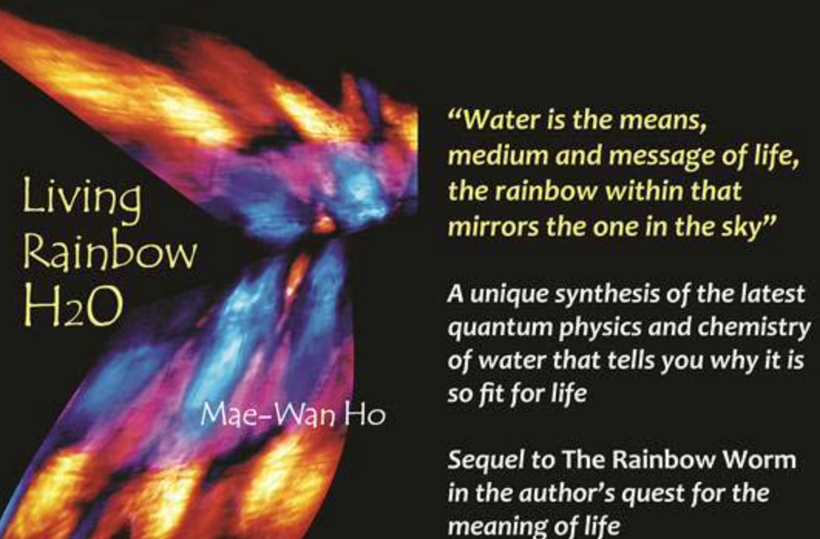
Last and definitely not least, water provides the electrons and protons to fuel the photosynthesis-respiration dynamo that spins inanimate molecules into living organisms out of pure sunlight. Water is truly the medium, message and means of life.
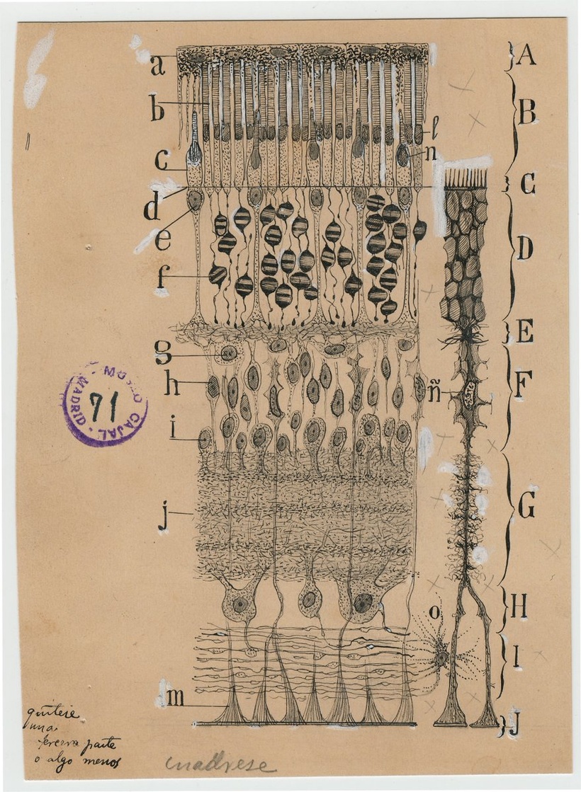
Consider our vision. Many of the photoreceptors in our eye have microtubules in them as you saw in the video above and the picture above. What do we know about how vision operates today? Even today there is the great mystery of how human vision operates: Vivid pictures of the world appear before our mind’s eye, yet the brain’s visual system receives very little information from the world itself to see. Much of what we “see” we conjure in our heads. A lot of the things you think you see you’re actually making up in a quantum reconstruction in your brain. How does it happen?
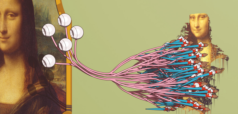
Microtubules seem to be key to these abilities.
HOW DOES THE HUMAN IMMUNE SYSTEM GATHER INFORMATION TO DO WHAT THEY CAN?
T cells activate to become killer cells and then attack infected cells and cancer with remarkable ferocity. This seems to happen magically. When we use the term magical, you can bet a quantum mechanical process is operational behind the scenes. This includes an ability to dramatically change its metabolism in many ways to alter its shape, speed of reproduction and ability to survive in any environment, even without oxygen.
How does a cell capture information from the environment to alter its physiological behavior?
Global cooling seems to be associated with increases in geomagnetic field intensity whereas global warming is associated with decreased geomagnetic field intensity. It appears magnetic fields changes in our environments determine how CO2 is handled in the atmosphere and seas. This information appears to be captured in water networks in the body and transmitted to our cells via microtubule formation.
Since mitochondria make CO2 from oxidative metabolism, what might the effect be on mitochondrial function during an infection in the body?
In the human body, carbon dioxide is formed from the metabolism of carbohydrates, fats, and amino acids, in a process known as cellular respiration which occurs in mitochondria. While cellular respiration is notable for being a source of ATP, it also generates the waste product, CO2. The body gets rid of excess CO2 by breathing it out via the lungs and trachea which have abundant cilia and microtubules. However, CO2 in its normal range from 38 to 42 mm Hg plays various roles in the human body. We have chemoreceptors that pay attention to CO2 levels. They regulate the pH of blood, stimulates breathing, and influences the affinity hemoglobin has for oxygen (O2). Fluctuations in CO2 levels are highly regulated and can cause disturbances in the human body if normal levels are not maintained.
CO2 retention is known as hypercapnia. Hypercapnia is usually due to hypoventilation or increased dead space in which the alveoli are ventilated but not perfused. The microtubules in those poorly perfused alveoli collect information from the cytoarchecture and deliver the information in quantum mechanical fashion via water networks linked to the tensgrity system in a cell’s microarchitecture.
In a state of hypercapnia or hypoventilation, there is an accumulation of CO2 in the blood. The increased CO2 causes a drop in pH, leading to a state of respiratory acidosis. The chemoreceptor reflex is important in allowing the body to respond to changes in pO2, pCO2, and pH. Chemoreceptors can be categorized as peripheral or central. This links directly to how CSF flows around the vascular in the CNS. Peripheral chemoreceptors are positioned in the carotid and aortic bodies and in the brain’s blood vessels. Central chemoreceptors are located near the ventrolateral surfaces of the medulla. While peripheral chemoreceptors are sensitive to changes in mostly O2 and CO2 and pH to a lesser degree, central chemoreceptors are sensitive to changes in pCO2 and pH.
The glomus cells in the carotid and aortic bodies detect states of hypoxia, hypercapnia, and acidosis. On the other hand, central chemoreceptors do not detect states of hypoxia. They detect a change in PCO2 very rapidly because CO2 diffuses through the blood-brain barrier (BBB) and into the CSF easily where a massive array of microtubules are present.
On the other hand, central chemoreceptors take longer to detect a change in arterial pH because H+ does not cross the BBB. When a state of hypercapnia is introduced, central chemoreceptor activity is increased. As a result, the sympathetic outflow to the vasculature is increased via the brainstem nuclei, and efforts are made to increase the respiratory rate.
DOES THE SUN/EARTH INFORM YOUR BLOOD AND IMMUNE SYSTEM HOW TO ACT?
The magnetic-field effect on CO2 solubility in humans is twice as large, therefore a geomagnetic field variation on Earth can modulate the carbon exchange between atmosphere and ocean. Your microtubules can and do sense that change. A 1% reduction in magnetic dipole moment may release up to ten times more CO2 from the surface ocean than is emitted by subaerial volcanism. This implies it does it in your body as well. How might this affect mitochondrial function in your immune cells given what biology has found out about CO2 physiology?
Most do not know that T cells are inherently magnetic because of the amount of ferritin (iron) they contain. This is why theses cells become “magnetically frozen” to pathogens EMF signals that they react too. This is done via molecular resonance. This action magnetically locks them into their target pathogen and the fever reaction in the hyporthalamus results and this leads to better T cell killing of the invader pathogen. UV solar exposure unlocks the T cells magnetic grip and allows the cells to limit the cytotoxic damage to the hosts organs.
Note, most viral deaths have been in immunocompromised humans which were tied to uncontrollable cytotoxic storms causing organ destruction. In other words, their own immune cells caused an unrelenting hurricane of damage to organs. Also recall that most deaths in the last two years are linked to humans with significantly lowered Vitamin D levels. Note, vitamin D in cells is only made from UV light exposure of the skin. The skin also has a massive microtubule array with deep connections to the tensgrity system of the organism. Microtubules seem to deliver the information in our environment to our skin to inform our immune cells how to operate throughout the body.
Can T cells work well on a planet with competing magnetic field signals? It appears they cannot operate properly.
DO IMMUNE CELLS USE MICROTUBLE SYNAPSES TO TRANSFER DATA ABOUT CO2 in WATER?
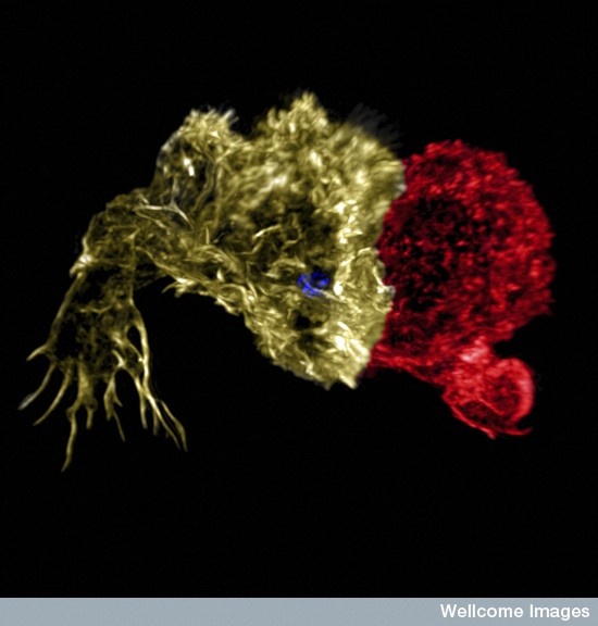
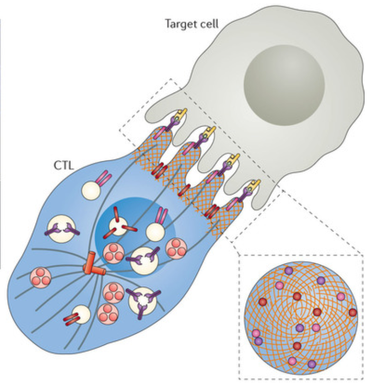
A major T cell tactic is direct contact with an offending cell by forming a synapse between the two cells. This immune synapse is sometimes called a cytolytic synapse, meaning killing of a cell. Mechanisms at this synapse are very complex and involve rapid elaborate remodeling of the scaffolding inside the cell. Most of these involve microtubules which have been implicated to be one of the mechanisms Nature uses to inject quantum mechanically operations to cells.
Microtubule proteins are able to produce electromagnetic fields and have been shown to play an important role in memory formation, and learning in various cell lines. Therefore, microtubules have the potential to be affected by exogenous electromagnetic fields.
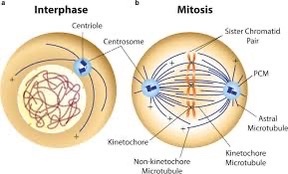
Despite very brief contact times, microtubules and actin in both cells undergo many different alterations. Structures are built with scaffolding molecules to rapidly form a temporary connection and then a very secure connection that allows multiple rapid actions. In fact, unlike other types of synapses, centrosomes are brought right up to the membrane to direct the action. Centrosomes are vital structures that coordinate all microtubule structures as well as the spindles in cell division. They organize microtubules in the cell and I believe this is done topographically via electric and magnetic fields that map out the body plan of the organism. Robert O. Becker was the first researcher to link electric and magnetic field polarization to body plan construction during his wound healing experiments.
Centrosomes are composed of two centrioles arranged in an orthogonal configuration, is an indispensable cellular organelle for mitosis. 130 years after its discovery, the structural-functional relationship of centrosome is still obscure. We have clues in the literature that the centrosome might act as an electromagnetic generator in cells to direct energy and information transfer. (below)
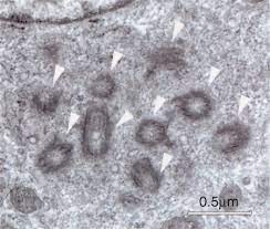
Centrosomes are known to coordinate complex activity such as cell division, where they drag two sets of chromosomes in different directions to form two daughter cells. Centrosomes are also used to build and coordinate the microtubule highways that provide very elaborate transport along the long neuronal axon in humans. You can see this in the cochlea and in the rod photoreceptors in the eye. The only other time that a centrosome is directly at a cell’s surface is when cilia are formed. Cilia provide both movement and the extremely elaborate signaling machine called the primary cilium. Cilia line many organs where fluid must travel, such as the secretions in the lung and the cerebrospinal fluid in the brain.
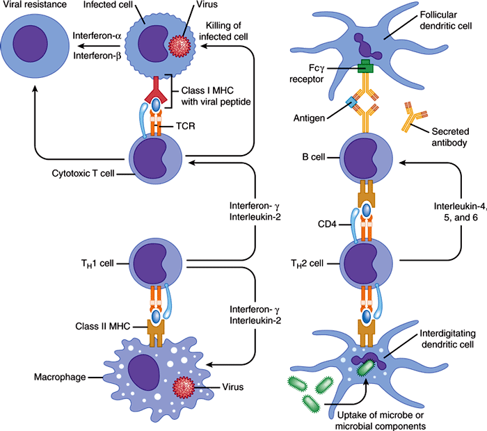
Killer T cells come from CD8+ T cells, which are originally small and quiet cells that travel about looking for problems. When they see a problem molecule in the groove on the surface of the presenting cell, they stop at the cell. This molecule from inside the cell is placed in the groove so the T cell will know if the cell is normal or infected. It is called a MHC or major histocompatibility antigen. If it is abnormal or infected it the molecule integrin is stimulated to stick out from the T cells to grab onto the other cell. Activation produces a dramatic change in the status of the T cell that produces an army of killer cells over several days with many granules filled with molecules that are weapons. Some of these granule toxins cut holes in cells (perforin) and some (granzymes) cut proteins in cells leading to cell death. Killer cells become much bigger and develop a very complex internal architecture with scaffolding molecules.
Upon electromagnetic induction activation, a large activation cluster is formed right at the synapse, called central cSMAC (or supra molecular activation cluster). This cluster includes many special receptor molecules and a complex large machine called the microtubule-organizing center (or MTOC). MTOC is a type of centrosome where the minus ends of microtubules are anchored and from which the plus ends of microtubules extend.
Microtubules appear to be self-assembled linear hollow circular tubes with inner and outer diameters of 17 and 25 nm, respectively, growing from the centrosome in the center of the cell towards its membrane. I believe the cSMAC and MTOC are electric generators that actually cause microtubule assembly via the electromagnetic force.
Cells can contain multiple MTOCs wherever microtubules are being used. Another cluster is formed called distal dSMAC, which is away from the synapse at the back of the cell. These two clusters work together in the processes that occur. This appears to be how immune cells are transfering information in the environment to cells. I believe they are using electromagnetic spectrum to convey this message.
Why do I believe this. I covered this in my Quantum Brain blog years ago. The work of Frolich is critical in this prediction.
The cellular electromagnetic field should propagate to all parts of the biological system. the centrosome is key in this action. It has to be coherent (the same frequencies, form, and phase) within the specific region: in the cell, in the tissue, and in the whole biological system. Coherence in time is also an important condition for this to happen. Proper circadian timing makes this possible. If time keeping is innaccurate quantum coherent mechanisms become impossible.
Oscillations of different generating ‘antennae’ must be controlled by signals delayed by one or more periods of oscillations. The cellular electromagnetic field should be generated by energy-supplied structures which are distributed inside the cell with a convenient density, are of linear geometry resembling macroscopic antenna systems. Moreover, they should be electrically polar to react to electric fields. The generating system must be strongly nonlinear for spectral energy transfer. Why? Fröhlich predicted this would be a REQUIREMENT FOR the living state. Based on these assumptions, microtubules have been predicted as generators of coherent vibrations and electromagnetic field. Most recently this was done by Pokorný et al.
The synapse first forms in the CD8 T cell and then is altered as the cell becomes a cytotoxic killer cell. At first it helps with the changeover to killer cells. In the killer cell, it is prepared to send the attack granules within minutes.
Some have speculated that microtubules form a quantum computer that directs cellular activity in the immune system. While there is no proof of this, it is interesting that many extremely important and vastly complex activities that use microtubule machines, centrosomes and spindles seem to be heavily involved in information processing in cells. This includes transport along the axon, spindles for cell division, primary cilium and moving cilia. There is no doubt that microtubules appear to operate as an intelligent LEGO like system vital to the life of many cells.
SUMMARY
Biological activity is conditioned by a continuous energy/information supply and transformation, system ordering at various length scales, controlled processes including chemical reactions, and a massive information transfer. Microtubules seems to be at this nexus. All these abilities life contains cannot be facilitated purely by diffusion or chemical interactions acting at the length scale of several nm as we are taught in a biochemistry book. A fast, efficient physical mechanism acting at large length scales should be involved. This is the scale of the electromagnetic force.
Biological systems SHOULD be described as open dissipative structures with a dynamic stability sustained by exchange of matter, energy, and information based on a special organized structure–solitons and generation of electromagnetic field. Microtubules are a cog in this nanomachine. The organization of atoms in them is the key to really understanding how life operates electromagnetically.
CITES
https://www.researchgate.net/publication/272648139_Illuminating_Water_and_Life
https://www.ncbi.nlm.nih.gov/pmc/articles/PMC8348406/
https://pubmed.ncbi.nlm.nih.gov/32763375/
https://pubmed.ncbi.nlm.nih.gov/11567874/
https://tbiomed.biomedcentral.com/articles/10.1186/1742-4682-9-26
https://bmcmolcellbiol.biomedcentral.com/articles/10.1186/s12860-019-0224-1
https://www.mdpi.com/1422-0067/22/15/8215
Pokorný J., Jelínek F., Trkal V., Lamprecht I., Hölzel R. Vibrations in microtubules. J. Biol. Phys. 1997;23:171–179. doi: 10.1023/A:1005092601078.

