This autoimmune disease is linked to a POOR connection the sun and Earth. This is tied to your choices and lifestyle. It may be an unpleasant message for some to hear but before you can get well you must know the things you control and can change to get a reversal. Read this hyperlink if you think it is not true.

^^^^this is wise if you have MG.

^^^^do not be this type of primate if you have MG or want to avoid it in a 5G world.
OCULAR MYASTHENIA:
Over two-thirds of all patients with myasthenia gravis (MG) begin with symptoms relating to their vision. Overall, the ratio of affected females to males in generalized MG is 3:2 or higher. In ocular myasthenia, men are more frequently affected, especially after the age of 40. In addition, the average age of onset for generalized myasthenia is 33 years, while that of ocular MG is 38 years. The ocular motor system may be especially vulnerable to MG since it cannot adapt rapidly to variable weakness. The most common symptoms seen in patients with ocular MG are diplopia (double vision), ptosis (droopy eyelids), and incomplete eye closure. Compared to other involved skeletal muscles, only slight weakness of the extraocular muscle may cause diplopia and visual disturbances to occur. These symptoms occur due to the weakness of the muscle that controls eyeball and eyelid movement. Light sensitivity due to sluggish pupils may occur in some patients. Symptoms are frequently influenced by environmental, emotional, and physical factors. Some of these factors include bright sunlight, extreme temperature, emotional stress, illness, surgery, menstruation, and pregnancy, among others. Symptoms tend to be worse at the end of the day because of mitochondrial fatigue.
Symptoms
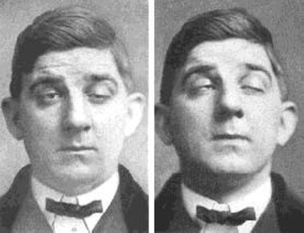
Diplopia or double vision results when the eyes cannot be focused as desired due to the weakness of one or more of the extraocular muscles which control eye movement. This most often occurs when looking up or to the side. To compensate for the weakness, the patient may tilt his/her head or turn their face to allow the stronger eye to work. For example, if the muscle which allows the eye to look upward is weak, the patient could tilt their head back to look up. Ptosis (to-sis) is the drooping of one or both eyelids, also due to muscle weakness. Fluttering or twitching of the eyelid may occasionally be seen. If both eyelids are droopy, one may be worse than the other.
Nystagmus or constant involuntary repeated movement of the eyeball in any direction may also occur in one or both eyes.
Diagnosis
The edrophonium (Tensilon) test is the first-line test for diagnosis of MG. The Tensilon test consists of injecting a small amount of medication edrophonium intravenously. If the patient has MG the ocular muscle weakness, the ptosis, and general muscle weakness and/or nystagmus will improve dramatically for a short period of time. In recent years an ice test is being more widely used. This is when ice is applied to the eyes, after a short period of time; the eyes will have an improvement of ocular symptoms. Usually, a blood test called acetylcholine receptor antibody titer (AChR Ab) is ordered as well. Additional blood work may include other antibody studies, thyroid profile, and a sedimentation rate.
Tests
A test to check for fatigue and weakness of the eye muscle which may be done by the examining doctor includes attempting to open the eyes while the patient tries to hold them shut, sometimes called a “peek sign”. This may result in one or both eyes opened, and the patient appears to “peek” at the examiner.
The “sleep test”, which is based on the tendency for MG symptoms to improve following rest, may be used in small children and patients who have allergies or sensitivity to anticholinesterase drugs such as Tensilon. The patient is placed in a quiet, darkened room and instructed to close their eyes for 30 minutes. The patient is photographed and eye movement is measured before and after the rest. The test is considered positive if there is an improvement in the ptosis and/or eye movement (motility) following the 30 minute rest period.
The morning/evening comparison test is similar in concept to the sleep test. The patient is photographed, and the ptosis and ocular motility are compared at different times during the day. Old photos are very helpful to determine how long the patient has had drooping of the upper eyelid.
The “Ice Test” is a simple test for ocular MG in patients who have ptosis. A surgical glove filled with ice is held against the droopy eyelid for several minutes. In ocular MG the patient can open his/her eye normally for a short period after the ice is removed. This shows the clinician this disease is related to inflammation/deuterium fraction (low pH of cell water), poor semiconductive currents in the electron tunneling along the inner mitochondrial membrane.
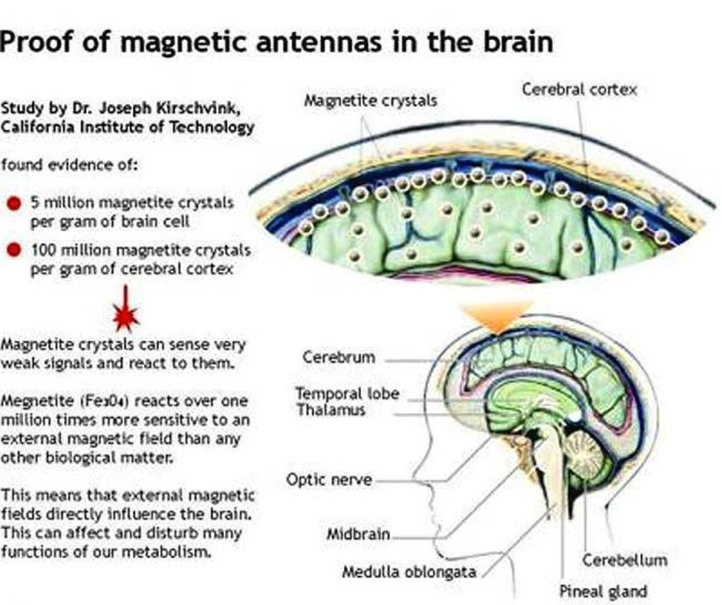
Electromagnetic radiation in the environment is capable of driving the disease.
”A significant change in pH of water (0.5-1.5 unit) was induced by a 30-irradiation at a frequency of 49, 50.3, 51.8, or 53 GHz, when the initial pH value was 6.0 or 8.0, but not 7.5. These results indicate the changes in the properties of water and its role in the effects of EMI of extremely high frequency. ”
The dielectric properties of water make it a sponge for electromagnetic radiation; particularly in microwave frequency range because of matching resonance. This link to nnEMF is rarely made in MG cases by clinicians. Anytime someone gets a new diagnosis I always ask them how much he or she use his or her cell phone for work and pleasure and if they hold it up to the side of their eyes. I also ask if they live close to an airport, TV or radio stations, or a Doppler radar installation. I also ask about military bases or HAARP installation in the area. It is well known that certain portions of the spectrum of light outside of the visible spectrum of light cause acidification of water. This is true in the atmosphere, oceans, and in our cells. We seem to be oblivious to these links.
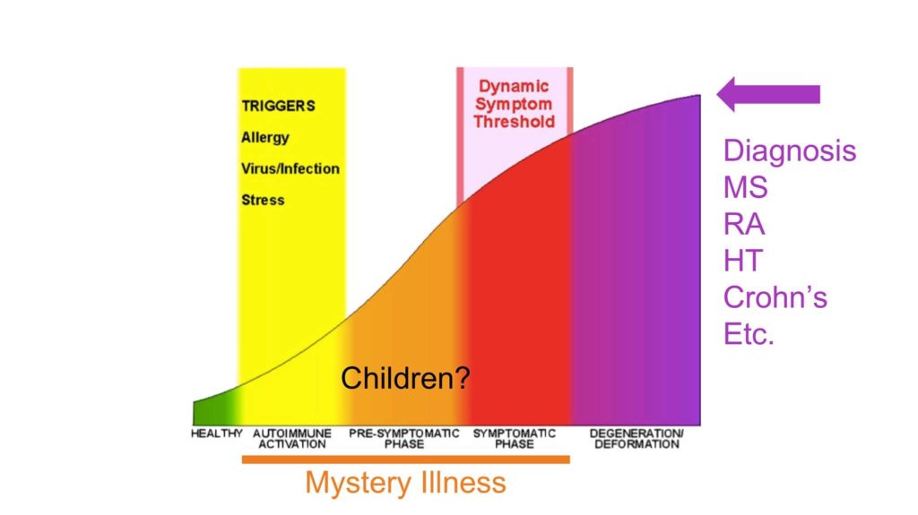
“The absorption of electromagnetic radiation by water depends on the state of the water. The absorption in the gas phase occurs in three regions of the spectrum. Rotational transitions are responsible for absorption in the microwave and far-infrared, vibrational transitions in the mid-infrared and near-infrared. Vibrational bands have rotational fine structure. Electronic transitions occur in the ultraviolet regions. Liquid water has no rotational spectrum but does absorb deeply in the microwave region. Many mystery illnesses are due to the changing proton concentration in cells. We need to realize often this begins way before we meet the diagnostic criteria for most of these immune activated diseases. The reason it is missed is that allopathic and functional medicine are poor at understanding how the dielectric constant of water changes as the environment varies.
MORE SYMPTOMS
”Another simple test for ptosis is the “fatigue” test. This consists of having the patient look at an object held up by the examiner in front of the patient. After a short period of time, the eyelid(s) will droop in the person with ocular MG.
An MRI or CT scan of the head is often done to rule out other possible causes of the patient’s symptoms. Additional testing may also be needed to confirm the diagnosis. Myasthenia Gravis 101: The adenine nucleotide transporter 1 (Ant1) gene encodes an inner mitochondrial membrane protein that transports ADP into mitochondria and ATP from mitochondria to the cytosol. Mutations within Ant 1 have been shown to produce a syndrome of chronic progressive external ophthalmoplegia (CPEO) in humans. Myesthenia, however, appears more related to ANT 2 defects. extraocular muscles have much higher levels of ANT 2 mRNA compared to the limb muscles. ANT 2 is a nonskeletal muscle isoform previously described in the human heart. Its presence in the extraocular muscles may explain the lack of effects of ANT 1 loss, and it was the first documented difference between extraocular muscle and limb muscle mitochondria in the literature. Many textbooks blame MG on the release of acetylcholine into the synaptic cleft of skeletal muscles but they seem to forget that this process is electrically controlled by charged subatomic particles that control the release of calcium in mitochondrion that is the foundation of neurotransmitter release. Things like deuterium, fluoride, and bromine inside of cells doing things to mitochondria ruin the protein and light communication inside of cells because it disrupts the dielectric constant of cell water. Things inside our mitochondria have to move fast and these things slow reactions down because there is not enough energy in the mitochondria of muscle afflicted by MG. Below is an example how deuterium is tightly controlled by the location of substrates in the matrix.
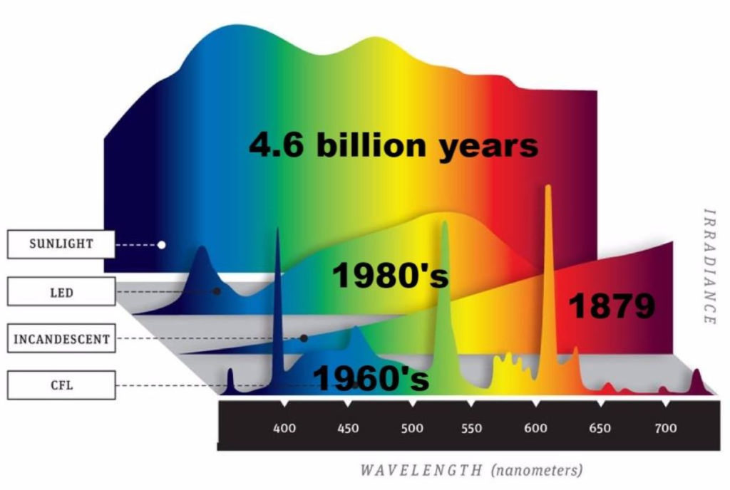
Sunlight connects with mitochondria wirelessly via the blood. People with MG need to understand you can develop symptoms of this disease with a lack of sunlight or a problem inside your mitochondria because the first steps in porphyrin synthesis occur inside mitochondria. Myasthenia gravis with thymoma has been associated with pure red blood cell aplasia. This is a complete knockout, but I have a sense that porphyrin synthesis is very slowed in MG patients. This is why many people with MG have other connective tissue disorders linked to and cannot tolerate some drugs. It is also why many people are photosensitive to light and they stay out of the sun. I believe this sense is linked to something in the skin that increases thermal sensitivity tied to dopamine creation in the muscle and skin. That link is to defects in tyrosine kinase receptors in the skin that affects dopamine synthesis because of an excessive antibody load in the blood plasma. The key defect in MG is the antibody production by B cells, and its collection in the blood, but these antibodies cause collateral damage few clinicians seem to be linking together even today.
The porphyrias are genetic disorders of heme metabolism, caused by a defect in an enzyme responsible for the synthesis of the heme molecule, which in turn is necessary for the production of hemoglobin, myoglobin, and cytochromes in the matrix. MG gravis can have a version of this disease but it is cause post translationally by things like deuterium/halogens in the mitochondrial matrix that ruin water flows and heme synthesis. Heme is an oxygen carrier, and is essential for aerobic respiration and adenosine triphosphate (ATP) production via the electron transport chain. Cytochrome production is necessary for the metabolism of multiple drugs within the body, most notably through the cytochrome P450 system, and also mediates the removal of some toxic substances which is why some with MG cannot tolerate some drugs. Muscles have heme requiring proteins in myoglobin, hemoglobin and the cytochromes in their mitochondria. The heme biosynthetic pathway starts in the mitochondria, with the production of d-aminolevulinic acid from glycine and succinyl-CoA, which is catalyzed by the pyridoxine-dependent enzyme d-aminolevulinic acid synthase (ALAS). This is the rate-limiting step in heme production and is the site of feedback inhibition, with inhibition of ALAS by heme, the end product of this pathway.
So if these steps of heme synthesis are slowed or sticky inside of mitochondria FOR ANY reason, your red blood cells will not be able to turn over as fast, your P450 system used for detox, and your myoglobin in muscles can be affected. Hemoglobin is a heme porphyrin and it is a light ferry that brings oxygen and light to mitochondria. If RBC’s is defective because of poor heme synthesis they should and may not carry as much oxygen and light from your skin to the muscles with MG. There is no one cause of this autoimmune condition. Many physicians overlook the connection of MG to RBCs diseases. When people do poorly in sunlight they should be tested for porphyrin issues because this links it to poor mitochondrial function in these MG patients. I believe it is a lot more common than people realize because there is a STRONG link of MG to low vitamin D levels and poor oxygen delivery to muscles. Vitamin D levels control how immune cells can function and this can affect the number of antibodies made to the Acetylcholine receptor and tyrosine kinase receptors in muscles. Sunlight is normally a tyrosine kinase inhibitor.

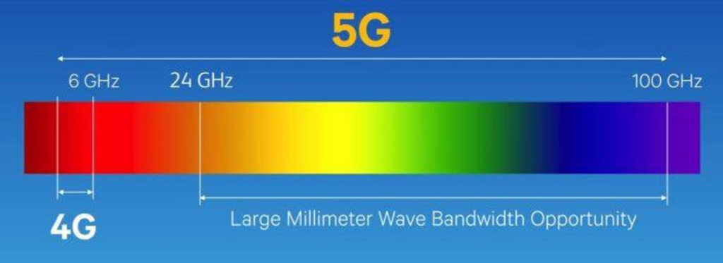
This implies that MG can act as another severe allergy to nnEMF that impairs the release of acetylcholine in skeletal muscle. I fully expect MG and CPEO to increase DRAMATICALLY in incidence and prevalence in a 5G world which will use RF/microwaves to generate massive electric fields on the surfaces of things in the ionosphere and on the Earth’s surface. People live in this environment today and it will increase as technologic “progress” is fueled by the economy. Clinicians need to be ready for these expectations.
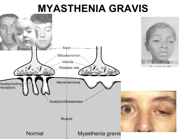
The ability of muscles to perform aerobic work depends on their mitochondrial volume density, with the assumption that the composition of these organelles is fairly constant across muscle types and mammalian species. One of these components is the electron transport chain, a series of multimeric complexes (complexes I–IV, plus the ATP synthase which is sometimes called complex V) in the inner mitochondrial membrane responsible for most of the aerobic ATP generation. Recently, it was found that the extraocular muscle mitochondria have lower content or lower activity of some enzyme complexes of the electron transport system, causing them to respire at slower rates. This means that the EOM have a lower basal metabolic rate than most other tissues in the body. This is puzzling given that the extraocular muscles are constantly active and aerobic capacity was predicted to be elevated, given their high mitochondrial content. This is an important teaching point. Longevity and health are tied to lower basal metabolic rates. Deuterium in the matrix or cytosol is capable of altering the metabolic rate of tissues.
These findings are not explained by differences in the ultrastructure of extraocular muscle mitochondria: the surface area of their inner membrane is comparable to values reported for other skeletal muscle. Furthermore, the differences are not generalized or systematic: complex II content and activity, and complex III content are similar in mitochondria from triceps surae (a limb skeletal muscle) and extraocular muscle. Complexes I and IV give a more puzzling result: their activities are lower, but their content is higher in the extraocular muscle mitochondria. These are multimeric protein complexes, and differential expression of isoforms of some subunits has been described in skeletal muscle and other tissues. Therefore, the content of some electron transport chain complexes (I, IV, and V) and the subunit composition of some others (I and IV) may not be the same in the extraocular muscles compared to limb muscles and may explain why these muscles are damaged preferentially in autoimmune diseases.
This demonstrates that the metabolic divergence between extraocular and limb muscles includes major differences in the composition and basic function of their respective mitochondrial populations. Intrinsic differences in mitochondrial structure and function may explain the susceptibility of the extraocular muscles to some hereditary and acquired mitochondrial myopathies such as MG and CPEO and related syndromes. For example, the extraocular muscles present the most severe age-dependent loss of mitochondrial respiratory complex activity among muscles. There is a significant increase in the number of fibers with cytochrome c oxidase defects in the extraocular muscles of humans and other primates, even when compared to other highly aerobic muscles such as the diaphragm and heart. This can be at least partially explained by mitochondrial DNA mutations, presumably due to reactive oxygen species generated during elevated mitochondrial respiration or present as part of a more generalized cellular oxidative stress.
Treatment
Reconnect with Nature!!!
Both ocular and generalized myasthenia gravis patients tend to have remissions and exacerbations at regular intervals, so that the medications that are used for treatment may need to be changed accordingly. Medications may include cholinesterase inhibitors such as Mestinon, steroids such as Prednisone, or other immunosuppressants used alone or in combination.In addition to medications, other options which may be used are plasmapheresis or IVIG therapy. These treatments offer only a temporary improvement and repeated treatments are necessary to sustain the effect. While thymectomy (removal of the thymus gland) is often recommended for patients with generalized MG, it is rarely used in purely ocular MG unless a thymoma is suspected.
In those people whose ocular myasthenia is not adequately controlled by medicine, several aids are available. In many patients, simply wearing a patch over one eye will eliminate the double vision. The patient may alternate the eye patch from one eye to the other to avoid eye strain. Another method is wearing one contact lens which is opaque (can’t be seen through). This may also be changed periodically from one eye to the other. Ask the ophthalmologist how often this should be done. Special prism glasses may also help to correct double vision in some cases.
When both eyelids droop, taping one eyelid open may improve vision. The use of non-allergenic tape such as paper tape is suggested. Once again, alternating eyelids is recommended to prevent eye strain. Another way to hold one or both eyelids open is to have ptosis bars or eyelid crutches attached to the eyeglasses. These are thin, flexible wires which attach at the bridge of the nose and are free-floating at the other end or attached to the frame near the hinges. The wires rest against the eye socket and hold the eyelid open like a brace or crutch. These devices must be carefully fitted and adjusted by an ophthalmologist. Most patients report that the eyes adjust to the bars after a period of time. And they are much more convenient than taping the lids open. Since the eyes can’t blink normally, dryness and irritation may result. It is important to remove the glasses frequently to allow the eyes to close so that they can be moistened. Some MG patients have advised that every ten minutes or so is a good guideline. Artificial tear drops and forced blinking may also help. If the patient has double vision in addition to drooping eyelids, one eye should be allowed to close and the other be held up by the ptosis bar.
Remember this wisdom does not grow in every clinicians garden. You need to think differently about this disease if you have it and want to avoid it. Mitochondriacs do not settle for half truths on any disease.

CITES:
http://www.ncbi.nlm.nih.gov/pubmed/17969925
https://en.wikipedia.org/wiki/Electromagnetic_absorption_by_water
https://www.researchgate.net/post/Relationship_between_pH_and_conductivity2

